Worldwide, over two million people are affected by spinal cord injury, which is a heterogeneous condition, depending on the cause and the severity of the injury. Injury can be caused by contusion, compression and laceration, of which contusion as a result of fracture dislocation of the spine is the most common (Ref. Reference Oudega1). Trauma to the spinal cord leads to primary loss of cells at the site of injury. This initiates various inflammatory responses, which together with other cytotoxic events, leads to secondary damage and formation of fluid-filled cysts, demyelination and degeneration of axons at the site of injury (Ref. Reference Kwon, Borisoff and Tetzlaff2). Repair of spinal cord injuries requires regeneration of damaged axons or stimulation of the growth of spared axons at the site of injury to allow possibility of any functional recovery. The failure of cut axons in the spinal cord to regenerate or sprout has many causes. These can be categorised into extrinsic inhibitory effects and the intrinsically low ability of central nervous system (CNS) axons to regenerate.
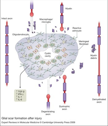
Formation of a glial scar and the presence of various inhibitory molecules at the site of injury lead to the extrinsic inhibition of axon growth, which prevents any substantial regrowth and sprouting of the axons following injury. Reactive gliosis is an injury-induced change in the behaviour of cells at the site of injury, primarily by astrocytes and meningeal cells, which ultimately culminates in the formation of glial scar tissue at the site of injury (Ref. Reference Silver and Miller3). Reactive gliosis is associated with proliferation and hypertrophy of astrocytes and oligodendrocyte precursors, with invasion of fibroblastic meningeal cells following the disruption of the blood–brain barrier (BBB) (Refs Reference Silver and Miller3, Reference Fawcett and Asher4). Reactive gliosis and glial scar formation are both important in the early stages of injury. They limit further damage by suppressing inflammation and preserve function by establishing a boundary between damaged tissue and salvageable tissue (Refs Reference Okada5, Reference Nakamura6, Reference Faulkner7). Depletion of reactive astrocytosis after injury prevents re-establishment of the BBB and is associated with increased leukocyte infiltration, severe demyelination and increased cell death (Ref. Reference Faulkner7). Experiments have shown that interaction of invading meningeal cells with reactive astrocytes is important in the formation of a new glia limitans and resealing of the BBB (Refs Reference Faulkner7, Reference Bundesen8, Reference Abnet, Fawcett and Dunnett9). However, although glial scar formation is very important for tissue preservation, it acts as a physical (Ref. Reference Windle, Clemente and Chambers10) and chemical barrier to axon regeneration (Refs Reference Kwok11, Reference Niclou, Ehlert and Verhaagen12). Within the glial scar, there is upregulation of various inhibitory factors capable of blocking axonal growth (Ref. Reference Fawcett and Asher4). There is no single trigger to the formation of glial scars. Instead, there are many cytokines and chemokines involved, including TGF-β (TGFB) (Ref. Reference Asher13), interferon-γ (IFNG) (Ref. Reference Yong14), interleukin (IL)-1 (Ref. Reference Giulian15) and IL-6 (IL6) (Refs Reference Okada5, Reference Nakamura6), which are released at the site of injury as a result of inflammation and immune cell infiltration. This then leads to astrocytic hypertrophy, proliferation and upregulation of inhibitory factors, such as chondroitin sulphate proteoglycans (Ref. Reference Silver and Miller3). The other major sources of inhibitory molecules for axon regeneration in the damaged spinal cord are myelin and myelin debris, which also have an important role in preventing axonal regeneration and growth (Refs Reference Grados-Munro and Fournier16, Reference McGee and Strittmatter17).
For axons to regenerate successfully, they must be able to initiate the process of regrowth followed by a dynamic process of continuation to reach the necessary target for synapse formation. However, axons vary greatly in their intrinsic regenerative ability, with most CNS axons being poor regenerators. Soon after injury, an axon has to undergo a series of cell-autonomous changes at the injury site. The first step is the sealing of the plasma membrane at the cut end, followed by disintegration of the cytoskeletal elements, retraction of axon from the injury site, formation of a growth cone, regrowth to the target area, cessation of growth and synaptogenesis. The ability of axons to complete this process varies. It is dependent on the neuronal type (peripheral neurons regenerate better than central neurons), age (embryonic and neonatal neurons regenerate better than adult neurons) and distance of site of injury from the cell body (axonal injuries closer to the cell body regenerate better). In addition, there are also changes in the pattern of gene expression, which modulate the responses to axonal injury.
To achieve efficient axonal regeneration and functional recovery, a combined approach will be essential, in which the external environment of the axon is made more permissive by (1) promoting the factors supporting regeneration and (2) blocking the external elements inhibiting regeneration. In addition, the intrinsic ability of axonal outgrowth must be manipulated to enhance the vigour of the regenerative response.
Extrinsic factors
Modification of the extrinsic environment for promoting axonal regeneration has progressed significantly in the past two decades. Some of these interventions work by removing the inhibitory elements within the CNS and glial scar, and others by promoting and creating a more permissive environment to support regeneration and sprouting of the axons.
Myelin inhibitors
Myelin is a major inhibitory component of the CNS and many studies have examined the mechanism of inhibition of axons by myelin inhibitors. One myelin-associated molecule, Nogo-A (RTN4) was the first inhibitor to be isolated (Refs Reference Caroni and Schwab18, Reference Caroni and Schwab19). Application of the Nogo-A-blocking monoclonal antibody IN-1 in vitro (Ref. Reference Caroni and Schwab18) and in vivo inhibits the effect of the Nogo-A isoform and leads to substantial axonal sprouting in the injured adult CNS (Ref. Reference Schnell and Schwab20). Other monoclonal antibodies, mostly targeting the extracellular N-terminus of the molecule, also block the inhibitory effects of Nogo-A on axons, and promote axon regeneration in the CNS. Anti-Nogo antibodies have also been shown to promote the sprouting of corticospinal axons in marmosets (Ref. Reference Fouad, Klusman and Schwab21) and macaque monkey (Ref. Reference Freund22) after spinal cord injury. A humanised version of the anti-N-terminus Nogo monoclonal antibody is currently undergoing clinical trials in acute spinal cord injury (Ref. Reference Blight and Tuszynski23).
Inhibition of Nogo has been attributed to two separate domains on the molecule. One is a 66-residue loop (Nogo-66) located between two putative transmembrane portions of Nogo (Ref. Reference Chen24) and the other is at the N-terminus (Ref. Reference Hu and Strittmatter25). Nogo-66 interacts with a glycosyl phosphatidylinosital (GPI)-linked receptor – the Nogo receptor (NgR or RTN4R) – that is expressed in various neurons (Ref. Reference Fournier, GrandPre and Strittmatter26). Introduction of NgR into cells that normally do not respond to this myelin inhibitor confers sensitivity to Nogo. Both p75 (NGFR) (Ref. Reference Wang27) and Lingo-1 (LINGO1) (Ref. Reference Mosyak28) have been identified as coreceptors for NgR. A truncated peptide comprising residues 1-40 of Nogo (NEP1-40) has been shown to act as a competitive antagonist for the binding of Nogo-66 to NgR. Application of NEP1-40 in spinal cord injury models in rats has led to increased corticospinal sprouting with significant functional recovery (Ref. Reference GrandPre, Li and Strittmatter29).
It is important to remember that Nogo is, however, only one of many inhibitors present within the CNS. Knockout mice produced to study the effect of Nogo after spinal cord injury have provided variable and conflicting results on regeneration of corticospinal tracts (Refs Reference Zheng30, Reference Simonen31). There are three Nogo isoforms, and some authors have proposed that the lack of regeneration in studies targeting only one or two of these can be explained by a compensatory increase in the remaining isoforms, and some studies have been confounded by axon-labelling artefacts (Ref. Reference Kim32). A recent report reassessed the effect of Nogo by removing most of the previous confounding factors, using a reliable tracing dye and deleting all Nogo isoforms, in addition to reanalysing the mouse line previously reported to promote regeneration (Ref. Reference Lee33). The authors conclude that deletion of all Nogo isoforms and other confounding factors did not lead to any corticospinal tract regeneration. Therefore, the removal or neutralisation of this molecule alone is not sufficient to significantly promote axon regeneration (Ref. Reference Lee33), and might not provide any measurable therapeutic effect.
Other myelin-associated inhibitory molecules include myelin-associated glycoprotein (MAG) and oligodendrocyte myelin glycoprotein (OMG). MAG is a transmembrane protein belonging to the immunoglobulin superfamily and is present in both the CNS and peripheral nervous system (PNS) (Ref. Reference Martini34). MAG inhibits axon growth from adult dorsal root ganglion (DRG) and cerebellar granule cells (Refs Reference Mukhopadhyay35, Reference McKerracher36), through interaction with the NgR and gangliosides (Refs Reference Schnaar and Lopez37, Reference Mehta38, Reference Domeniconi39). OMG is a GPI-linked protein that is abundant in the CNS. It causes growth-cone collapse in many neuronal populations (Refs Reference Mikol and Stefansson40, Reference Kottis41). Remarkably, both of these molecules have also been shown to act through the NgR receptor. A polyclonal antibody to NgR, a soluble version of NgR and a dominant-negative NgR mutant are all capable of blocking inhibition of neurite outgrowth by MAG (Ref. Reference Domeniconi39). A separate study identified NgR as an OMG receptor and showed that expression of NgR in DRG neurons confers sensitivity to OMG (Ref. Reference Wang42).
Engagement of NgR with myelin ligands has been associated with activation of RhoA and downstream pathways, which is known to inhibit neurite extension (Refs Reference Niederost43, Reference Yamashita44). Identification of the same receptor complex for three major myelin inhibitors, Nogo, MAG and OMG, therefore opens up a new therapeutic avenue that is capable of intercepting several myelin-associated inhibitors at once. However, the mechanism of action of the N-terminus of Nogo is not clearly understood, although recent studies suggest a role in the disruption of integrin function (Ref. Reference Hu and Strittmatter25).
Chondroitin sulphate proteoglycans
Proteoglycans are a major inhibitory component of the glial scar (Ref. Reference Gallo, Bertolotto and Levi45). Astrocytes, the main cell type of the glial scar, are capable of producing four classes of proteoglycans, including heparan sulphate proteoglycan, dermatan sulphate proteoglycan, keratan sulphate protoglycan and chondroitin sulphate proteoglycan (CSPG) (Ref. Reference Johnson-Green, Dow and Riopelle46). CSPGs consist of a core protein to which one or more unbranched glycosaminoglycan (GAG) chains are attached (Ref. Reference Johnson-Green, Dow and Riopelle46). The chondroitin sulphate GAG chains are sulphated polysaccharides made of repeating disaccharides of glucuronic acid and N-acetylgalactosamine (Refs Reference Silver and Miller3, Reference Crespo47). Various studies have shown that mature reactive astrocytes upregulate several proteoglycans that contribute to the inhibition of axonal outgrowth (Refs Reference Dou and Levine48, Reference McKeon, Hoke and Silver49). One of the major tools for studying proteoglycans is the bacterial enzyme chondroitinase ABC derived from Proteus vulgaris. This enzyme digests the CSPG GAG chains into disaccharides (Ref. Reference Crespo47). In vivo application of this enzyme has been shown to be effective in reversing the inhibitory nature of CSPGs. Application of chondroitinase ABC to the brain after nigrostriatal tract lesioning promotes regeneration of dopaminergic neurons back to their targets (Ref. Reference Moon50). Intrathecal application of the enzyme after spinal cord injury in rats has also been shown to enhance regeneration and sprouting of the axons, and functional recovery in several models of injury, including contusion injury (Refs Reference Caggiano51, Reference Bradbury52). Transgenic expression of chondroitinase under the control of the GFAP promoter permitted regeneration of sensory axons after dorsal rhizotomy (Ref. Reference Cafferty53). These studies with chondroitinase ABC demonstrated that one of the major proteoglycan inhibitory components is the sulphated GAG chains on CSPG.
Further work supporting the role of GAG chains in the inhibition of axonal growth came from studies that interfered with enzymes involved in GAG synthesis, such as polymerases and sulphotransferases (Refs Reference Yada54, Reference Kitagawa55). Treatment of inhibitory astrocytes with siRNA targeting chondroitin-polymerising factor (CHPF) reduced their inhibitory nature, facilitating axon growth from cerebellar granule neurons (Ref. Reference Laabs56).
Together, these and other experiments show that GAG chains have an important role in inhibiting the regeneration of axons. The mechanism by which CSPGs mediate the inhibitory effect is not clearly understood, and to date, no specific receptors for CSPGs have been identified. However, accumulating data suggest that CSPGs mediate their inhibitory effect via disruption of integrin function on axons (Refs Reference McKeon, Hoke and Silver49, Reference Zhou57, Reference Smith-Thomas58), and therefore prevent axon growth cones from responding to the growth-promoting extracellular cues in the microenvironment.
Decorin
Although CSPGs are established as one of the main inhibitory components of glial scar, not all CSPGs are considered inhibitory. One of the exceptions to this is decorin. Decorin (DCN) is a small leucine-rich dermatan- or chondroitin-sulphate proteoglycan found naturally in many tissues. Decorin is known to inhibit the activity of three isoforms of TGF-β, which are involved in astrocytosis and promoting the reactive astrocyte response at the site of injury (Ref. Reference Yamaguchi, Mann and Ruoslahti59).
Continuous infusion of decorin in acute stab injuries of spinal cord in rats leads to a significant reduction in the astrocytic response, macrophage accumulation and deposition of the CSPGs, such as neurocan (NCAN), NG2 (CSPG4), phosphacan (PTPRZ1) and brevican (BCAN) in scar tissue (Ref. Reference Davies60). The mechanism is probably related to the ability of decorin to block the effects of TGF-β and EGFR (epidermal growth factor receptor) activation, which are involved in reactive astrocytosis (Ref. Reference Santra, Reed and Iozzo61). The modification of the environment of the injured cord leads to an increase in neurite outgrowth from microtransplanted sensory neurons at the site of injury (Ref. Reference Davies60). Decorin also appears to have direct effects on neurite outgrowth and branching in axons (Ref. Reference Minor62). How decorin promotes neurite outgrowth is yet to be elucidated.
Semaphorins
Semaphorins are a large family of guidance molecules that are either secreted or membrane bound. Vertebrates express secreted class III, transmembrane (Class IV–VI) and GPI-linked (Class VII) semaphorins. Semaphorins have important roles in development, including midline guidance (Ref. Reference Zou63), through their ability to repel axons by interacting with their receptors – neuropilins and plexins (Ref. Reference Raper64). The best studied semaphorin in the context of spinal cord injury is semaphorin-3A (SEMA3A), which is produced by meningeal fibroblasts of the glial scar (Refs Reference Pasterkamp, Anderson and Verhaagen65, Reference Pasterkamp, Giger and Verhaagen66). Treatment of spinal cord injury with a specific pharmacological inhibitor of SEMA3A, xanthofulvin or SM-216189 (Ref. Reference Kikuchi67), leads to significant regeneration of axons, Schwann cell infiltration and functional recovery (Ref. Reference Kaneko68). Semaphorins are therefore a possible target with the potential to improve outcome after spinal cord injury.
Ephs and ephrins
Eph receptor tyrosine kinases and their ligands have many roles in development and axonal pathfinding (Ref. Reference Klein69). The Eph family has 16 receptor members in vertebrates (EPHA1–EPHA10 and EPHB1–EPHB6) and they interact with nine separate ephrin ligands (Ref. Reference Goldshmit, McLenachan and Turnley70). Both Eph receptors and ephrins are subdivided into class A and B, depending on their binding affinity: EPHA receptors bind mainly to class A ephrins and EPHB receptors interact with class B ephrins. The exceptions to this rule are EPHA4 and EPHB2, which can be activated by both classes of ephrins (Ref. Reference Gale71). Class A ephrins attach to the membrane via a GPI link, whereas class B are transmembrane molecules with a highly conserved C-terminal tail (Refs Reference Goldshmit, McLenachan and Turnley70, Reference Torres72). Although Ephs are generally assumed to be the receptors and the ephrins their ligands, it has also been shown that ephrins themselves are capable of acting as receptors after engagement with the corresponding Eph receptor, leading to reverse signalling (Ref. Reference Kullander and Klein73). Ephs and ephrins have been shown to be involved in the repulsion of growth cones in different neuronal types in vitro (Refs Reference Gao74, Reference Kullander75). EphrinB3 (EFNB3)-knockout mice show abnormal axonal guidance at the midline of the spinal cord, suggesting a role for EPHA4 in mediating the inhibition of corticospinal axons from passing the EFNB3-positive midline during development (Ref. Reference Benson76).
Several studies have indicated the importance of Ephs and ephrins in the prevention of axon regeneration after spinal cord injury. Several Eph receptors are upregulated after injury, including EPHA3, EPHA4, EPHA6, EPHA8 and EPHB3 (Refs Reference Willson77, Reference Miranda78). In addition, some ephrins are upregulated at the site of injury by astrocytes (Ref. Reference Bundesen8). After injury, there is accumulation of EPHA4 in the stumps of injured corticospinal neurons, with the complementary upregulation of EFNB2 in astrocytes (Ref. Reference Fabes79). In addition, injury-induced upregulation of EPHA4 has been shown to be linked to reactive gliosis after injury. EPHA4-knockout mice show significant reductions in astrocytosis and glial scar formation after spinal cord injury. Astrocytes from EphA4 –/– mice do not respond to interferon-γ or leukaemia inhibitory factor (LIF) (Ref. Reference Kliffen80), which are both involved in inducing reactive astrocytosis after injury (Ref. Reference Goldshmit81). Two groups have demonstrated that EPHA4-mediated inhibition has an important role in preventing regeneration after spinal cord injury. EphA4 –/– mice show robust regeneration and functional recovery after spinal cord injury (Ref. Reference Goldshmit81). In addition, application of an EPHA4-blocking peptide after corticospinal cord injury in adult rats preserved corticospinal tract axons and sprouting after injury, with significant functional recovery in paw control, a function that is specific to the corticospinal tract pathway (Ref. Reference Fabes82).
Various mechanisms of action have been described in different cell types; however, a common theme emerging from various studies is the role of ephrins and Ephs in modulating integrins. Ephs and ephrins have been shown to mediate various integrin-mediated functions, such as adhesion, migration and axonal elongation (Refs Reference Lawrenson83, Reference Miao84). In addition, repulsive ephrins and Ephs have been shown to mediate their effect by activating RhoA and downstream inhibitory pathways involved in inhibition of neurite outgrowth (Refs Reference Lawrenson83, Reference Yamaguchi85). Ephrins and Ephs are therefore one of the most promising targets for intervention in spinal cord injury. More specific tools will allow a better understanding and targeting of this class of molecules.
Polysialic acid
Polysialic acid (PSA) is a cell-surface glycan and a linear polymer of N-acetylneuraminic acid in α2-8 linkage. It is added to glycoproteins, particularly to neural cell adhesion molecule (NCAM), as a post-translational modification in the Golgi complex by two polysialytransferase enzymes (Ref. Reference Rutishauser86). PSA, when present on the cell surface, has the general effect of preventing cell adhesive interactions because of its large hydrated volume, which serves to modulate cell interactions and the distance between cells (Ref. Reference Rutishauser86). PSA–NCAM expression is higher in embryonic brain than in adult brain, but in the adult CNS, it continues to be present in areas of high plasticity and neurogenesis (Ref. Reference Finne87). During development, PSA–NCAM is involved in processes such as cell migration, axonal guidance and targeting (Ref. Reference Kiss88). In the adult brain, PSA is expressed in specific regions such as the hippocampus (Ref. Reference Becker89) and hypothalamus (Ref. Reference Hoyk, Parducz and Theodosis90), areas that are specifically associated with structural plasticity. Its presence is also necessary for the migration of progenitor cells from the subventricular zone to the olfactory bulb (Ref. Reference Hu91). After injury, the most persistent axonal growth occurs in close apposition to a small subpopulation of astrocytes that express PSA–NCAM after injury (Ref. Reference Dusart92).
These results all led to the idea that forced expression of PSA on NCAM might improve axon regeneration and sprouting, by shielding the axons from negative cues and allowing positive cues to take over. Various studies have examined the effect of forced expression of polysialyltransferase to increase PSA–NCAM. This strategy has led to some regrowth and sprouting of corticospinal tract axons (Ref. Reference El Maarouf, Petridis and Rutishauser93) in a dorsal column crush lesion model. It induces a 20-fold increase in axons growing into the lesion cavity when combined with a peripheral conditioning lesion, which primes the neurons (Ref. Reference Zhang94). Increased PSA-NCAM on Schwann cells greatly increases their migration into a spinal cord injury by providing a substrate for axonal regeneration (Refs Reference Zhang94, Reference Zhang95). As a result of the difficulty in manipulating PSA–NCAM itself, mimetic peptides have been produced that have been used to improve recovery in mouse spinal cord injury (Ref. Reference Marino96).
Neurotrophic factors
Many neurotrophic factors are released in the normal spinal cord, and many of these are up- or downregulated after injury. The neurotrophins nerve growth factor (NGF), brain-derived neurotrophic factor (BDNF), neurotrophin-3 (NTF3) and glial-derived neurotrophic factor (GDNF) all promote the regrowth of populations of axons. Efficient regenerative ability of the PNS is partly dependent on the ability of Schwann cells to secrete growth factors, supporting axon elongation after nerve injury. CNS axons also respond to exogenously introduced neurotrophic factors. Different studies have shown the effectiveness of genetically engineered cells that secrete growth factors, or matrices containing trophic factors, in promoting the regeneration and sprouting of the injured axons (Ref. Reference Lu and Tuszynski97).
Some of the trophic factors that are capable of promoting axon growth after injury include NGF (Ref. Reference Ramer, Priestley and McMahon98), BDNF (Ref. Reference Kobayashi99), NTF3 (Ref. Reference Schnell100), NTF4, NTF5 (Ref. Reference Blesch101) and GDNF (Ref. Reference Blesch and Tuszynski102), with different factors slightly favouring the outgrowth of different populations of neuronal axons. An example of a specific-acting neurotrophic factor is artemin (ARTN). Artemin is a member of the GDNF family of neurotrophic factors that has been shown to favour neurite extension in DRG neurons and it prevents the loss of C-fibres following injury (Ref. Reference Bennett103). Virus-mediated transfection of neurons to express artemin has been shown to promote neurite extension by DRG cells, both in vitro and following dorsal hemisection lesions in vivo (Ref. Reference Zhou104). The effects of artemin are mediated through several pathways involving transcriptional changes and upregulation of genes necessary for neurite extension (Ref. Reference Park and Hong105), and activation of Src-family kinase activity (Ref. Reference Paveliev, Airaksinen and Saarma106).
The use of trophic factors is therefore likely to be one of the essential components of any combinatorial therapeutic strategy that is used to promote axon regeneration at the site of injury. Overall, to improve regeneration of the axon, the surrounding microenvironment must be rendered more permissive, either by promoting positive cues or by reducing the negative cues (Fig. 1).
Glial scar formation after injury. Injury to the central nervous system is accompanied by the formation of a glial scar and upregulation of inhibitory molecules, such as TGF-β, IFN-γ, IL-1 and IL-6, posing a physical and chemical barrier. Inflammatory cells and immune cells surround the central cavity, in addition to being present throughout the site of injury. Meningeal fibroblasts are mainly present at the core of the lesion, disrupting the blood–brain barrier. Oligodendrocytes are dispersed throughout the scar. Astrocytes, the main cellular component in the mature glial scar, undergo proliferation and hypertrophy in response to injury. Damaged axons at the site of injury undergo Wallerian degeneration and demyelination. Cut axons attempting to regenerate face the hostile environment of the scar, which contains inhibitory factors, such as chondroitin sulphate proteoglycans and myelin debris and are unable to mount an efficient regenerative response or penetrate the dense glial scar, and form dystrophic axons.

Macrophages and inflammation
Immune cells have a pivotal role in injury and repair. Although inflammation is traditionally considered to be harmful to the surrounding tissues (Ref. Reference Block, Zecca and Hong107), increasing evidence demonstrates the beneficial role of inflammation following injury. Crucial evidence came from studies investigating the role of activated macrophages in spinal cord injury repair (Ref. Reference Blesch101), with some results demonstrating the ability of macrophages to promote regeneration by secreting growth factors and removing debris from the site of injury (Refs Reference Hashimoto108, Reference Dougherty, Dreyfus and Black109).
In an elegant series of experiments, it was shown that macrophages infiltrating through an opened BBB have a crucial role in promoting recovery by expressing IL-10 (IL10) and acting as local anti-inflammatory agents at the site of injury, leading to functional recovery. This task cannot be performed by activated resident microglia (Ref. Reference Shechter110). CSPGs, which are discussed earlier in this review, have also been shown to interact with inflammatory cells, such as macrophages and microglia, in the acute phase of injury. Interactions with CSPG are mediated via the CD44 receptor and modulate the release of insulin-like growth factor-1 (IGF-1) from the activated microglia and macrophages (Ref. Reference Rolls111). Therefore, removal of CSPGs immediately following injury has adverse effects, because of disruption of immune-mediated recovery, whereas delayed removal of CSPGs preserves the beneficial effects of inflammation at the site of injury (Ref. Reference Rolls111). Inflammation can thus be harnessed to promote recovery. This demonstrates that careful attention should be given to the temporal and dynamic nature of an injury and injury-mediated mechanisms during treatment.
Intrinsic factors
There is little merit in rendering the external environment of the axon more permissive for regeneration if the cut axons themselves make no attempt to regenerate. Unfortunately, many damaged axons in the spinal cord appear to make no regenerative response after they have been cut, and some regenerate only feebly. In this part of the review, we shall describe some of the most important intrinsic factors that influence the ability of axon to regenerate.
Peripheral versus central regenerative ability
Axons from the PNS generally have a superior ability to regenerate compared with those of the CNS. This could be a result of the evolution of a complex nervous system in higher animals, such as mammals. PNS axons are more prone to injury from daily life events such as contusions, cuts and other injuries; whereas those of the CNS are protected by a bony skull and vertebral column as well as cerebrospinal fluid, which together absorb most of the external shocks. The reduced regenerative ability could alternatively have evolved as a protective mechanism to prevent excess sprouting and the possibility of miswiring within the complex CNS circuits, which would have detrimental consequences to the animal. However, there are some distinct factors, both extrinsic and intrinsic, which differentiate the PNS and CNS in their regenerative ability. Understanding these factors has important implications for finding new therapeutic approaches to injuries and disorders involving axonal degeneration.
Superior PNS regeneration is due partly to the permissive environment created by Schwann cells and partly to the absence of the inhibitory molecules, which are always present within the CNS. There are also molecular differences in the response of PNS and CNS axons to axotomy. For example, cut PNS axons activate regeneration-associated genes such as JUN, GAP43 (growth-associated protein-43) and genes involved in the JAK–STAT3 pathway (Refs Reference Raivich112, Reference Skene113, Reference Qiu114), whereas injury to CNS axons (such as those of Purkinje cells) does not result in activation of these genes (Ref. Reference Rossi, Buffo and Strata115). There are two main phases of axon regeneration that we address. The first is the rapid production of a new axon growth cone at the site of axotomy. The second is the continued growth of the axon through the damaged CNS.
Calcium
Calcium has a very important role in axon growth guidance and its local elevation in axoplasm is necessary for the rapid formation of a growth cone at the end of cut axons (Refs Reference Ziv and Spira116, Reference Chierzi117). After axotomy, there is a brief influx of calcium into the axon until the membrane reseals. The calcium has two main effects on the process of growth cone regeneration. First, it is essential for membrane sealing after axonal transection (Refs Reference Xie and Barrett118, Reference Yoo119). After this, calcium plays an important role in the rearrangement of cytoskeletal proteins required for the formation of the growth cone. This cytoskeletal reorganisation results in the formation of a locus for microtubules to polymerise and extend into a new growth cone, as well as creating a compartment in which membrane vesicles remain trapped and are readily available for the advancing growth cone (Ref. Reference Spira120). The importance of influx of calcium on the assembly and formation of growth cones from the extracellular medium has also been demonstrated in mammalian neurons in vitro (Refs Reference Chierzi117, Reference Chu and Tator121). Calcium probably has several roles. It is involved in the activation of kinases such as protein kinase A (PKA) and extracellular signal-related protein kinases (ERKs), which in turn can induce cytoskeletal reorganisation and axonal protein synthesis (Ref. Reference Chierzi117). Influx of calcium also activates proteolytic machinery involving calpains. Calpain has been shown to be involved in degradation of the submembrane protein spectrin, promoting decoupling of cytoskeletal elements from plasma membrane and rearrangement of microtubules and neurofilaments, which is necessary for axonal growth cone formation (Refs Reference Spira120, Reference Gitler and Spira122).
cAMP and conditioning lesions
Cyclic AMP (cAMP) is a key intracellular signalling molecule, so it is not surprising that it influences axonal regeneration. Raising the cAMP level in neurons allows them to overcome the inhibitory effects of myelin and MAG (Ref. Reference Cai123). In vivo, a stronger regenerative response can be obtained by elevated levels of cAMP in retinal ganglion cells (Refs Reference Chierzi117, Reference Li124), and in the damaged spinal cord (Refs Reference Qiu125, Reference Qiu, Cai and Filbin126). The effect of cAMP on regeneration is mediated through various mechanisms. Elevated cAMP levels promote the regenerative ability of central axons by increasing growth factor receptor translocation from the cytoplasm to the cell membrane (Ref. Reference Meyer-Franke127). cAMP also induces activation of PKA and the subsequent activation of CREB transcription factor. This pathway has been shown to be responsible for mediating the neuritogenic effects of neurotrophic factors (Ref. Reference Gao128). One of the products of CREB-mediated gene upregulation is arginase I (ARG1). It stimulates synthesis of polyamines, which are capable of overcoming the effects of inhibitors such as myelin, by hydrolysing arginine to urea and ornithine (Ref. Reference Lange129).
One setting where cAMP has a role in enhancing regeneration is in conditioning lesions. Conditioning is a process by which the regenerative ability of CNS axons can be enhanced after injury by priming the neuron with an initial injury. In other words, the first injury to the nerve leads to the priming of the neuron, which then leads to better axonal growth in the subsequent injury. This effect has been well studied in DRG axons, which have a peripheral and a central branch. Studies have shown that lesions to the peripheral branch of DRG axons, condition the neuron into a growth-promoting mode. A subsequent injury to the central axon of the DRG is followed by enhanced regenerative ability compared with the nonconditioned state. The timing of this conditioning lesion is important, with the best results obtained when peripheral axotomy is performed 1 week before the injury to the central funiculus; no beneficial effect is seen when central axons are injured before the peripheral branch (Ref. Reference Neumann and Woolf130). The growth-promoting action of conditioning lesions has been mostly attributed to changes within the cell body of the DRG neurons, involving an increase in cAMP concentration in the perikarya in the first days after injury (Ref. Reference Qiu125). Further details of enhanced regeneration mediated by conditioning lesions have emerged that involve changes such as upregulation of arginase I (Ref. Reference Cai131) and IL-6 (Ref. Reference Cao132). Conditioning lesions in the peripheral branch of DRGs also upregulate other regeneration-associated genes (Ref. Reference Domowicz133), including GAP43 (Ref. Reference Schreyer and Skene134) and activating transcription factor-3 (ATF3) (Ref. Reference Seijffers, Allchorne and Woolf135). GAP43 protein is expressed mostly during development, and is downregulated in the adult (Ref. Reference Skene113); however, it can be upregulated following injury, especially in the PNS, leading to a successful regeneration response. CNS axons show a reduced ability to upregulate GAP43 in response to injury (Ref. Reference Skene113). Exogenous expression of ATF3 in DRG axons promotes neurite outgrowth on permissive substrates (Ref. Reference Seijffers, Allchorne and Woolf135), but it remains unclear whether ATF3 overexpression is sufficient to overcome the inhibitory effects of myelin.
Pathways associated with increased regeneration
In addition to cAMP and calcium, various other signalling pathways involved in regeneration have been identified. ERK (Refs Reference Atwal136, Reference Liu and Snider137) and Akt (Refs Reference Atwal136, Reference Markus, Zhong and Snider138) are two kinases that are activated in response to various neurotrophic factors. Akt also mediates the formation of colateral branches in response to local exposure to neurotrophins (Refs Reference Markus, Zhong and Snider138, Reference Gallo and Letourneau139). Both ERK and Akt also promote growth cone motility and regeneration after axonal injury in adult DRG axons (Ref. Reference Chierzi117).
Following the acute phase of axon injury and rearrangement of cytoskeletal elements, a chronic phase is activated. This phase involves upregulation of various genes, which are commonly referred to as regeneration-associated genes (Ref. Reference Domowicz133), such as GAP43 and ATF3, as mentioned earlier. However, another set of genes known to be upregulated after injury include those involved in the JAK–STAT3 pathway, through which the neuritogenic effect of cytokines such as LIF and IL-6 is mediated. IL-6 administration has been shown to be sufficient (though not necessary) for axons to overcome the inhibitory effects of MAG and myelin, both in vitro and in vivo (Ref. Reference Cao132). Inhibition of the JAK–STAT3 pathway blocks the IL-6-mediated effect on axon regeneration (Ref. Reference Cao132). However, it should also be noted that IL-6 is a potentially harmful proinflammatory cytokine, which is upregulated after CNS injury, contributing to increased cell death (Ref. Reference Gadient and Otten140).
Pathways associated with inhibition of axon regeneration
In addition to pathways involved in promoting axon regeneration, some pathways have been shown to be responsible for the inhibition of axon growth and signalling downstream of inhibitors such as CSPGs and myelin. One such signalling pathway is via the Ras-related small GTPase Rho. Rho functions as a molecular switch of various cellular processes by shuttling between the inactive, GDP-bound form and the active, GTP-bound form (Ref. Reference Ishizaki141). Studies have shown that Rac and Cdc42 Ras-related GTPases generally promote neurite outgrowth, whereas Rho inhibits neurite elongation (Ref. Reference Yamaguchi85). Commonly used agents to inhibit the Rho pathway are the ribosylating enzyme C3 and the Rho kinase inhibitor Y27632 (Ref. Reference Mukhopadhyay35). Various studies have demonstrated that inhibiting the Rho pathway allows axonal and neurite outgrowth from primary cortical neurons (Ref. Reference Dergham142), retinal ganglion cells (Ref. Reference Monnier143) and DRG neurons (Ref. Reference Borisoff144) on inhibitory substrates including myelin, CSPG and aggrecan, and in doing so, have demonstrated the role of the Rho pathway in mediating the inhibitory effects of these molecules.
In addition to in vitro studies, various in vivo studies have shown the effectiveness of Rho and ROCK (Rho-associated protein kinase) inhibitors in promoting regeneration and neurite outgrowth. Application of C3 or Y27632 in vivo to mice with a dorsal crush of the spinal cord affects axonal growth and promotes motor recovery, with an increase in forelimb–hindlimb coordination (Ref. Reference Dergham142). Similarly, application of a high dose of Y27632 to cervical dorsal column transections in rats leads to an increase in axonal growth distal to the lesion and functional recovery compared with animals receiving a low dose or no treatment (Ref. Reference Chan145). Owing to the important role of the Rho–ROCK pathway in mediating the inhibitory effect of CSPGSs (Ref. Reference Monnier143) and other inhibitory molecules such as myelin (Ref. Reference Fournier, Takizawa and Strittmatter146) and ephrins (Ref. Reference Wahl147), Rho inhibitors have been an important target in the development of pharmacological treatments for spinal cord injury in humans (Ref. Reference Blight and Tuszynski23).
Axonal protein synthesis and degradation
Early events in regeneration are membrane sealing, retraction of the axon from the injury site and formation of a new growth cone. Although retraction involves degradation of cytoskeletal elements such as microtubules, growth cone formation involves de novo synthesis of a variety of proteins, including microtubules, neurofilaments and actin. The two separate but inter-related processes of protein degradation and synthesis are important for these steps to occur. One of the earliest indications that axons are able to synthesise proteins in situ after injury came from the evidence that the earliest regenerative sprouting at the axon tips occurs within a day of axonal injury, suggesting that growth cone formation is independent of the cell body reaction (Ref. Reference Sjoberg and Kanje148). Accordingly, various studies have showed that inhibition of protein synthesis in peripheral nerves attenuates their ability to regenerate (Refs Reference Gaete, Kameid and Alvarez149, Reference Verma150). Further evidence has now emerged that one of the key differences between peripheral and central axons is the level of protein synthesis and degradation machinery in the axons. Comparison between DRG and retinal ganglion cell axons has shown strict correlation between the ability of axons to generate a new growth cone and the presence of proteins involved in protein synthesis, such as the ribosomal protein P0 and phosphorylated translation factor eIF-4E (EIF4E) (Refs Reference Verma150, Reference Twiss and van Minnen151, Reference Zheng152).
In addition to providing proteins for sustaining axon regeneration, local protein synthesis has also been shown to have a role in retrograde signalling, as seen for importin-β (KPNB1), which is necessary for upregulating gene expression after injury (Ref. Reference Hanz153). As well as protein synthesis machinery, protein degradation machinery is also crucial for the axon regeneration process (Ref. Reference Verma150).
Membrane-associated factors for regeneration
In addition to the axoplasmic factors, there are many molecular events on the membrane of the axon that modulate the ability of axons to regenerate. Of recent interest is the role of lipid-rich domains consisting of cholesterol and saturated sphingolipids, commonly known as ‘rafts’. These rafts are small, highly dynamic, liquid-ordered phases of membrane that ‘float’ in a liquid-disordered phase of the membrane. Through a series of elegant experiments, it has been shown that these lipid-rich domains are important in growth cone guidance (Ref. Reference Guirland154). There is also evidence for a role of the different lipid components of ‘rafts’ in aspects of axon growth. Disruption of cholesterol-rich domains specifically attenuates BDNF-induced attraction, whereas disruption of ganglioside-rich rafts inhibits the ability of axons to elongate in the presence of BDNF (Ref. Reference Guirland154). This suggests a dichotomous role for membrane lipids, with cholesterol-rich regions affecting growth cone guidance and ganglioside-rich regions affecting axonal elongation. Little is known about the role of cholesterol in axon regeneration. However, gangliosides have been the focus of great interest in the past few years. Gangliosides are saturated glycosphingolipids that may be conjugated to one or more sialic acid residues. Axons are enriched with only a few of the various types of ganglioside that the body can produce; these include GM1, GD1a and GT1b (Ref. Reference Ledeen155). During normal development, ganglioside biosynthesis is essential for axonogenesis (Ref. Reference Harel and Futerman156). Different gangliosides have been shown to have different effects on axon growth process. Both GD1a and GT1b act as functional receptors of MAG (Ref. Reference Vyas157), which has been shown to inhibit regeneration of axons in CNS. Sialidase, which digests these gangliosides to GM1, promotes regeneration in vivo in mammalian models of brachial plexus avulsion injuries (Ref. Reference Yang158). By contrast, antibodies against GM1 gangliosides inhibit neurite outgrowth (Refs Reference Wu, Nakamura and Ledeen159, Reference Spirman160), supporting a role of this ganglioside in promoting axon regeneration. Most of the downstream effects of GM1 ganglioside have been shown to be via the potentiation of the effects of neurotrophins on the Trk family of neurotrophin receptors. Owing to its acute neuroprotective and long-term regenerative effects in several experimental models (Refs Reference Ferrari161, Reference Rybak, Ginzburg and Yavin162, Reference Schneider, Schroeder and Rothblat163), GM1 ganglioside has been used in a multicentre acute spinal cord injury study (Ref. Reference Geisler164). Although the first phase was considered to be highly successful, it failed in the subsequent stages. However, it may be envisaged that providing GM1 ganglioside exogenously would have different effects on the membrane when compared with the endogenous production. This is supported by findings that the metabolic pathways of endogenous and exogenous gangliosides in cerebellar granule cells differ (Ref. Reference Riboni165). Thus, interest is now shifting to the ganglioside metabolic pathways and ways to manipulate the cells to convert various other ‘neuronal’ gangliosides such as GD1a and GT1b gangliosides into GM1 gangliosides using specific enzymes, such as neuraminidase-3 (NEU3). However, it remains to be seen whether this strategy can overcome the failures of initial studies in spinal cord injury repair where exogenous GM1 gangliosides were used as therapeutic approach.
Another family of membrane-associated proteins involved in regeneration are the integrins. These are transmembrane receptors that are functionally active when present as heterodimers (consisting of an α- and a β-subunit). In neurons, it has been shown that the integrin activation state and change in conformation of the integrin structure greatly influences neurite outgrowth in culture. Activated integrins promote high-affinity binding of integrins to their ligands, which promotes regeneration of axons, even under inhibitory and nonpermissive conditions (Refs Reference Neugebauer and Reichardt166, Reference Lemons and Condic167). There is also much indirect evidence for involvement of integrins in regeneration following axonal injury. For example, a number of integrin subunits, including α4, α5, α6, α7 and β1 are upregulated in sensory neurons after injury (Refs Reference Yanagida, Tanaka and Maruo168, Reference Wallquist169, Reference Gardiner170).
Furthermore, upregulation of integrins after injury to the PNS is associated with successful regeneration (Refs Reference Ekstrom171, Reference Plantman172). The increase in the vigour of axon growth after preconditioning lesions, which promote regeneration of DRG axons, has been shown to be at least partly attributable to the upregulation of α5 (Ref. Reference Gardiner170) and α7 integrins (Ref. Reference Ekstrom171). Moreover, neurons from integrin-α7-deficient transgenic animals show impaired axon regeneration (Ref. Reference Ekstrom171). More recently, it has been shown that lentivirus-mediated overexpression of integrins not normally expressed by neurons allows better interaction with the available ligands and promotes regeneration and functional recovery (Ref. Reference Andrews173). Overall, these data support a role for integrins as key intrinsic players in regeneration and axonal outgrowth following injury.
Clinical implications
Advances in the identification of factors controlling axon regeneration has opened new avenues for the development of therapeutic strategies after spinal cord injury. Various clinical trials for the treatment of acute spinal cord injury are currently in progress or recently completed. The largest trial is to test safety and efficacy of the myelin inhibitor neutralising antibody anti-Nogo. Clinical trials of anti-Nogo antibody following acute spinal cord injury have completed Phase I and are anticipated to start Phase II soon (Ref. Reference Blight and Tuszynski23). In addition to changing the extracellular environment, other clinical trials aimed at the manipulation of the intrinsic ability of the axons to regenerate are currently evaluating the effect of the Rho kinase inhibitor Cethrin® in acute spinal cord injury lesions (Ref. Reference Blight and Tuszynski23). With the identification of more-specific targets for improving regeneration, more therapeutic compounds are likely to enter clinical trials in the future, increasing the prospect for treatments for patients suffering from spinal cord injury.
A common theme that has emerged in various interventions aimed at improving axonal regeneration and nerve repair has been the superior outcome of using combinatory treatments rather than single-agent treatments (Ref. Reference Lu and Tuszynski97). The importance of combinatory studies and investigating the possible combination strategies is twofold. First, because of the complex and multifactorial nature of spinal cord injury, combinatory treatments are more likely to be able to tackle and overcome the various regenerative obstacles present within the CNS environment, leading to a better regenerative outcome. Second, not all combinatory elements are suitable for use together. Therefore, further studies on the use of combinatory treatments are necessary to determine strategies that can be used together to improve the prospects for regeneration in spinal cord injury. Despite this need, the field of spinal cord injury repair has advanced exponentially within the past two decades, revealing increasingly more detail about the mechanisms of CNS regeneration.
Acknowledgements and funding
We are grateful to Dr Jessica Kwok for her input in this review. We would also like to thank the peer reviewers for their time and constructive suggestions.