Complications of cardiac catheterisation and balloon angioplasty include aneurysm, arterial dissection, and restenosis.Reference Ovaert, Benson, Nykanen and Freedom 1 A cardiac mass in children could represent infective endocarditis, thrombus, or tumour.Reference Becker 2 , Reference Parker 3 We present a rare case in which a mass on the aortic valve was thought to be induced by catheter manipulation 2 years earlier. Fibromyxoid excrescence of the aortic valve was diagnosed after surgical resection. Diagnosis of this rare entity is based on both the unique morphological features and results of immunohistochemical examination.
Case report
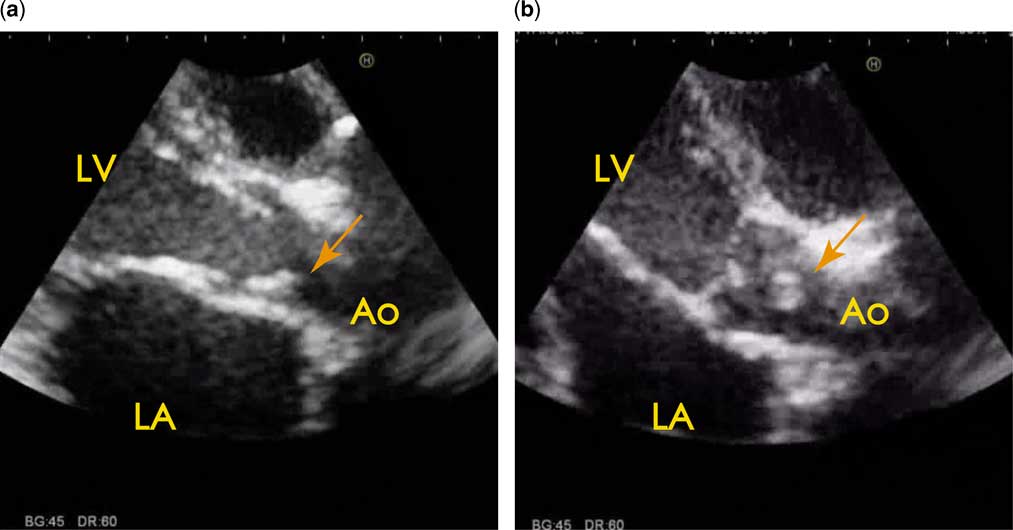
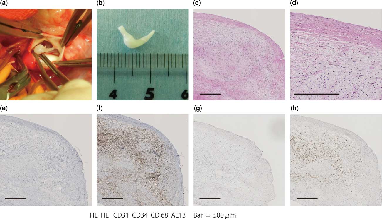
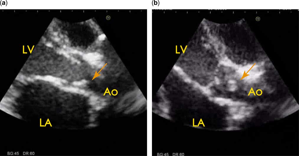
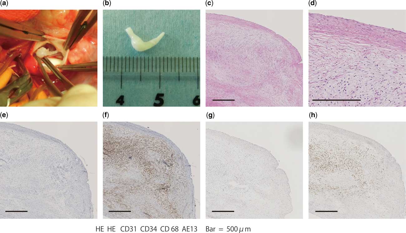
The patient was a 2-year-old boy who had undergone balloon dilatation for simple aortic coarctation with a pressure gradient of 47 mmHg at 1 month of age. After treatment, no aortic dissection or aneurysm was observed, aortic coarctation improved, and the pressure gradient at the stenosed region showed almost complete normalisation. A mobile mass adhering to the non-coronary cusp of the aortic valve was first observed on transthoracic echocardiography during routine examination at 2 years of age (Supplementary Video S1). The mass measured 9.5×3.0 mm (Fig 1). Although the systemic condition was stable, surgical resection was promptly performed because of the risk of embolism. Complete resection of the mass was performed and no subsequent trauma to or avulsion of the aortic valve was observed, rendering valvuloplasty unnecessary. Histopathological examination of the mass revealed myxomatous changes, including abundant extracellular matrix, and a partially cystic void (Fig 2). Hyperplasia of mildly atypical spindle cells was also observed. No evidence of atypia or other malignant changes was apparent. On immunostaining, interstitial cells showed positive staining for CD34 and negative results for CD31. The absences of mesothelial cells stained with AE1/AE3 and cytokeratin 5/6 and CD68-positive histiocytic cells excluded the possibility of mesothelial/monocytic incidental cardiac excrescence.Reference Boroumand, Sotoudeh Anvari, Movahedi and Yazdanifard 4 , Reference Censi, Dell’Amore, Conti and Lorenzini 5 The present findings suggested a reactive proliferative lesion, presumably resulting from physical stimulation during catheter manipulation. Fibromyxoid excrescence of the aortic valve was therefore diagnosed. The post-surgical course was uneventful.
Transthoracic echocardiography. ( a , b ) Echocardiographic images of systolic ( a ) and diastolic ( b ) phases. Parasternal long-axis view shows a mobile mass on the non-coronary cusp of the aortic valve (arrow). Ao=ascending aorta; LA=left atrium; LV=left ventricle.
Features of the cardiac mass. ( a ) Intra-operative findings showing the mass adhering to the non-coronary cusp. ( b ) Macroscopic appearance of the excised mass. ( c , d ) Haematoxylin and eosin staining shows myxomatous changes, including abundant extracellular matrix, and partially cystic void. No evidence of atypia or other malignant changes is apparent. ( e , f ) On immunostaining, interstitial cells show no staining for CD31 ( e ), but display positive results for CD34 ( f ). ( g ) No CD68-positive histiocytic cells are present. ( h ) Similarly, no mesothelial cells staining positively for AE1/AE3 are seen. Bars in Panels C-H: 500 μm.
Discussion
To diagnose masses adhering to the aortic valve, differentiation between tumours, thrombi, verrucae, aortic dissection, and aortic valve avulsion is necessary.Reference Becker 2 , Reference Parker 3 , Reference Shim, Lee and Wi 6 Myxoma, papillary fibroelastoma, and mesothelial/monocytic incidental cardiac excrescence were considered pre-operatively in the present case, but pathological findings led to a diagnosis of reactive proliferative lesion, that is, “fibromyxoid excrescence of the aortic valve” resulting from aortic valve trauma during catheter manipulation. In the examination during cardiac catheterisation, the guidewires and catheters were inserted into the left ventricle through the aortic valve. The development of the mass in this case offers a warning that manipulation of catheters and guidewires should be performed as gently as possible, to minimise injury to the valve. No atypia or malignant changes were observed. Mesothelial/monocytic incidental cardiac excrescence was excluded based on the immunohistochemical findings.Reference Boroumand, Sotoudeh Anvari, Movahedi and Yazdanifard 4 , Reference Censi, Dell’Amore, Conti and Lorenzini 5 Mesothelial/monocytic incidental cardiac excrescence is a benign finding of a mixture of cuboidal mesothelial cells, histiocytes, adipocytes, inflammatory cells, and fibrin, commonly found incidentally in cardiac chambers, valves, and the pericardial sac after cardiac catheterisation.Reference Boroumand, Sotoudeh Anvari, Movahedi and Yazdanifard 4 , Reference Censi, Dell’Amore, Conti and Lorenzini 5 Although the histopathogenesis of that lesion remains unclear, some theories have been proposed. Mesothelial/monocytic incidental cardiac excrescence may represent a reactive lesion resulting from inflammation, mechanical irritation, or tumour. From this perspective, the pathology in the present case might have been similar to mesothelial/monocytic incidental cardiac excrescence, although the immunohistological findings were definitively different. As the present proliferative mass formed and developed in the aortic valve several years after catheterisation, follow-up observation of catheterised patients should include all cardiovascular regions in addition to evaluation of the originally treated region.
Acknowledgement
The authors gratefully acknowledge the help rendered by Dr Hatsue Ishibashi-Ueda, Division of Pathology, National Cardiovascular Center for the pathological interpretation.
Conflicts of Interest
None.
Supplementary material
To view supplementary material for this article, please visit http://dx.doi.org/10.1017/S1047951114000092.