INTRODUCTION
Oweniids are gregarious organisms which build tubes of various consistencies; they are selective in the use of the materials for the construction of the tube, which are concise and they make difficult the removal of the worms. In general, the tube is formed by grains of sand, foraminiferans and sponge spicules. The form and ornamentation of the tube is a useful tool to separate some species (Parapar, Reference Parapar2003a, Reference Paraparb). The body is elongate, cylindrical, with few segments and reduced parapodia. There are five genera of Oweniidae: Galathowenia Kirkegaard, Reference Kirkegaard1959, Myriochele Malmgren, 1867, Myrioglobula Hartman, 1967, Myriowenia Hartman, 1960 and Owenia delle Chiaje, 1842; and about 40 valid species. Despite its modest number of species, it has been the subject of several regional studies (Blake, Reference Blake and Hutchings1984, Reference Blake, Blake, Hilbig and Scott2000; Milligan, Reference Milligan, Uebelacker and Johnson1984; Nielsen & Holthe Reference Nilsen and Holthe1985; Imajima & Morita, Reference Imajima and Morita1987; Martin, Reference Martín1989; Fiege et al. Reference Fiege, Kröncke and Barnich2000; Parapar, Reference Parapar2001, Reference Parapar2003a, Reference Paraparb, Reference Paraparc, Reference Parapar2006; Koh et al. Reference Koh, Bhaud and Jirkov2003; Martin, et al. Reference Martin, Koh, Bahud, Dutrieux and Gil2006). Records of the family from the Mexican coasts are few and only five species are known; thus, Fauchald (Reference Fauchald1972) reports three species, Myriochele gracilis, M. heeri and Galathowenia pygidialis (as Myriochele pygidialis), all found along the western coast of the Baja California Peninsula; Blake (Reference Blake, Blake, Hilbig and Scott2000) reports G. pygidialis for the northwest Baja California Peninsula and Kudenov (Reference Kudenov and Brusca1980) reported Owenia collaris and O. fusiformis from the Gulf of California. Note however, that Koh et al. (Reference Koh, Bhaud and Jirkov2003) have concluded that O. fusiformis is limited to the Mediterranean Sea; thus the records for the eastern Pacific should be revised. In a recent work, Villalobos-Guerrero (Reference Villalobos-Guerrero, de León-González, Bastida-Zavala, Carrera-Parra, García-Garza, Peña-Rivera, Salazar-Vallejo and Solís-Weiss2009) synthesized the knowledge about the family, emphasizing the species of Tropical America, including some species reported for California and the northern end of the Gulf of Mexico. Sene-Silva (Reference Sene-Silva2002) published a study on the phylogeny of Oweniidae, reporting that Myriochele and Galathowenia were paraphyletic, however his study was based on a limited number of species, and characters taken from literature; in addition four of the six species of Myriochele used in that analysis are currently considered members of Galathowenia. Blake (Reference Blake, Blake, Hilbig and Scott2000) and Parapar (Reference Parapar2006) demonstrated that the morphological differences based on the anterior region are sufficient to retain Myriochele and Galathowenia as separate genera.
Galathowenia was established by Kirkegaard (Reference Kirkegaard1959) for G. africana from the west coast of Africa. The validity of the genus was discussed by Parapar (Reference Parapar2003). The consequences of Parapar's findings have been that the number of its species in the genus has increased, including species transferred from Myriochele. In this study a species of Galathowenia from Tamiahua Lagoon, Gulf of Mexico, is described; a key to all Galathowenia species is included.
MATERIALS AND METHODS
The study area is situated on the eastern coast of México, in the northern part of Veracruz State. The Tamiahua Lagoon has a few openings to the open sea, and seasonally it is affected by the inflow of fresh water transported by rivers or heavy rains. Material was collected with an Eckman dredge from sand or mud bottoms. Specimens were fixed in a 10% formalin–seawater solution, and preserved in 70% ethanol. Type material has been deposited in the Polychaete Collection of the Universidad Autónoma de Nuevo León (UANL), the Natural History Museum of Los Angeles County (LACM-AHF), Zoologisches Museum und Institut, Hamburg Universität (ZMH), Muséum National d'Histoire Naturelle, Paris (MNHN) and The Natural History Museum, London (BMNH).
TYPE MATERIAL
Tamiahua Lagoon, Veracruz, México. Holotype (UANL 7425) and 9 paratypes (UANL 7426), 10 paratypes (LACM-AHF), 10 paratypes (ZMH), 10 paratypes (MNHN), 10 paratypes (BMNH), Station T-08C, 21°38′47′′N, 97°31′14′′W, 2.1 m depth, 23 February 2002. Collected by Jesús Angel de León González
NON-TYPE MATERIAL
Tamiahua Lagoon, Veracruz, México; 11 April 1999, Station T-01B (24 specimens), 21°42′01′′N 97°38′08′′W, 3 m depth; 7 March 2000, Station T-02A (10 specimens), T-02B (3 specimens), T-02C (5 specimens), 21°42′01′′N 97°35′54′′W, 2.75 m depth; 19 August 2000, Station T-02B (3 specimens), T-02C (2 specimens), 21°42′01′′N 97°35′54′′W, 2.75 m depth; Station T-04C (1 specimen), 21°42′01′′N 97°32′52′′W, 2.75 m depth; Station T-12A (1 specimen), 21°36′10′′N 97°25′59′′W, 4.1 m depth; 25 November 2000, Station T-02C (4 specimens), 21°42′01′′N 97°35′54′′W, 2.5 m depth; Station T-04A (1 specimen), 21°42′01′′N 97°32′52′′W, 1.5 m depth; 12 July 2001, Station T-02C (5 specimens), 21°42′01′′N 97°35′54′′W, 2.5 m depth; Station T-04A (7 specimens), Station T-04C (6 specimens), 21°42′01′′N 97°32′52′′W, 1.5 m depth; Station T-07A (1 specimen), 21°38′47′′N 97°34′01′′W, 3.5 m depth; Station T-12A (7 specimens), Station T-12C (5 specimens), 21°36′10′′N 97°25′59′′W3.75 m depth; Station T-13B, (1 specimen), 21°32′09′′N 97°35′54′′W, 2 m depth; Station T-14A (3 specimens), Station T-14C (1 specimen), 21°32′09′′N 97°35′54′′W, 2 m depth; Station T-15A (2 specimens), Station T-15B (2 specimens), 21°32′09′′N 97°30′47′′N, 2.5 m depth; Station T-16A (5 specimens), Station T-16B (5 specimens), Station T-16C (4 specimens), 21°32′09′′N 97°27′14′′W, 2.5 m depth; 23 February 2002, Station T-01A (5 specimens), Station T-01B (6 specimens), Station T-01C (3 specimens), 21°42′01′′N 97°38′08′′W, 2.5 m depth; Station T-02A (1 specimen), Station T-02B (100 specimens), Station T-02C (234 specimens), 21°42′01′′N 97°35′54′′W, 2 m depth; Station T-03A (15 specimens), Station T-03B (15 specimens), Station T-03C (23 specimens), 21°42′01′′N 97°34′19′′W, 2.5 m depth; Station T-04A (26 specimens), Station T-04B (48 specimens), Station T-04C (32 specimens), 21°42′01′′N 97°32′52′′W, 1.8 m depth; Station T-05A (3 specimens), Station T-05C (6 specimens), 21°38′47′′N 97°39′13′′W, 2 m depth; Station T-07A (4 specimens), Station T-07B (1 specimen), Station T-07C (1 specimen), 21°38′47′′N 97°34′01′′W, 2.6 m depth; Station T-08A (14 specimens), Station T-08B (39 specimens), 21°38′47′′N 97°31′14′′W, 2.1 m depth; Station T-09A (2 specimens), Station T-09B (3 specimens), Station T-09C (5 specimens), 21°36′10′′N 97°34′39′′W, 2 m depth; Station T-11A (3 specimens), Station T-11B (3 specimens), Station T-11C (12 specimens), 21°36′10′′N 97°32′09′′W, 3 m depth, 23 February 2004, Station 12A (10 specimens), Station 12B (28 specimens), Station 12C (8 specimens), 21°36′10′′N 97°25′59′′W, 3 m depth; Station 13A (8 specimens), Station 13B (8 specimens), Station 13C (5 specimens), 21°32′09′′N 97°35′54′′W, 1 m depth; Station T-14A (1 specimen), 21°32′09′′N 97°32′48′′W, 2.5 m depth; Station 15A (9 specimens), Station 15B (3 specimens), 21°32′09′′N 97°30′47′′W, 2 m depth; Station T-16C (2 specimens), 21°32′09′′N 97°27′14′′W, 2 m depth. Collected by Jesús Angel de León-González
DESCRIPTION
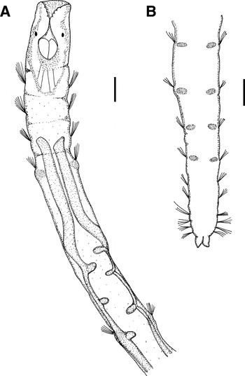
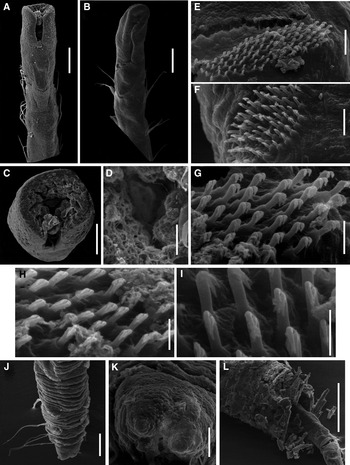
Holotype complete, body short, slender and cylindrical, 12 mm long, 0.2 mm wide, 25 chaetigers (number of chaetigers is constant in all specimens studied), three thoracic chaetigers with relative lengths 1:1:1, and 22 abdominal ones, chaetiger 5 longer, posterior end slightly tapering. Body transparent, greenish anteriorly, with two slim discreet lines of pigment. Prostomium truncate, mouth terminal, with lips slightly crenulate, forming a mid-ventral slit (Figures 1A & 2A). With mouth closed anterior end rounded with mouth located ventrally (Figure 2B). Inner part of prostomium apparently ciliated (Figure 1A). A pair of rounded brownish eyespots, located at ventro-lateral sides of head (Figure 2C, D).
Galathowenia kirkegaardi. (A) Anterior end in ventral view; (B) posterior end in ventral view. Scale bars: 0.1 mm.

Galathowenia kirkegaardi. Scanning electron micrographs: (A) anterior end in ventral view, with open mouth; (B) anterior end in ventral view, with closed mouth; (C) anterior end in frontal view; (D) detail of anterior end; (E) abdominal neuropodia of median chaetigers; (F) abdominal neuropodia of posterior chaetigers; (G) neuropodial uncini from anterior abdominal parapodia; (H) neuropodial uncini from middle abdominal parapodia; (I) neuropodial uncini from posterior abdominal parapodia; (J) posterior end in ventral view; (K); posterior end in frontal view; (L) detail of tube. Scale bars: A–B, 0.1 mm; C, 50µm; D–F, 15 µm; G–I, 5 µm; J, 50 µm; K, 15 µm; L, 300 µm.

Thorax consisting of three equal-sized chaetigers with notopodia only armed with capillary chaetae, smooth basally, from middle region with densely regular pointed scales arranged in spiralling bands. A constriction at transition between thorax and abdomen (Figure 1A). Abdominal chaetigers bi-ramus. Abdominal chaetigers 1–4 elongate (= 4–7 body chaetigers). Notopodial abdominal capillary chaetae similar to thoracic notochaetae; neurochaetae include numerous long-shafted, bidentate, hooked, unicini in ventrolateral position. Neuropodial tori from anterior and middle abdominal chaetigers longer than wide, with numerous uncini (85–90) arranged in dense field formed of oblique rows of uncini (Figure 2E), uncini of posterior chaetigers (60–65) with rows forming oval field (Figure 2F). Hooks with an antero-ventral orientation at approximately 45°, with two long, nearly equal slender teeth, hooks of anterior parapodia with one tooth slightly above second (Figure 2G). Some hooks of middle region with teeth placed almost one over the other (Figure 2H). Posterior hooks with almost superimposed teeth (Figure 2I). Tori of right side of body with the left tooth of the hooks longer than right one (Figure 2H), and tori on the left side of body with hooks with an inverse relationship to the teeth (Figure 2I). Pre-pygidial chaetigers with long capillary notochaetae, and small tori of neuropodial uncini in each segment (Figure 2J). Posterior end distally tapered, with two conical pygidial lobes (Figures 1B & 2K).
Tube slender, cylindrical, stiff, formed by three layers; a membranous inner lining, a middle layer formed by sponge spicules, and an external layer coated with white sand grains of diverse diameters and colours, mostly white and yellow, with only a few black ones. The anterior part of the tube covered densely with sand grains, middle and posterior less dense, with sponge spicules matrix visible (Figure 2L).
ECOLOGICAL NOTES
Muddy bottoms, with high percentage of clay particles.
DISCUSSION
Galathowenia currently includes 12 species separated on only a few morphological features between them; there are however differences in geographical and bathymetrical distribution of the different species. These 12 species can be divided into two groups by the presence or absence of eyes. Species having eyes include: G. kirkegaardi sp. nov., G. africana Kirkegaard, Reference Kirkegaard1959 from West Africa (Nigeria and Congo, 32–42 m), G. eurystoma (Caullery, 1944) from the East Indies (32–1570 m), G. haplosoma (Gibbs, 1972) from the Cook Islands (sublittoral in a hypersaline lagoon in Aitutaki Atoll in 1–5 m), and G. oculata (Zachs, 1922) from the Boreo-Arctic White Sea (12–2500 m). Only G. kirkegaardi sp. nov. and G. haplosoma have been reported from coastal lagoons, all other species are from deeper water in open ocean environments.
The nature of the posterior end appears to be useful to separate species based on the number of anal cirri; thus, G. kirkegaardi sp. nov., together with G. haplosoma and G. oculata have two pygidial lobes or cirri, G. africana lacks pygidial lobes, and the condition in G. eurystoma is not mentioned in the description. Based on the illustration of G. oculata by Nilsen & Holthe (Reference Nilsen and Holthe1985: 24), the relative length of the thoracic chaetigers are 2:2:1, not 1:1:1 as mentioned in the text for this species; in the same way, in G. africana this relationship is 1:2:2, just as shown by Kirkegaard (Reference Kirkegaard1959: 68, figure 17b); G. kirkegaardi sp. nov. as well as G. eurystoma and G. haplosoma all have this ratio between the thoracic chaetigers as 1:1:1. As for the elongated chaetigers it is difficult to determine how large the extension is, because for all species a significant lengthening of the segment is present from first abdominal chaetiger (4th).
Another characteristic is the external material included in the tube walls; thus, G. africana builds with very small, not overlapping sand grains (Kirkegaard Reference Kirkegaard1959: 67), G. eurystoma uses sponge spicules, G. haplosoma uses fine coralline sand grains, G. oculata has oblong sand grains, and G. kirkegaardi sp. nov. has both white sand and sponge spicules.
Galathowenia kirkegaardi is the only species in this group with a constriction between thorax and abdomen.
Etymology
This species is named in honour of the late J.B. Kirkegaard, who originally recognized and described Galathowenia, but as much for his extensive and very important contributions on polychaete systematics.
KEY FOR THE GALATHOWENIA SPECIES
1. Prostomium with a pair of reddish eye-spots…………2
– Without eye-spots…………6
2. With constriction between thorax and abdomen…………G. kirkegaardi sp. nov.
– Without body constriction…………3
3. Relative length of thoracic chaetigers 1:1:1…………4
– Relative length of thoracic chaetigers different…………5
4. Tubes built of sponge spicules, sublittoral, found between 32 and 1570 m depth…………G. eurystoma
– Tubes built with sand grains, in hypersaline lagoon, in 1–5 m depth…………G. haplosoma
5. Relative length of thoracic chaetigers 1:2:2, without anal lobes G. africana
– Relative length of thoracic chaetigers 2:2:1, with two blunt anal lobes…………G. oculata
6. With thoracic constriction…………7
– Thoracic constriction not seen…………10
7. Relative length of thoracic chaetigers 1:2:1…………8
– Relative length of thoracic chaetigers otherwise…………9
8. Four to six abdominal chaetigers elongated G. scotiae
– Two abdominal chaetigers elongate…………G. fragilis
9. Relative length of thoracic chaetigers 1:2:4; 4th to 8th chaetigers elongated…………G. longicollaris
– Relative length of thoracic chaetigers 1,5:1:1; 5th to 7th chaetigers elongated…………G. pygidialis
10. Abdomen with 13–15 chaetigers, relative length of thoracic chaetigers 2:2:1, with two blunt anal lobes…………G. australis
– Abdomen with 28–29 chaetigers; 4th–6th abdominal chaetigers elongated…………11
11. Thorax with two segments; pygidium with ten short lobes and one larger dorsal lobe…………G. joinvillensis
– Thorax with three segments; with two large dorsal anal lobes and two low lateral ones…………G. piltzi
ACKNOWLEDGEMENTS
The authors would sincerely like to thank Julio Parapar, Sergio I. Salazar-Vallejo and two anonymous referees for their valuable comments and useful suggestions. The scanning electron micrographs illustrating this paper were made with the technical skill of Ariel Arturo Cruz-Villacorta, former head of the SEM Laboratory of the Centro de Investigaciones Biológicas del Noroeste, S.C. Financial support was provided by grant H-029 from the Comisión Nacional para el Conocimiento y Uso de la Biodiversidad (CONABIO).