In the past decade, the peri-operative mortality of the arterial switch operation in neonates with transposition of the great arteries is reported to be considerably low.Reference Losay, Touchot and Serraf 1 – Reference Thrupp, Gentles and Kerr 9 Even in low birth weight infants, arterial switch operation was performed with acceptable morbidity and mortality;Reference Chen, Chen and Chang 10 however, long-term outcomes of translocated coronary arteries still remain one of the most crucial issues.Reference Losay, Touchot and Serraf 1 – Reference Thrupp, Gentles and Kerr 9 , Reference Tsuda, Imakita and Yagihara 11 , Reference Haas, Wottke and Poppert 12 In fact, most peri-operative or late deaths result from coronary obstructive events. Sudden late death secondary to acute myocardial infarction has been reported in 1–2% of hospital survivors after arterial switch operation and it usually occurs within 6 months after the operation.Reference Losay, Touchot and Serraf 1 , Reference Prandstetter, Hofer and Lechner 2 , Reference Haas, Wottke and Poppert 12 Losay et alReference Losay, Touchot and Serraf 1 reported no deaths after 5 years in 1095 survivors. Coronary arterial obstruction was reported to be caused by intimal thickening, thrombosis, coronary kinking, stretching, twisting, extrinsic compression, or by an intramural coronary artery.Reference Prandstetter, Hofer and Lechner 2 , Reference Tsuda, Imakita and Yagihara 11 – Reference Veltman, Beeres and Kalkman 16 There have been few reports on the long-term morbidity or mortality associated with coronary obstructive disorders after arterial switch operation in patients with transposition of the great arteries.Reference Losay, Touchot and Serraf 1 – Reference Williams, McCrindle and Ashburn 4 Furthermore, there is little information on the chronological changes in coronary stenosis after arterial switch operation. In the present study, we focussed on the long-term outcome and chronological change of translocated coronary arteries using follow-up coronary angiography and myocardial scintigraphy.
Patients and methods
The observational study period was from January, 1981 to June, 2010. Between January, 1981 and June, 2009, a total of 202 patients with transposition of the great arteries underwent arterial switch operation at the National Cerebral and Cardiovascular Center in Japan. Patients with double-outlet right ventricle and subpulmonic ventricular septal defect were excluded from the study. Of the 202 patients, 110 underwent arterial switch operation during the neonatal period (median age, 9 days; with a range from 2 to 30 days). The surgical technique for arterial switch operation at the National Cerebral and Cardiovascular Center was standard and has been previously described.Reference Tsuda, Imakita and Yagihara 11 They were observed for >6 months after the arterial switch operation. A total of 83 children had transposition of the great arteries with an intact ventricular septum, and 27 had ventricular septal defect including four aortic arch obstructive anomalies. Coronary artery variation was classified according to Shaher’s classification: 78 children (72%) had type 1, 16 had type 2, five had type 3, seven had type 4, two had type 5, one had type 7, and one had type 9. In all, three late deaths (2.7%) occurred that ranged from 314 to 396 days after surgery. In one case, the autopsy revealed a coronary obstruction, and the other two patients died of infection and an unknown cause, respectively. The remaining 107 patients were followed-up until June, 2010. The observational period ranged from 9 months to 23 years with a median of 11 years.
A total of 106 of 107 patients (99%) underwent angiography 1 month to 14 years (median, 1.0 year) after arterial switch operation. Subsequent follow-up angiography was performed in 59 of those ranging from 1 to 4 times. In our institution, the coronary arteries of infants <1-year old are evaluated by aortography with appropriate angulation, whereas selective coronary angiography are added if necessary in children who are ⩾1-year old and <5-years old. Both aortography and selective coronary angiography are used in children who are ⩾5-years old. A coronary obstruction was semi-quantitatively evaluated as 0, 25, 50, 75, 90, or 99% stenotic or totally occluded based on the American Heart Association classification. Myocardial perfusion was evaluated by SPECT with (99mTc) tetrofosmin with dipyridamole loading and at rest. Myocardial scintigraphy was performed during the same hospitalisation; two independent examiners semi-quantitatively evaluated myocardial perfusions on each segment. Evaluation of the ventricular septum was excluded because of enrolment of children with ventricular septal defects. The severity of the myocardial perfusion was graded into four degrees from mild, moderate, severe, to complete defect. A more than mild perfusion defect with dipyridamole loading was regarded as a significant finding in the present study. Written informed consent was obtained from all patients or patients’ guardians before catheter examinations.
Results
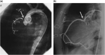
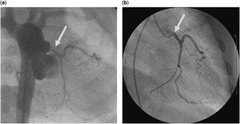
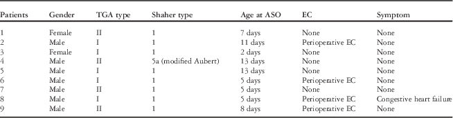
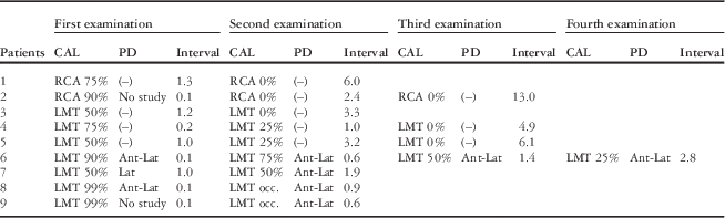
The patients’ characteristics are shown in Table 1. Although peri-operative coronary artery events such as ST depression or hypokinesis of the left ventricular wall were found in four children, only one child had a congestive heart failure-related symptom. Angiography showed coronary arterial involvement in nine (8.4%) patients at 1–16 months after the arterial switch operation. The coronary lesions are summarised in Table 2. The coronary lesions were detected at all ostia, at the right coronary artery in two patients, and at the left main trunk in seven patients. Right coronary artery stenosis in two patients improved over time. In patient 1, the proximal right coronary artery showed 75% stenosis 15 months after the arterial switch operation, but the stenosis could not be detected at the 6-year follow-up examination. Similarly, although 90% stenosis of the right coronary artery orifice was present 1 month after the arterial switch operation in patient 2 (Fig 1a), it disappeared at both the 2-year and the 13-year follow-up angiographies (Fig 1b). In patients with left main trunk lesions, the degree of stenosis improved in four children, did not change in one child, and progressed to complete occlusion in two children based on follow-up angiography. In patient 3, the left main trunk stenosis was 50% 14 months after the arterial switch operation, but it disappeared at the 3-year follow-up angiography. In patients 4, 5, and 6, the left main trunk stenosis at the initial angiography was regarded as 75, 50, and 90%, respectively; however, it regressed to 25% at 12 months and to no stenosis at 4 years in patient 4, 25% at 3 years and to no stenosis at 6 years in case 5, and 75% at 7 months, 50% at 16 months, and 25% at 33 months in case 6 (Fig 2a and b). On the contrary, severe 99% obstruction in patients 8 and 9 deteriorated to total occlusion at 10 and 7 months after the arterial switch operation, respectively. In patient 7, left main trunk stenosis did not change during the observation period.
Chronological change of coronary stenosis on angiography in case 2: ( a ) 90% stenosis (arrow) was shown at the orifice of the right coronary artery 2 months after the arterial switch operation; ( b ) Stenosis could not be detected by follow-up angiography (arrow) 13 years after the arterial switch operation.

Chronological change of coronary stenosis on angiography in case 4: ( a ) 75% stenosis (arrow) was shown at the orifice of the left main trunk 2 months after the arterial switch operation; ( b ) Obvious stenosis could not be detected by follow-up angiography (arrow) 4.9 years after the arterial switch operation.

Patients’ profile with coronary artery involvements.

ASO=arterial switch operation; EC=episode of cardiac event associated with coronary artery; TGA=transposition of the great arteries
Coronary involvements by angiographic assessment and myocardial scintigraphy.

Ant=anterior wall; CAL=coronary arterial lesions evaluated by angiography; Interval=period from arterial switch operation (year); Lat=lateral wall; LMT=left main trunk; PD=perfusion defect evaluated by myocardial scintigraphy; RCA=right coronary artery
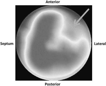
The results of myocardial scintigraphy with (99mTc) tetrofosmin in children with coronary arterial involvement are shown in Table 2. In two right coronary artery lesions, myocardial scintigraphy did not show any perfusion defect. In seven patients with left main trunk lesions, myocardial scintigraphy showed a perfusion defect in five out of six (83%) with left main trunk stenosis ⩾75% and in four out of four patients with left main trunk stenosis ⩾90% (Fig 3). In contrast, patients 1–5 who showed angiographical regression of coronary arterial lesions over time had no perfusion defects on myocardial scintigraphy at any time during the study period. In cases 1, 6, and 7, perfusion defect was somewhat fill-in at rest by re-injection of an isotope. In cases 8 and 9, perfusion defect did not change at rest by re-injection of the isotope.
Myocardial scintigraphy in case 9. A moderate perfusion defect was shown at the antero-lateral wall (arrow).

Discussion
Coronary arterial obstruction in the early stage was associated with intimal proliferation, the intramural coronary artery, re-implanted coronary deformation such as stretching, twisting, and kinking, and extrinsic compression.Reference Pasquali, Hasselblad and Li 14 – Reference Veltman, Beeres and Kalkman 16 Therefore, most of the hospital or early deaths after arterial switch operation were directly or indirectly associated with the surgical manipulation as coronary re-implantation;Reference Losay, Touchot and Serraf 1 however, long-term chronological changes of lesions in the translocated coronary arteries could be affected by coronary artery growth, a change in the relationship between an adjacent organ and the coronary artery, or intimal proliferation or regression. Angiography was widely recognised as the gold standard to evaluate coronary arterial lesions after an arterial switch operation in patients with transposition of the great arteries; however, it could have a limitation in demonstrating the precise morphology of the coronary arteries because of the size of small infants and peculiarities of the coronary anatomy in transposition of the great arteries.Reference Tsuda, Imakita and Yagihara 11
In the present study, angiographical regression of ostium stenosis of the transplanted coronary artery was observed in six out of nine patients. The “coronary artery stenosis” on angiography can include coronary kinking, stretching, or twisting, which can be changed over time. These aetiologies could have the potential to be resolved with body growth. When the “coronary artery stenosis” is due to intimal proliferation, coronary artery growth might overcome intimal proliferation. When the aetiology of the stenosis is extrinsic compression by glue or haematoma at the time of surgery, angiographical findings could be relieved because of its absorption. The “coronary artery stenosis” on angiography should be considered not always equal to actual coronary stenosis. Therefore, the indication for surgical or catheter intervention should depend not only on angiographic findings but also on the presence of myocardial ischaemia detected by myocardial scintigraphy or other imaging modalities. Although coronary stenosis progressed in some patients, in the present study, two coronary arteries with 99% stenosis with delayed angiographic enhancement of the distal coronary artery progressed to total occlusion. There was obvious myocardial ischaemia on myocardial scintigraphy within 1 year after the arterial switch operation in those patients. Thus, the aetiology of stenosis in those two patients was thought to be progressive intimal proliferation.Reference Bartoloni, Bianca and Patanè 17 , Reference El-Segaier, Lundin and Hochbergs 18 Surgical repair or catheter intervention should have been considered for the rescue of ischaemic myocardium without delay in those patients.Reference El-Segaier, Lundin and Hochbergs 18 Ostial angioplasty or mammary artery grafting could be applied with acceptable mortality.Reference Legendre, Losay and Touchot-Kone 8 Meanwhile, the management of regressive coronary artery but persistant myocardial perfusion defect, as in patient 6, may have been due to viability of the myocardium as well as the degree of the myocardial perfusion defect and angiographic findings. Previous studies have reported that early sudden death associated with coronary involvements is not rare.Reference Losay, Touchot and Serraf 1 – Reference Thrupp, Gentles and Kerr 9 Considering both these facts and our observation together, severe stenosis with myocardial ischaemia could progress to total occlusion within 1 year after arterial switch operation and could cause sudden death, but “coronary artery stenosis” on angiography without myocardial ischaemia might not mean actual stenosis and can be be improved. Indication of surgery or catheter intervention for these patients should be carefully considered.
The morbidity of the coronary stenosis after arterial switch operation was previously reported to be 3–7.8%.Reference Losay, Touchot and Serraf 1 – Reference Thrupp, Gentles and Kerr 9 , Reference Tanel, Wernovsky and Landzberg 13 , Reference Bonnet, Bonhoeffer and Piechaud 19 In the present study, the morbidity was 8.4%, which was a little higher compared with those studies. The morbidity of coronary involvements depends on the frequency and scrutiny of the coronary arteries on angiography. The patients after arterial switch operation are insensitive to chest pain because of the disturbance of the cardiac nerves adjacent to the great arteries by the surgical procedure.Reference Bonnet, Bonhoeffer and Piechaud 19 Therefore, symptoms related to myocardial ischaemia are not reliable. In the present study, all patients but one routinely underwent post-operative coronary angiography at least once whether or not there were symptoms of myocardial ischaemia, and coronary arterial assessment was always carried out even when the purpose of angiography was for some other reason, such as pulmonary arterial stenosis. The diagnosis of an ostial lesion can be sometimes missed based on selective coronary angiography, because the insertion of the catheter tip into the coronary arteries could result in insufficient enhancement of the ostial segment. Aortography adjacent to the target coronary artery could be helpful to detect ostial stenosis only if appropriate angiographic angulation can differentiate the proximal portion of the coronary artery from the aortic sinus of valsalva. Thus, the combination of selective coronary angiography and appropriate angulated aortography should be adopted to avoid misdiagnosis as much as possible. Enhanced CT could also be useful to evaluate coronary arterial lesions induced by extrinsic compression;Reference Ou, Khraiche and Celermajer 15 , Reference Veltman, Beeres and Kalkman 16 however, it could be difficult to detect ostial stenosis because of limited spatial resolution and motion artefacts in small children. Echocardiography is another modality, but it is extremely difficult to detect coronary ostial lesions by conventional 2D echocardiography. Coronary flow reserve evaluated by Doppler echocardiography can detect these lesions and might be a promising methodology in older children.Reference Aburawi, Carlsson and Berg 20 Although MRI has been used to detect myocardial ischaemia in patients with arterial switch operation, the usefulness of this imaging modality has not been clearly established in the paediatric population.Reference Manso, Castellote and Dos 21 In contrast, myocardial scintigraphy could be useful to evaluate obstructive coronary lesions in patients with arterial switch operation, although the influence of open-heart surgery itself on the myocardial perfusion scan cannot be eliminated.Reference Hayes, Baker and Kakadeker 22 – Reference Kuehn, Vogt and Schwaiger 24 In the present study, myocardial ischaemia was detected by scintigraphy in five out of six (83%) patients with left main trunk stenosis ⩾75% and in four out of four (100%) with left main trunk stenosis ⩾90%. All five children whose coronary arterial lesions angiographically regressed over time had no myocardial perfusion defects throughout the study. In some cases, discrepancy between the angiographic findings and the scintigraphic findings was revealed. The aetiology of the discrepancy was unclear. Coronary stenosis on angiography might not be always equal to the actual stenosis. Ostium of the translocated coronary artery after arterial switch operation was morphologically varied. Therefore, the coronary artery may not be as stenotic as it appears on the angiography with certain angle. Furthermore, scintigraphic assessment in small children might have the limitation of sensitivity. Angiographical assessment alone could have limitations for detecting myocardial ischaemia. Consequently, a combination of myocardial scintigraphy and angiography could be a useful modality to determine the necessity for surgical or catheter intervention and expect prognosis.
Study limitations
The retrospective nature of this study is an important limitation. The number of patients with coronary arterial lesions was still small. Further accumulation of experiences is necessary to reveal the relationship between coronary angiography and myocardial scintigraphy in these patients.
Conclusions
The present study showed chronological changes in the translocated coronary arteries after the arterial switch operation. Careful observation is necessary during follow-up even when patients have no symptoms of myocardial ischaemia. A combination of angiographic assessment and myocardial scintigraphy could provide us with useful information for further interventions.
Acknowledgement
None.
Financial Support
This research received no specific grant from any funding agency, commercial, or not-for-profit sectors.
Conflicts of Interest
None.
Ethical Standards
The authors assert that all procedures contributing to this work comply with the ethical standards of the relevant national guidlines on human experimentation and with the helsinki Declaration of 1975, revised in 2008.