Introduction
The cultivable microorganisms found on exposed surfaces of unmanned and manned spacecraft have been well characterized (Favero Reference Favero1971; Dillon et al. Reference Dillion, Gavin, Roark and Trauth1973; Taylor Reference Taylor1974; Puleo et al. Reference Puleo, Fields, Bergstrom, Oxborrow, Stabekis and Koukol1977; Venkateswaran et al. Reference Venkateswaran, Satomi, Chung, Kern, Koukol, Basic and White2001; Schuerger et al. Reference Schuerger, Mancinelli, Kern, Rothschild and McKay2003; Schuerger Reference Schuerger and Cockell2004; and the papers cited within these studies). The microbial bioloads isolated from early unmanned spacecraft launched in the 1960s ranged between 1×10⁴ to 2×10⁸ viable microorganisms per vehicle (Favero Reference Favero1971; Dillon et al. Reference Dillion, Gavin, Roark and Trauth1973). During the Viking missions, terminal heat-sterilization protocols just prior to launch reduced the probability of launching viable bioloads to 2×10⁻⁴ spore-forming bacteria per lander (Puleo et al. Reference Puleo, Fields, Bergstrom, Oxborrow, Stabekis and Koukol1977). However, more recently, modern Mars landers and rovers such as Mars Pathfinder, Spirit, and Opportunity have been launched with an estimated total bioload of not more than 3×10⁵ viable aerobic spores per vehicle (Rummel Reference Rummel1989; Crawford Reference Crawford2005). The most common fungal genera recovered from these robotic spacecraft were Alternaria, Aspergillus, Bipolaris, Candida, Cladosporium, Fusarium, Mucor, Penicillium, and Trichoderma; the most common bacterial genera were Acinetobacter, Alcaligenes, Bacillus, Corynebacterium-Brevibacterium group, Flavobacterium, Micrococcus, Staphylococcus, and Streptococcus (see the reviews by Taylor (Reference Taylor1974), Schuerger et al. (Reference Schuerger, Mancinelli, Kern, Rothschild and McKay2003) and Schuerger (Reference Schuerger and Cockell2004)). In addition, several yeast, actinomycete, and fastidious unidentified mould and bacterial species have been recovered from robotic spacecraft. More recently, two new reports have expanded the species list of recovered microorganisms from spacecraft to include several non-cultivable species (Venkateswaran et al. Reference Venkateswaran, Satomi, Chung, Kern, Koukol, Basic and White2001; La Duc et al. Reference La Duc, Nicholson, Kern and Venkateswaran2003).
Disinfection and sterilization protocols for spacecraft include a wide diversity of technologies that seek to reduce the viable bioloads of vehicles to acceptable levels prior to launch. Microbial decontamination procedures currently include dry-heat, wet-heat at high pressure, gaseous sterilization of components, and chemical surface sterilants, depending on the spacecraft component and phase of assembly (Pflug Reference Pflug1971; Venkateswaran et al. Reference Venkateswaran, Chung, Allton and Kern2004; Kempf et al. Reference Kempf, Chen, Kern and Venkateswaran2005). In addition, UV irradiation of sun-exposed surfaces on Mars may also contribute to reductions in viable bioloads on spacecraft during sol 1 after landing (Schuerger et al. Reference Schuerger, Mancinelli, Kern, Rothschild and McKay2003, Reference Schuerger, Richards, Newcombe and Venkateswaran2006; Newcombe et al. Reference Newcombe, Schuerger, Benardini, Dickinson, Tanner and Venkateswaran2005). However, all of these sterilization protocols have constraints on their effectiveness and utility, and cannot be used under all circumstances during prelaunch processing of spacecraft components or post-landing operations.
Recently, several reports have suggested the use of non-thermal atmospheric plasmas for sterilization of spacecraft surfaces (Panikov et al. Reference Panikov, Paduraru, Crowe, Ricatto, Christodoulatos and Becker2002; Mogul et al. Reference Mogul, Bol'shakov, Chan, Stevens, Khare, Meyyappan and Trent2003; Bol'shakov et al. Reference Bol'shakov, Cruden, Mogul, Rao, Sharma, Khare and Meyyappan2004). Non-thermal plasmas can be generated in a variety of gaseous environments including He, N2, Ar, air, CO2, O2, H2O2, or combinations of the above (Laroussi et al. Reference Laroussi, Alexeff and Kang2000; Laroussi Reference Laroussi2002; Moisan et al. Reference Moisan, Barbeau, Crevier, Pelletier, Philip and Saoudi2002; Philip et al. Reference Philip, Saoudi, Crevier, Moisan, Barbeau and Pelletier2002; Choi et al. Reference Choi, Han, Baik, Lee, Han, Park, Lee, Song and Lim2006). Each of these types of non-thermal plasmas can generate a diversity of free radicals (O−, O2−, O3−, OH−), charged particles and molecules (e −, CO2+, N2+, NOx), charged cellular membranes, and UV irradiation; all of which can contribute to the inactivation of microorganisms (Laroussi et al. Reference Laroussi, Alexeff and Kang2000; Laroussi Reference Laroussi2002; Choi et al. Reference Choi, Han, Baik, Lee, Han, Park, Lee, Song and Lim2006). The inactivation kinetics of non-thermal plasmas are extremely fast, achieving several log reductions in microbial populations in periods of times measured in seconds to tens of seconds (Laroussi et al. Reference Laroussi, Alexeff and Kang2000, Reference Laroussi2002; Panikov et al. Reference Panikov, Paduraru, Crowe, Ricatto, Christodoulatos and Becker2002; Mogul et al. Reference Mogul, Bol'shakov, Chan, Stevens, Khare, Meyyappan and Trent2003; Bol'shakov et al. Reference Bol'shakov, Cruden, Mogul, Rao, Sharma, Khare and Meyyappan2004; Choi et al. Reference Choi, Han, Baik, Lee, Han, Park, Lee, Song and Lim2006). Generally, greater than six-log reductions in microbial populations can be achieved within 10–30 min of exposure to the plasmas. Fungal cells generally require slightly longer exposures for full inactivation probably due to the generally larger pigmented cells when compared to bacteria (Efremov et al. Reference Efremov, Adamiak, Blochin, Dadashev, Dmitriev, Gryaznova and Jusbashev2000). The most aggressive inactivation kinetics were reported by Garate and Evans (Reference Garate and Evans1998) for Escherichia coli and Bacillus subtilis in which greater than 1010 cells or spores, respectively, were inactivated within 15 min under a pulsed radio frequency (RF) driven plasma. In addition, non-thermal plasmas have been shown to physically degrade vegetative cells and endospores of a diversity of microorganisms by directly etching and eroding cell walls, membranes, cytosol, and DNA (Philip et al. Reference Philip, Saoudi, Crevier, Moisan, Barbeau and Pelletier2002; Choi et al. Reference Choi, Han, Baik, Lee, Han, Park, Lee, Song and Lim2006). In a recent study by Mogul et al. (Reference Mogul, Bol'shakov, Chan, Stevens, Khare, Meyyappan and Trent2003), experiments with a low-power O2 plasma indicated that 100% of cells of Deinococcus radiodurans were physically degraded and removed from sample coupons within 45 min. In two studies (Philip et al. Reference Philip, Saoudi, Crevier, Moisan, Barbeau and Pelletier2002; Choi et al. Reference Choi, Han, Baik, Lee, Han, Park, Lee, Song and Lim2006), spores of three bacteria were ruptured within a few tens-of-seconds exposure to O2 plasmas. In general, the breakdown products of bacterial cells under non-thermal plasmas are CO2, CO, N2, CH4, H2O, and OH− (Moisan et al. Reference Moisan, Barbeau, Crevier, Pelletier, Philip and Saoudi2002; Mogul et al. Reference Mogul, Bol'shakov, Chan, Stevens, Khare, Meyyappan and Trent2003), but these often depend on the gas admixtures used in the tests. Lastly, non-thermal plasmas can degrade and remove biogenic signature molecules such as nucleic acids, lipids, and proteins present on metal surfaces (Mogul et al. Reference Mogul, Bol'shakov, Chan, Stevens, Khare, Meyyappan and Trent2003).
The advantages of non-thermal atmospheric plasmas for the sterilization of spacecraft surfaces include low temperatures at target sites, rapid inactivation kinetics of exposed microbial cells, physical degradation and removal of microbial cells, physical removal of organic biosignature molecules, and short exposure times for the materials. As part of a long-range goal to develop a low-temperature atmospheric plasma system for sterilizing spacecraft surfaces, the objectives of this study were to (a) determine the biocidal effects of an oxygen–helium atmospheric plasma on spores of the common spacecraft contaminant, B. subtilis, and (b) characterize the effects of these plasmas on several spacecraft materials at exposure times and power settings that were biocidal against B. subtilis.
Materials and methods
Plasma generation and sample exposure
Six spacecraft materials were used for the spore adhesion experiments: aluminium 6061 (henceforth Al-6061), polytetrafluoroethylene (PTFE), polycarbonate, Saf-T-Vu (a polyvinylchloride material used in solid rocket booster joint enclosures), Rastex (a PTFE woven cloth used in Shuttle orbiter radiator protective covers), and Herculite 20 (used in launch service processing). Coupons of each material were ultrasonically cleaned for 10 min each in acetone, methanol, and isopropanol, and finally rinsed in DI water. The coupons were allowed to air dry in a positive pressure sterile hood until used.
The atmospheric pressure glow-discharge (APGD) plasma treatment used in the current study was optimized on Saf-T-Vu, Herculite 20, and Rastex with different gases (O2, air, O2+H2O, and He+O2) at 47±3% RH and 25°C. Helium+O2 produced the most significant effect, determined by monitoring the increase in the O:C ratio on the surface by X-ray photoelectron spectroscopy (XPS) and by contact angle measurements (Trigwell, Schuerger, Calle, unpublished). The Al-6061, PTFE, and polycarbonate were treated by the optimized protocol for inclusion in the bacterial assays discussed below. For this study, a Surfx Technologies Atomflo 1500R plasma source was used. Helium and O2 were delivered to the head at a flow rate of 60 l min−1 at 275 kPa in the ratio 98% He and 2% O2. The Atomflo head requires a carrier gas, typically helium, to sustain the plasma discharge in the reactive head. The plasma is generated by RF power, generally 150 W at 27.12 MHz. Up to 2% volume of a reactive gas (e.g., O2, H2, or N2) is used to produce the reactive species that may include free radicals (O−, O2−, O3−, OH−), charged particles and molecules (e −, CO2+, N2+, NOx), charged cellular membranes, and UV irradiation (Laroussi et al. Reference Laroussi, Alexeff and Kang2000; Laroussi Reference Laroussi2002; Choi et al. Reference Choi, Han, Baik, Lee, Han, Park, Lee, Song and Lim2006). The coupons were exposed to a plasma measuring 150×3 mm2 emitted from a slit in the plasma head at a distance of 4 mm. The head was scanned at a speed of 15 mm s−1 across the coupons so that total exposure times could be precisely calculated. Coupons for experimental data presented in Fig. 1 and Fig. 2 were treated for 6 min. All other coupons were treated for various time periods up to 5 min, as described below.
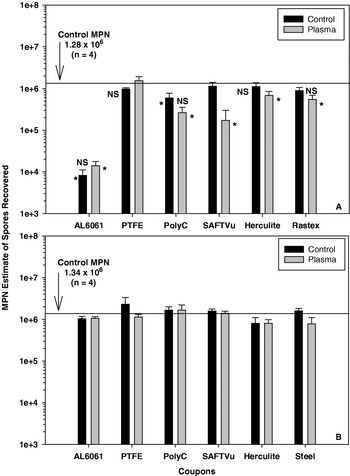
Retention of spores of B. subtilis HA101 on six spacecraft materials treated with a He+O2 APGD plasma. (A) Results from MPN assays of six spacecraft materials (aluminum 6061, polytetrafluoroethylene (PTFE), polycarbonate (PolyC), Saf-T-Vu, Rastex, and Herculite 20) extracted with sterile deionized water only. Spores of B. subtilis were retained strongly by aluminum 6061 exposed or unexposed to the APGD plasma for 6 min. (B) Results from MPN assays with a duplicate set of six spacecraft coupons, but extracted with silica sand added to the extraction tubes. Spores of B. subtilis were recovered equally from all coupons regardless of APGD plasma treatment. NS=no significant differences between plasma-exposed or -unexposed coupons for individual materials; asterisks (*) identify treatments significantly different from the control MPN samples that are represented by horizontal lines in each graph; results based on ANOVA and protected least-square mean separation tests (P⩽0.05; n=4; bars are standard errors of the means).

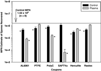
Retention of spores of B. subtilis HA101 on six spacecraft materials (aluminum 6061, polytetrafluoroethylene (PTFE), polycarbonate (PolyC), Saf-T-Vu, Rastex, and Herculite 20) incubated at 100% relative humidity at 30°C for 7 days. Three materials (Al-6061, polycarbonate (PolyC), and Saf-T-Vu) exhibited lower numbers of recovered spores from plasma-treated coupons, and three materials (PTFE, Herculite, and Rastex) exhibited higher numbers of recovered spores from plasma-treated coupons compared to untreated coupons. All plasma-exposed and -unexposed treatments were significantly different from each other for individual materials; asterisks (*) identify treatments significantly different from the control MPN samples that are represented by the horizontal line in the graph; results based on ANOVA and protected least-square mean separation tests (P⩽0.05; n=8; bars are standard errors of the means).

Microbiological procedures
Coupons of all six materials were treated as above, and then transferred to a microbiological laboratory to evaluate how spores of the bacterium, B. subtilis HA101, would adhere to the treated materials. Spores of B. subtilis were applied to the treated upper surfaces of each coupon at the rate of ≈1.5×10⁶ viable spores per coupon. Two sets of treated coupons were processed according to the following procedures. First, in order to estimate how ‘sticky’ the materials were after plasma treatment, spores were applied as suspensions in 100 μl drops per coupon, air dried at 25°C for 24 h, and then processed with an enhanced Most Probable Number (MPN) assay (Schuerger et al. Reference Schuerger, Mancinelli, Kern, Rothschild and McKay2003, Reference Schuerger, Richards, Newcombe and Venkateswaran2006). In brief, treated coupons were placed in 50 cc plastic conical centrifuge tubes containing 20 ml of sterile deionized water (SDIW), agitated for 2 min with a vortex mixer, serial diluted, and 20 μl suspensions of each spore dilution dispensed into individual wells in a 96-well microtiter plate containing 180 μl each of a liquid spore medium ideal for growing B. subtilis (Schuerger et al. Reference Schuerger, Mancinelli, Kern, Rothschild and McKay2003). The MPN assays were incubated at 30°C for 24 h, and read to estimate the numbers of spores recovered from coupons. Second, in order to boost the recovery rates of the spores from the coupons, a second set of coupons were similarly prepared and exposed to the He–O2 plasma field, doped with spores of B. subtilis HA101, and processed with the MPN assay (as described above), except that 1 g of pre-heat sterilized silica sand (10–20 mesh) was added to each 50 cc extraction tube. The silica sand was found to be very successful in improving the recovery rates of spores of seven Bacillus spp. from aluminium coupons in a previous study by Schuerger et al. (Reference Schuerger, Richards, Newcombe and Venkateswaran2006). The silica sand acts as a mild abrasive to dislodge spores from surfaces during the vortex-mixing step, without causing reductions in viability. Based on the work reported by Schuerger et al. (Reference Schuerger, Mancinelli, Kern, Rothschild and McKay2003, Reference Schuerger, Richards, Newcombe and Venkateswaran2006) the detection limit of the current MPN assay was estimated as approximately 10 endospores per coupon.
It was hypothesized that APGD plasma treated materials might inhibit the long-term survival of B. subtilis spores on the different materials in humidified ambient environments. However, if spores of B. subtilis were found to germinate and grow on the plasma-treated surfaces, then the use of plasmas to modify the surface characteristics of materials might not be useable for spacecraft applications within humidified environments. Thus, a new set of plasma-treated coupons were doped with spores of B. subtilis (as described above), placed within 6×6×2.5 cm plastic chambers maintained at 30°C and 100% relative humidity for 7 days, and assayed for numbers of surviving spores with the enhanced MPN assay (Schuerger et al. Reference Schuerger, Mancinelli, Kern, Rothschild and McKay2003, Reference Schuerger, Richards, Newcombe and Venkateswaran2006). After incubation, the MPN assays were used to estimate the numbers of viable spores recovered from coupon surfaces. When the plasma-treated and B. subtilis doped coupons were placed within the plastic incubation containers, the coupons were kept elevated above the liquid water by use of polystyrene petri dishes. Thus, the spore-doped coupons could be maintained at 100% RH without condensation forming on the coupon surfaces.
A third experiment was designed to test the hypothesis that the APGD plasmas can also rapidly inactivate and physically remove spores of B. subtilis on aluminium coupons. Aluminium (grade 6061) coupons measuring 1×2.5 cm2 were heat sterilized for 24 h at 130°C, and cooled to room temperature. Spores of B. subtilis were applied to the upper surfaces of coupons by pipetting 200 μl of a spore suspension containing ≈2×10⁶ viable spores to each coupon. The spore suspensions were dried at 25°C for 24 h. Doped aluminium coupons were then aseptically transferred to the APGD plasma lab and exposed to the He–O2 plasma for 0, 10, 20, 30, 40, 50, 60, 120, and 300 s. The APGD plasma settings were the same as in the previous experiments. The plasma-exposed coupons were then transferred back to the microbiology laboratory in which they were assayed using the enhanced MPN procedure described above.
In order to determine whether the spores were being physically degraded and removed from the coupons, a parallel set of aluminium coupons were prepared as above, and exposed to the He–O2 plasma field for 0, 30, 60, and 300 s. One-half of these samples were imaged at 3000× (effective resolution=0.1 μm) with a high-resolution video microscopy system (model VH-7000, Keyence Corporation of America, Woodcliff Lake, NJ, USA). The second half of the samples were coated with gold and imaged with a scanning electron microscope (SEM; model LEO1455, Leo Electron Microscopy, Inc., Thornwood, NY, USA). The SEM images were collected digitally with the LEO1455 system. Using the high-resolution video microscopy system, estimates were made on the numbers of spores retained on the aluminium coupons after plasma treatment. The numbers of spores in 15, 30 μm2 regions of interest were counted while randomly scanning the aluminium coupon surfaces.
Statistical analyses
Statistical analyses were conducted with version 8.0 of the PC-based Statistical Analysis System (SAS) (SAS Institute, Inc., Cary, NC, USA). Data for Figs 1 and 5 were not transformed prior to analysis. Data for Figs 2 and 3 were power-transformed to induce homogeneity of variances among the various treatments. Data were subjected to analysis of variance (ANOVA) procedures (PROC GLM) followed by protected least-squares mean separation tests (P⩽0.05). All data are presented as untransformed means in the figures.
Results
Compared to control MPN numbers of viable spores (i.e. MPN assays of spore inoculum prior to application to coupons), spores of B. subtilis adhered to Al-6061 significantly stronger than all other materials tested, regardless of whether the aluminium coupons were plasma-treated or not (Fig. 1(A)). In addition, spores adhered to aluminium surfaces regardless of the morphological features present, i.e. spores showed no preference for grooves, pits, or ridges. Saf-T-Vu was the only material in which spores of B. subtilis adhered more aggressively to plasma-treated coupons when compared to non-plasma treated coupons; all other materials exhibited no significant differences between plasma and non-plasma treated coupons. Although the reductions of adhered spores were not as dramatic as aluminium coupons, plasma-treated Saf-T-Vu, Herculite, and Rastex coupons retained spores slightly more aggressively than the non-plasma treated materials when compared to control MPN numbers of viable spores. Interestingly, when these assays were repeated with the addition of 1 g of pre-sterilized silica sand added to the extraction tubes, all materials exhibited equal levels of recovered spores regardless of plasma treatment (Fig. 1(B)). These results suggest that some of the plasma-treated materials tested here became slightly more aggressive in retaining spores, but that this adhesion was not strong enough to prevent the extraction of the spores with the silica sand. Furthermore, the silica sand was used in all subsequent assays to assure that most viable spores (i.e. above the detection limit of 10 spores per coupon) were removed from the coupons. The ability of the added silica sand to enhance spore removal from coupons has been reported previously (Schuerger et al. Reference Schuerger, Richards, Newcombe and Venkateswaran2006).
Compared to control MPN assays, incubation of spore-doped coupons in the 100% RH environments resulted in a significant decrease in recovered spores from all materials except Rastex and PTFE plasma-treated coupons and SAF-T-Vu non-plasma treated coupons (Fig. 2). The most dramatic difference between untreated and plasma-treated coupons was observed for the material Saf-T-Vu in which there was a two log decrease in the spores recovered from the plasma-treated coupons versus the non-plasma treated samples. Other materials in which significant decreases were observed for the plasma-treated coupons, compared to untreated coupons, were Al-6061 and polycarbonate. In contrast, the plasma-treated coupons for PTFE, Herculite, and Rastex all exhibited higher numbers of recovered spores compared to the untreated coupons. Thus, three materials (Al-6061, polycarbonate, and Saf-T-Vu) exhibited lower numbers of recovered spores from plasma-treated coupons, and three materials (PTFE, Herculite, and Rastex) exhibited higher numbers of recovered spores from plasma-treated coupons compared to untreated coupons.
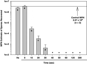
The rate of inactivation of viable spores of B. subtilis by the APGD plasma was extremely rapid (Fig. 3). The D value (the time required to reduce the population of a microorganism by one log) for the inactivation of spores was estimated to be 5–7 s under the set of operating parameters for the He–O2 plasma tested here. Greater than six log reductions in the numbers of recovered viable spores were observed at 40 s. One of the controls built into this assay was a test to determine whether the He–O2 gas flow alone (i.e. the APGD plasma turned off) could have contributed to the loss of spores from the Al-6061 coupons. Results indicated that although there was a very slight decrease in the numbers of recovered spores from a set of aluminium coupons exposed to the He–O2 gas flow alone, this difference was not statistically different from the untreated controls (Fig. 3; P>0.05).
Recovery of spores of B. subtilis HA101 on aluminum 6061 coupons exposed for various times up to 300 s (5 min) to a He+O2 APGD plasma. Loss of viability was noted in as short a time as 10 s; the minimum detection limit of the MPN assay was 10 viable spores per coupon. Letters depict significant differences among treatments as measured by ANOVA and protected least-square mean separation tests (P⩽0.05; n=6; bars are standard errors of the means); asterisks (*) indicate no recoverable spores from coupons.

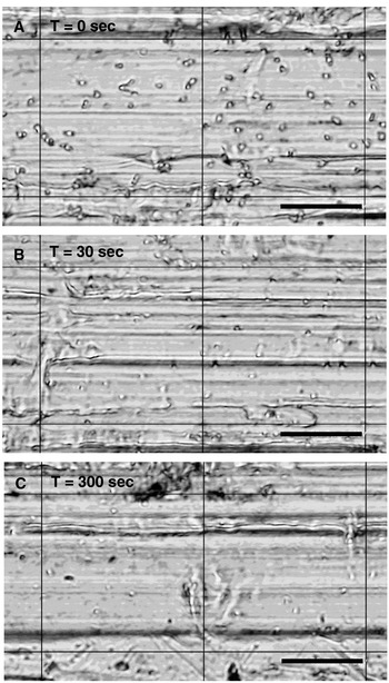
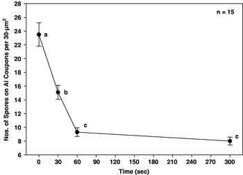
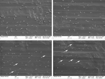
High-resolution video microscopy revealed that spores of B. subtilis were being removed at a rapid rate (Fig. 4) in close correlation to the loss of viable spores recovered from aluminium coupons (Fig. 5). The numbers of spores recovered after only 30 or 300 s exposures to the APGD plasma were 36% or 66%, respectively, lower than the untreated controls (Fig. 5). Direct observations of the spores on the aluminium coupons paralleled this observation very closely. Spores were obvious on all untreated control coupons, but were reduced in size and number on the coupons exposed for 30, 60, or 300 s to the APGD plasma. However, the resolution was not adequate in the high-resolution video microscopy images to directly evaluate the plasma damage on the spores themselves.
High-resolution video images of spores of Bacillus subtilis HA101 on aluminum 6061 coupons and exposed to the He+O2 APGD plasma for 0, 30, or 300 s. Numbers of spores exposed to the APGD plasma decreased dramatically over short time periods of 30–300 s. (Bars=15 μm.)

Numbers of spores of B. subtilis HA101 on aluminum 6061 coupons and exposed to the He+O2 APGD plasma for 0, 30, 60, or 300 s. Spore numbers decreased significantly for each time step between 0 and 60 s, but then remained flat between 60 and 300 s. Letters depict significant differences among treatments as measured by ANOVA and protected least-square mean separation tests (P⩽0.05; n=15; bars are standard errors of the means).

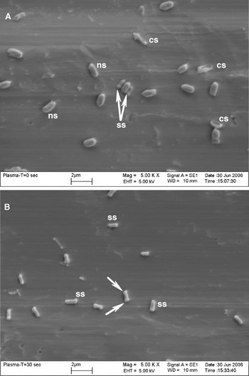
Scanning electron microscopy was used to examine the physical degradation of spore bodies on samples exposed to the APGD plasma for 0, 30, 60, or 300 s (Figs. 6 and 7). After only 30 s exposure to the plasma, all spores of B. subtilis were reduced by ≈50% in size, and many spores exhibited ruptured end walls (Fig. 7(B)). Furthermore, as the time of exposure was increased from 30 s to 300 s (5 min), spore density dropped significantly (Fig. 6(A) versus 6(D)), and residues of spores were observed on the aluminium coupons (arrows in Figs. 6(C) and 6(D)). Although biochemical data is lacking at this time, we interpret these residues as the remnants of spore adhesives that bound the spores to the coupons, or biofilms formed around the spores as they dried onto the coupon surfaces. Adhesion of Bacillus spores to surfaces has been shown to be mediated by the formation of biofilms (Faille et al. Reference Faille, Jullien, Fontaine, Bellon-Fontaine, Slomianny and Benezech2002). Spores of B. subtilis on untreated coupons exhibited a range of morphologies that included fully hydrated normal spores (ns), collapsed spores (cs), and a low number of small spores (ss) (Fig. 7). The smaller than normal spores in the untreated controls (Fig. 7(A)) were very similar to the shrunken spores observed in the plasma-treated samples (Fig. 7(B)). However, the densities of the small spores were much less than 1–2% of the total population on control coupons, which did not match the nearly 100% of the shrunken spores observed in all plasma-treated samples. Thus, although there might have been a natural population of smaller than normal spores in untreated controls, the shrunken spores in all plasma-treated samples were obviously created by the exposure of the normal spores to the APGD plasma.
Scanning electron microscopy images of spores of B. subtilis HA101 on aluminum 6061 coupons and exposed to the He+O2 APGD plasma for 0, 30, 60, or 300 s. The numbers of spores decrease over time, and the spores appear physically degraded by the plasma at all time steps of over at least 30 s. Arrows indicate locations that have lost spore bodies and at which only residual biofilm scars of the attached spores are visible.

Scanning electron microscopy images of spores of Bacillus subtilis HA101 on aluminum 6061 coupons and exposed to the He+O2 APGD plasma for 0 or 30 s. (A) Although a few small sized spores (ss) and a few collapsed spores (cs) are visible in the T=0 treatments, exposure to the He+O2 APGD plasma for as little as 30 s significantly degrades spore bodies of B. subtilis. (B) Plasma-damaged spores exhibit degraded terminal ends (arrows) and appear very different as compared to normal cells (ns).

Discussion
The search for evidence of extinct or extant life on Mars remains a key goal of the Mars Exploration Program (Garvin et al. Reference Garvin, Figueroa and Naderi2001; Morrison Reference Morrison2001; Des Marais et al. Reference Des Marais2003). Over the next decade several rover and lander missions will renew the search for a martian biota begun with the Viking landers in 1976. However, unlike the Viking landers, these spacecraft are unlikely to be terminally heat-sterilized prior to launch due to budget and technology constraints (Debus Reference Debus2006). Thus, it becomes imperative that other methods be used to render spacecraft components free of both viable spores and biogenic signature molecules. Atmospheric pressure glow-discharge plasmas have been developed in the biomedical and electronics industries to both sterilize surfaces and remove biological materials from exposed components (Laroussi Reference Laroussi2002; Moisan et al. Reference Moisan, Barbeau, Crevier, Pelletier, Philip and Saoudi2002; Panikov et al. Reference Panikov, Paduraru, Crowe, Ricatto, Christodoulatos and Becker2002; Philip et al. Reference Philip, Saoudi, Crevier, Moisan, Barbeau and Pelletier2002; Mogul et al. Reference Mogul, Bol'shakov, Chan, Stevens, Khare, Meyyappan and Trent2003; Bol'shakov et al. Reference Bol'shakov, Cruden, Mogul, Rao, Sharma, Khare and Meyyappan2004), but few tests have been undertaken to determine whether APGD plasmas might be applicable to spacecraft materials (Mogul et al. Reference Mogul, Bol'shakov, Chan, Stevens, Khare, Meyyappan and Trent2003).
The He+O2 APGD plasma used in the current study was found to aggressively kill and physically degrade spores of the common spacecraft contaminant, B. subtilis. Greater than six orders of magnitude of spore inactivation was observed after only 40 s exposure, and bacterial cell walls were observed to be severely degraded physically by only 5 min of exposure to the APGD plasma. These results are very consistent with previously published reports using APGD plasmas to inactivate a wide diversity of microbial species (Kelly-Wintenberg et al. Reference Kelly-Wintenberg, Montie, Brickman, Roth, Carr, Sorge, Wadsworth and Tsai1998, Reference Kelly-Wintenberg, Hodge, Montie, Deleanu, Sherman, Roth, Tsai and Wadsworth1999; Efremov et al. Reference Efremov, Adamiak, Blochin, Dadashev, Dmitriev, Gryaznova and Jusbashev2000; Lerouge et al. Reference Lerouge, Wertheimer, Marchand, Tabrizian and Yahia2000; Roth et al. Reference Roth, Sherman, Gadri, Karakaya, Chen, Monite, Kelly-Wintenberg and Tsai2000; Laroussi et al. Reference Laroussi2002; Phillip et al. Reference Philip, Saoudi, Crevier, Moisan, Barbeau and Pelletier2002; Mogul et al. Reference Mogul, Bol'shakov, Chan, Stevens, Khare, Meyyappan and Trent2003; Choi et al. Reference Choi, Han, Baik, Lee, Han, Park, Lee, Song and Lim2006). The inactivation of microbial spores appears to be a combination of UV irradiation emitted by APGD plasmas and the generation of free radicals (O−, O2−, O3−, OH−), charged particles and molecules (e −, CO2+, N2+, NOx), and charged cellular membranes (Laroussi et al. Reference Laroussi, Alexeff and Kang2000; Laroussi Reference Laroussi2002; Choi et al. Reference Choi, Han, Baik, Lee, Han, Park, Lee, Song and Lim2006). However, the physical degradation of microbial cells and spores appears to be due primarily to the presence of free radicals and charged particles in the APGD plasmas (Lerouge et al. Reference Lerouge, Wertheimer, Marchand, Tabrizian and Yahia2000; Laroussi et al. Reference Laroussi2002; Phillip et al. Reference Philip, Saoudi, Crevier, Moisan, Barbeau and Pelletier2002).
A key advantage of non-thermal APGD plasmas for the sterilization of spacecraft components is that both inactivation of spore viability and the physical removal of cellular materials from surfaces can be achieved at room temperatures in relatively short periods of time. For example, greater than 60% of cellular materials in the current study were physically removed from aluminium coupons after only 5 min exposure to the APGD plasma. Moreover, in a few reports (Laroussi et al. Reference Laroussi, Sayler, Glascock, McCurdy, Pearce, Bright and Malott1999; Laroussi et al. Reference Laroussi2002; Mogul et al. Reference Mogul, Bol'shakov, Chan, Stevens, Khare, Meyyappan and Trent2003), nearly 100% of cellular material was removed from surfaces between 15 and 45 min exposures to APGD plasmas operated under similar conditions to the He+O2 plasma used here. In addition, biogenic molecular films have been degraded and removed by APGD plasmas over relatively long exposure times in excess of 15–30 min (Li et al. Reference Li, Belkind, Jansen and Orban1997). In many of these reports, and in the current study, the physical degradation of spores appears to occur first at the terminal ends of the spores, where cell walls appear to be ruptured, and it results in the eventual removal of the whole spore from the treated surface leaving only a residue of the biofilms used by spores to attach to materials.
A second key advantage of using non-thermal APGD plasmas to sterilize spacecraft components appears to be that only relatively minor changes in surface properties of some materials exposed to APGD plasmas are observed for the short exposure times (<15 min) required to achieve spore inactivation (Morra et al. Reference Morra, Occhiello and Garbassi1990; Gerenser Reference Gerenser, Strobel, Lyons and Mittal1994). However, caution must be used when applying plasmas to spacecraft surfaces until more is known about the precise effects that specific plasmas have on spacecraft materials. Surface modifications caused by APGD plasmas are possible with much longer treatments times than were used here and can occur because active species of the plasmas (O−, O2−, O3−, OH−, e −, CO2+, N2+, NOx) are either deposited directly onto the surfaces or alter surface chemistries (Laroussi et al. Reference Laroussi, Alexeff and Kang2000; Laroussi Reference Laroussi2002; Choi et al. Reference Choi, Han, Baik, Lee, Han, Park, Lee, Song and Lim2006). For example, collisions of the energetic species with the substrate surface results in the transfer of most of their energy to the surface atoms. As a result, substrate bonds can be broken, possibly making the surface more reactive; and depending upon the precursor gas used, certain new functional groups can be grafted onto the surface. Thus, plasmas can induce specific surface chemistries that affect the top few tens of nanometres but generally do not change the bulk properties of the materials. In some situations changes to surface chemistries may not be important (e.g., surface changes to structural components made of steel or aluminium), but in other situations surface changes might alter the operational functionality of the materials (e.g., solar panels, electronic boards, optics).
In the current study, the adhesion of spores of B. subtilis was observed to be slightly greater in plasma-treated materials, but this increased adhesive quality of the materials was not enough to prevent the full removal of spores once silica sand was added to the extraction tubes (Fig. 1). In contrast, when spores of B. subtilis were incubated for 7 days on plasma-treated materials (Fig. 2), three materials (Al-6061, polycarbonate, and Saf-T-Vu) exhibited lower numbers of recovered spores from plasma-treated coupons, and three materials (PTFE, Herculite, and Rastex) exhibited higher numbers of recovered spores from plasma-treated coupons, compared to untreated coupons. However, the effects were minor for all materials except Saf-T-Vu, which exhibited over a two orders of magnitude reduction in recovered spores after 7 days of incubation. Thus, the surface properties of these materials may have been slightly altered in ways such that recovery was either retarded or enhanced depending on the original surface properties of the materials.
There are two key applications of the APGD plasma technology relevant to future Mars surface missions. First, APGD plasmas could be used as prelaunch sterilization and cleaning systems that are likely to aggressively inactivate most terrestrial microorganisms and degrade a wide range of biofilms and biogenic molecular residues. The key limitation for prelaunch treatment of spacecraft materials is the general lack of data on how rapidly APGD plasmas might alter surface properties of spacecraft components. For example, Morra et al. (Reference Morra, Occhiello and Garbassi1990) showed for O2 plasma-treated PTFE that short treatment times such as 0.5 to 2 min did not have much effect on the surface morphology, but more than 15 min of treatment did induce the formation of a spongy-like surface due to extensive etching. However, treating the surface with an Ar plasma for the same time did not alter the roughness of the material. Gerenser (Reference Gerenser, Strobel, Lyons and Mittal1994) showed for N2 plasma-treated polyethylenetetraphthalate (PET) exposed for up to 30 s, and analysed by angle-resolved XPS, that the surface modifications were only as deep as 8 nm. Similar effects were observed for O2 plasma modified polyethylene (PE). Atomic force microscopy measurements on He plasma-treated acrylic polymer powder for 2 min showed an increase in the mean surface roughness from 13.6 to 23.2 nm (Sharma et al. Reference Sharma, Trigwell, Mazumder, Sims and Mittal2001). In a comparison between atmospheric and vacuum plasma treatments on several commercial polymers, Shenton and Stevens (Reference Shenton and Stevens2001) showed that the types of physical change are similar although the degree of alteration may vary. However, in all cases, longer treatment times appeared to induce damage to the subsurface, producing a weak boundary layer that may reduce adhesion properties.
Second, APGD plasmas might be used to sterilize and remove in situ organics during actual rover/lander operations on the martian surface. For example, if mission objectives were targeted to search for indigenous organics in the shallow subsurface niches (i.e. similar to the Phoenix lander), residual organics present on the sample handling equipment after sequential sampling at a given site might be removed between sampling events by exposure of the digging and collection equipment to APGD plasmas. Using an APGD plasma on a future Mars surface spacecraft is theoretically possible, but significant development is required to adapt terrestrial plasma systems for use within the rarefied and nearly pure CO2 atmosphere present on Mars.
Conclusions
The advantages of using non-thermal APGD plasmas for the sterilization of spacecraft surfaces include low temperatures at treatment sites, rapid inactivation kinetics of exposed microbial cells, physical degradation and removal of microbial cells, physical removal of organic biosignature molecules, and short exposure times for the materials. Results from the current study are consistent with a wide diversity of literature that indicates that APGD plasmas are very aggressive in both microbial inactivation and physical removal of cellular material, including biofilms, whilst at the same time not imparting dramatic changes to surface properties of materials during the short exposure times required for sterilization. The major disadvantage at this time is that inadequate research has been conducted on the effects of APGD plasmas on actual spacecraft materials. Coupons of spacecraft materials such as Saf-T-Vu, polycarbonate, PTFE, Aluminum 6061, and 316 stainless were treated with a He–O2 plasma for up to 6 min and relatively minor changes in surface properties were observed. However, numerous studies on APGD plasma treatment of polymers have shown that physical surface changes can occur with longer treatment times than those used here (>15 min). Thus, APGD plasmas hold promise in spacecraft cleaning and sterilization protocols required to meet planetary protection guidelines for Mars surface missions.
APGD plasmas can be used as a prelaunch cleaning and sterilization treatment on some spacecraft materials provided that the biocidal and cleaning times are shorter than those required to alter surface properties of materials. However, APGD alterations of some materials (e.g., structural components such as aluminium, titanium, composites, etc.) might not be important if the functionality of the materials is unaltered. In contrast, alterations to solar panels, electronics, and optics would negate the positive effects of sterilizing and cleaning these materials with APGD plasmas. Thus, additional study is required to better characterize the effects of APGD plasmas on spacecraft materials in order to better predict the optimum protocols for their use as prelaunch conditioning systems to mitigate residual bioloads or biofilms. Furthermore, APGD plasmas might be employed to burn-off indigenous organics on sample handling equipment on landers between digging operations at multiple sites in a martian landscape.
Acknowledgments
Research was supported by NASA grants from the Kennedy Space Center Discretionary program (#NNA05CS68G), and from the Mars Fundamental Research program (#NNX07A126G).