Case report
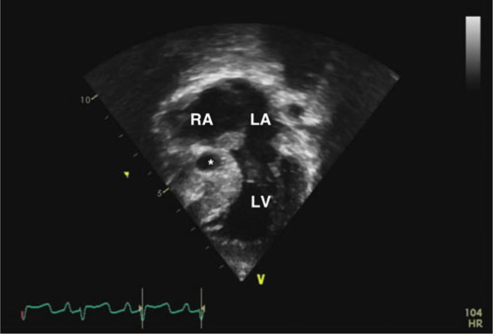
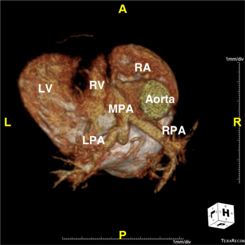
An 8-month-old male infant from a foreign country was admitted to our hospital for management of his CHD. He was noticed to be cyanotic shortly after birth and was diagnosed as having tricuspid atresia. He received oxygen therapy for 2 weeks without further treatment thereafter. At the time of admission to our hospital, he had oxygen saturation of around 75% on supplemental oxygen. Echocardiography revealed tricuspid atresia with situs solitus and concordant ventriculoarterial connections (Fig 1). The ventricular septum was intact, and the pulmonary valve was absent, with to-and-fro flow between the hypoplastic right ventricle and the main pulmonary artery (Supplementary video 1). The pulmonary blood flow was supplied by a small patent arterial duct. The right pulmonary artery was normal in size. However, there was juxtaductal left pulmonary arterial stenosis (diameter 4–5 mm). The atrial septal defect was restrictive, and the right atrium was severely dilated. The intact and hypertrophied ventricular septum bulged into the left ventricle (asymmetrical ventricular septal hypertrophy) without significant pressure gradient across the left ventricular outflow tract (Supplementary video 2). There was moderate degree of left ventricular diastolic dysfunction due to the left ventricular hypertrophy. Coronary-to-right ventricular fistulas were not observed. CT confirmed the findings of echocardiography and revealed multiple small collateral arteries supplying both lungs (Fig 2). After multidisciplinary discussion, the consensus operative plan was bidirectional cavopulmonary anastomosis.
Pre-operative echocardiography showing tricuspid atresia with a hypoplastic right ventricle (asterisk) and intact ventricular septum. LA = left atrium; LV = left ventricle; RA = right atrium.

Pre-operative CT showing a hypoplastic right ventricle and juxtaductal left pulmonary arterial stenosis. LPA = left pulmonary artery; LV = left ventricle; MPA = main pulmonary artery; RA = right atrium; RPA = right pulmonary artery; RV = right ventricle.

Surgery was performed under aorto-bicaval cardiopulmonary bypass. The arterial duct was divided. After cardioplegic arrest, atrial septectomy was performed. The main pulmonary artery was transected, and the cardiac stump was oversewn, thus completely excluding the right ventricle. No pulmonary valve tissue was observed through the transected main pulmonary artery. On beating heart, left pulmonary arterial angioplasty was performed using a flap of transected main pulmonary artery. Lastly, bidirectional cavopulmonary anastomosis was performed. Immediate post-operative mean pulmonary arterial pressure was around 12 mmHg, and oxygen saturation was around 85% with 50% inspired oxygen. Intraoperative transesophageal echocardiography revealed left ventricular outflow tract obstruction (peak velocity of 3.5 m/second) by the asymmetrical ventricular septal hypertrophy and dynamic left ventricular wall motion. Left ventricular systolic function was excellent and there was newly developed, moderate degree of mitral regurgitation. The patient was extubated at operating room. Post-operative course was uncomplicated, and the patient was discharged on the tenth post-operative day. Echocardiography before discharge revealed widely patent cavopulmonary anastomosis, non-restrictive atrial septal defect, mild degree of dynamic left ventricular outflow tract obstruction, and mild degree of mitral regurgitation.
Discussion
Tricuspid atresia is a CHD in which there is no or, rarely, an imperforate right atrioventricular connection. In the usual forms of tricuspid atresia, there is a hypoplastic right ventricle in the presence of a ventricular septal defect. When the ventricular septum is intact, the pulmonary valve is usually atretic. Tricuspid atresia with absent pulmonary valve and intact ventricular septum is an extremely rare cardiac malformation, historically associated with a poor prognosis.Reference Marin-Garcia, Roca, Blieden, Lucas and Edwards1–Reference Litovsky, Choy and Park3 Only a few cases with successful surgical palliation have been reported in the literature.Reference Mori, Ando, Satomi, Nakazawa, Momma and Takao2, Reference Forrest, Bini and Wilkinson4, Reference Lato, Gembruch and Geipel6–Reference Kasahara, Aeba and Yozu8 The present case shows that this rare CHD can be safely palliated in a staged approach towards the Fontan operation.
One of the characteristic morphologic features of this malformation is severely hypertrophied ventricular septum bulging into the left ventricular outflow tract (asymmetrical ventricular septal hypertrophy), which was also evident in our case.Reference Marin-Garcia, Roca, Blieden, Lucas and Edwards1–Reference Best, Vyas, Jaquiss and Sachdeva7 This can contribute to the development of left ventricular outflow tract obstruction, which is hazardous for single ventricle physiology. In our case, left ventricular outflow tract obstruction became obvious after the operation, probably due to volume-unloading nature of the bidirectional cavopulmonary anastomosis and dynamic left ventricular wall motion. Although the left ventricular outflow tract obstruction improved to mild degree at the time of discharge, judicious follow-up is mandatory for successful Fontan operation. Best et al reported “prophylactic” decompression of the right ventricle at the time of bidirectional cavopulmonary anastomosis by creating a ventricular septal defect to prevent the possibility of a hypertensive right ventricle bulging into the left ventricular outflow tract.Reference Best, Vyas, Jaquiss and Sachdeva7 Another characteristic morphologic feature of this malformation is the right ventricular dysplasia, in which large segments of the right ventricular myocardium are replaced with sinusoids and fibroelastic tissue.Reference Mori, Ando, Satomi, Nakazawa, Momma and Takao2, Reference Litovsky, Choy and Park3, Reference Lopes, Mennes, Delazoide, Iserin and Azancot5–Reference Kasahara, Aeba and Yozu8 This can cause variable degree of myocardial thinning and even aneurysmal change. A case with overt coronary-to-right ventricular fistulas has also been reported.Reference Kasahara, Aeba and Yozu8
Surgical management of the non-functional right ventricle in this malformation is controversial due to the rarity of this condition. Leaving the main pulmonary artery untouched and connected to the right ventricle is the simplest option, although this may cause suboptimal cavopulmonary circulation due to to-and-fro flow between the right ventricle and the pulmonary artery. Complete exclusion of the right ventricle at the time of bidirectional cavopulmonary anastomosis, as was performed in our case, is another option. However, there are concerns regarding the hazardous effects of the potentially hypertensive right ventricle on the left ventricle after complete exclusion of the right ventricle.Reference Best, Vyas, Jaquiss and Sachdeva7, Reference Kasahara, Aeba and Yozu8 Kasahara et al reported fenestrated exclusion of the right ventricle in a patient with this malformation and coronary-to-right ventricular fistulas.Reference Kasahara, Aeba and Yozu8 We believe that, in the absence of significant coronary-to-right ventricular fistulas, complete exclusion of the right ventricle can be safely performed in this malformation.
In summary, tricuspid atresia with absent pulmonary valve and intact ventricular septum is an extremely rare cardiac malformation. Although this was associated with a poor prognosis in the past, it can be safely palliated in a staged approach towards the Fontan operation in the present day. Surgical management of the non-functional right ventricle in this malformation may differ depending on the presence and severity of the coronary-to-right ventricular fistulas.
Supplementary material
To view supplementary material for this article, please visit https://doi.org/10.1017/S1047951119002749
Acknowledgements
None.
Financial Support
This research received no specific grant from any funding agency, commercial or not-for-profit sectors.
Conflicts of Interest
None.
Ethical Standards
This study was reviewed and approved by the Institutional Review Board of Seoul St. Mary’s Hospital.