In 1876, Sir James Paget published a scientific article describing cases of previously unrecognised chronic bone disease, which he called ‘osteitis deformans’ (Ref. Reference Roberts1). His study focused in particular on a man he had observed for over 20 years, who initially presented to St Bartholomew's hospital in London in 1854 with bone pain affecting the lower limbs. The patient subsequently went on to develop bowed and deformed legs, an enlarged skull, and eventually bone cancer in his left forearm, which ultimately led to his death. At that time Paget suggested that the patient was suffering from ‘a chronic form of inflammation of the bone’. While it is now realised that the condition Paget was describing does not simply represent bone inflammation, it is sobering to note that over 120 years elapsed before scientists and clinicians began to make significant inroads into understanding the aetiology of the condition we now know as Paget disease of bone (PDB).
Clinical aspects of PDB
Although PDB was not recognised as a clinical condition until the late 19th century, archaeological studies of skeletal remains over 500 years old show clear evidence of the disease at that time (Ref. Reference Rogers, Jeffrey and Watt2). It is now realised that this disorder represents the second most common metabolic bone disease in the UK, affecting as many as 1.6% of females and 2.5% of males over 55 years of age (Ref. Reference Cooper3). Notably, the prevalence is similarly high in other parts of Western Europe, Australia, New Zealand and North America, but much lower in areas such as Japan and Scandinavia (Ref. Reference Daroszewska and Ralston4). PDB is characterised by focal increases in bone turnover (remodelling), at so-called ‘pagetic lesions’, affecting one (monostotic) or more (polyostotic) sites throughout the skeleton (Ref. Reference Kanis5). The new bone formed in the lesions exhibits an abnormal architecture, which underlies the common symptoms such as bone pain, deformity, and susceptibility to pathological fractures – as well as deafness and osteoarthritis, which are evident in up to a third of those affected (Ref. Reference van Staa6). Osteosarcoma (as occurred in the case of Paget's patient) is the most serious complication and, although rare, most adult osteosarcomas occur in PDB patients (Ref. Reference Huvos7). These symptoms are associated with significant morbidity in PDB patients.
PDB as a disorder of bone remodelling
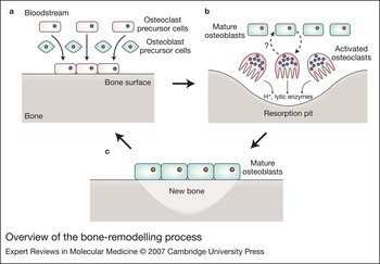
Bone mass in vertebrates is controlled by both osteoclasts (bone-resorbing cells, which are multinucleated cells derived from the monocyte–macrophage lineage) and osteoblasts (bone-forming cells, which are mononucleated and of mesenchymal origin). The concerted, opposing activities of these two cell types ensure bone is constantly remodelled or ‘turned-over’ (on a micro-scale at resorption pits or ‘lacunae’ throughout the skeleton) in a process essential for maintaining adult bone structure and function (Fig. 1).
Overview of the bone-remodelling process. Bone is subjected to constant remodelling throughout the skeleton. (a) Mononucleated osteoclast precursors are recruited from the blood stream to the bone surface. (b) Osteoclast precursors mature into multinucleated activated osteoclasts that resorb bone using a combination of acidification and lytic enzymes, generating resorption pits termed ‘lacunae’. Mature osteoblasts regulate osteoclast activity (dashed arrow) and likewise osteoclasts may regulate osteoblast activity. (c) Osteoclasts are replaced by mature osteoblasts, which form new bone matrix. Mineralisation of the newly formed bone restores the normal bone structure. Although bone formation and bone resorption are coupled in PDB, resorption initially exceeds formation; furthermore, in the later stages of disease, formation greatly exceeds resorption and sclerotic lesions develop.

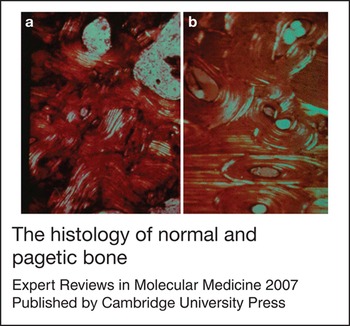
The primary cellular defect in PDB is thought to occur at the level of osteoclasts. In PDB, net osteoclastic activity is increased; pagetic lesions typically contain increased numbers of osteoclasts relative to normal bone, which are of increased size and contain more nuclei than normal osteoclasts (Ref. Reference Reddy8). The increase in osteoclast activity leads to secondary increases in osteoblast activity. Although bone formation and bone resorption are coupled in PDB, resorption initially exceeds formation, while in later stages formation greatly exceeds resorption and sclerotic lesions develop. As a result, the overall process of bone formation becomes both accelerated and disorganised, consistent with an elevation in markers of both bone formation and resorption, ultimately resulting in the abnormal bone structure that characterises the disorder (Fig. 2) (Ref. Reference Kanis5).
The histology of normal and pagetic bone. Images of (a) normal and (b) pagetic bone viewed under polarised light. Note the pagetic bone is of disorganised structure and demonstrates a so-called ‘mosaic pattern’. Reproduced with permission from The Paget Foundation (http://www.paget.org/).

Genetic factors and disease mechanisms
PDB and PDB-like syndromes
A genetic contribution to PDB is indicated by the findings that PDB remains prevalent in migrant populations originating from Western Europe (Refs Reference Daroszewska and Ralston4, Reference Gardner, Guyer and Barker9) and that there is a family history of PDB in up to 40% of patients (Refs Reference Siris10, Reference Sofaer, Holloway and Emery11, Reference Morales-Piga12). Familial PDB is usually inherited in an autosomal dominant manner (Refs Reference Morales-Piga12, Reference Hocking13, Reference Haslam14) with incomplete penetrance. At least seven susceptibility loci have been implicated (but not all confirmed) in classical PDB (Ref. Reference Daroszewska and Ralston4).
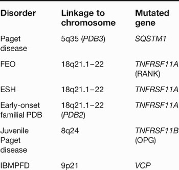
Several rarer PDB-like syndromes that are clearly genetic have also been identified, including familial expansile osteolysis (FEO), expansile skeletal hyperphosphatasia (ESH), early-onset familial PDB, and juvenile Paget disease (also known as juvenile hyperphosphatasia, which shares phenotypic overlap with classical PDB but is a distinct clinical entity). These conditions also involve a primary dysfunction of osteoclasts and show characteristic histological features similar to PDB, but they have an earlier age of onset as well as other subtle clinical differences. In addition, the disorder ‘inclusion body myopathy associated with PDB and frontotemporal dementia’ (IBMPFD) is characterised by (among other symptoms) early-onset PDB (Ref. Reference Layfield and Hocking15). It is largely through the identification of the genetic defects involved in PDB and these PDB-like syndromes that significant recent insights into disease mechanism at the molecular level have been made (Table 1).
Genetic defects identified in Paget disease of bone and related syndromes

Abbreviations: ESH, expansile skeletal hyperphosphatasia; FEO, familial expansile osteolysis; IBMPFD, inclusion body myopathy associated with PDB and frontotemporal dementia; PDB, Paget disease of bone; SQSTM1, sequestosome 1 gene; TNFRSF11A, tumour necrosis factor receptor superfamily, member 11a [encoding the ‘receptor activator of NF-κB’ (RANK)]; TNFRSF11B, tumour necrosis factor receptor superfamily, member 11b [encoding osteoprotegerin (OPG)]; VCP, valosin-containing protein.
Genetic defects
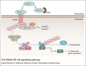
The most common genetic mutations found in classical PDB patients affect the SQSTM1 (sequestosome 1) gene, located on chromosome 5 at the PDB3 locus (Ref. Reference Watts16). This gene encodes the SQSTM1 protein (also known as p62), which has diverse functional properties in a variety of cell types (Ref. Reference Watts16). In osteoclasts, SQSTM1 appears to be an important component of an intracellular signalling pathway that leads to the activation of the nuclear factor κB (NF-κB) transcription factor (Ref. Reference Duran17). Specifically, SQSTM1 is one of the many scaffold proteins (Fig. 3) that act downstream of the ‘receptor activator of NF-κB’ (RANK) receptor, relaying extracellular signals to changes in osteoclast gene expression that are required for osteoclastogenesis as well as for the bone-resorbing activity of mature osteoclasts (Ref. Reference Teitelbaum and Ross18). RANK can be engaged by various ligands, but importantly is activated by RANK ligand (RANK-L), an osteoblast-derived cytokine that stimulates osteoclast activity (and also inhibits the ‘removal’ via apoptosis of osteoclasts). Indeed, there is emerging evidence that aberrant RANK-mediated NF-κB signalling in osteoclasts may underlie the disease process in PDB (see later). Several different SQSTM1 missense and truncating mutations have been identified over the past five years and these are found (in different populations) in up to 50% of familial and 10–20% of ‘sporadic’ (i.e. no family history) PDB cases (Ref. Reference Daroszewska and Ralston4). Notably, all of these mutations affect the same small region of the 440 amino acid SQSTM1 protein close to the C-terminus, which contains a ubiquitin-associated (UBA) domain (Refs Reference Laurin19, Reference Hocking20, Reference Johnson-Pais21, Reference Good22, Reference Beyens23, Reference Eekhoff24, Reference Falchetti25, Reference Hocking26, Reference Beyens27, Reference Rea28, Reference Collet29).
The RANK–NF-κB signalling pathway. RANK-L is an osteoblast-derived cytokine that stimulates osteoclast activity. RANK-L binding by the cell-surface receptor RANK promotes receptor trimerisation and recruitment of TRAF6 to the intracellular tail of RANK. TRAF6 uses its intrinsic ubiquitin-ligase activity to undergo autoubiquitination. SQSTM1, which complexes with aPKC, may use its UBA domain to regulate this ubiquitination of TRAF6. Assembly of a polyubiquitin chain on TRAF6 leads to activation of TAK1, a kinase that forms a complex with TAB2 and TAB3. TAK1 phosphorylates and activates the IKKβ subunit of a complex also involving IKKα and NEMO. This complex in turn phosphorylates IκB, which is subsequently ubiquitinated; in this case the polyubiquitin chain serves as a signal for the degradation of IκB by the proteasome complex, a process that may be regulated by VCP. The NF-κB transcription factor is then free from its inhibitor IκB and enters the nucleus to stimulate selective gene expression. Viral infection or exposure to environmental toxins such as arsenic may upregulate the expression of SQSTM1. Abbreviations: aPKC, atypical protein kinase C; IκB, inhibitor of κB; IKK, IκB kinase; NEMO, NF-κB essential modulator; NF-κB, nuclear factor κB; OPG, osteoprotegerin; RANK, receptor activator of NF-κB; RANK-L, RANK ligand; SQSTM1, sequestosome 1; TRAF6, TNF receptor-associated factor 6; Ub, ubiquitin; UBA domain, ubiquitin-associated domain; VCP, valosin-containing protein.

Although the other genes mutated in classical PDB remain to be identified, those involved in many of the rarer familial PDB-like syndromes have now been discovered (Table 1), and interestingly these provide further support for the idea that disordered osteoclast RANK–NF-κB signalling may be central to disease aetiology. For example, FEO, ESH and early-onset familial PDB are all caused by different mutations in the gene-encoding RANK (TNFRSF11A), the receptor that functions upstream of SQSTM1 in osteoclasts (Fig. 3) (Refs Reference Hughes30, Reference Whyte and Hughes31, Reference Nakatsuka, Nishizawa and Ralston32). By contrast, juvenile Paget disease is caused by deletions and missense mutations affecting a further gene, TNFRSF11B, which encodes a protein called osteoprotegerin (OPG) (Refs Reference Whyte33, Reference Cundy34). However, OPG is also connected to osteoclast RANK-mediated signalling, since it acts as a soluble ‘decoy receptor’ that binds to RANK-L and competes with its binding to RANK. Finally, IBMPFD is caused by mutations in the VCP (valosin-containing protein; also known as p97) gene (Ref. Reference Layfield and Hocking15), which encodes a protein that might also be connected to NF-κB signalling pathways (see later) (Ref. Reference Asai35).
RANK–NF-κB signalling pathway
RANK is a member of the tumour necrosis factor (TNF) receptor family (Ref. Reference Hsu36), which also includes the interleukin 1 (IL-1) receptor and Toll-like receptors, and plays a central role in osteoclast biology. RANK-mediated signalling is complex, but can potentially lead to the activation of several transcription factors – AP-1, NFATc1 and NF-κB – each of which regulates osteoclast activity or osteoclastogenesis (Ref. Reference Teitelbaum and Ross18). However, only the signalling pathway resulting in NF-κB activation is thought to involve RANK (and OPG) as well as SQSTM1, indicating this is the pathway that is likely to be commonly affected in PDB and the PDB-like syndromes.
Considerable insights into the molecular events that underlie RANK-mediated signalling have been made in recent years. When RANK is activated by RANK-L, the receptor undergoes trimerisation and its cytoplasmic tail then associates with TNF-receptor-associated factor (TRAF) adaptor protein TRAF6 (Ref. Reference Galibert37). The importance of the RANK–TRAF6 interaction in osteoclast homeostasis is clearly demonstrated by experiments using a cell-permeable peptide to block the binding of the two proteins, which results in inhibition of osteoclastogenesis in vitro (Ref. Reference Ye38). Upon activation by receptor binding, TRAF6 post-translationally modifies itself by ‘ubiquitination’: it uses its intrinsic ubiquitin-ligase (E3) activity (associated with its RING domain) to covalently conjugate multiple units of the small protein ubiquitin in a so-called Lys63-linked polyubiquitin chain (Ref. Reference Deng39). In fact, ubiquitination is a common modification used to regulate NF-κB signalling (Ref. Reference Chen40) and the resulting polyubiquitin chain effectively acts as a scaffold to establish new (signal-induced) protein–protein interactions required for the signalling pathway (Ref. Reference Chen40). Accordingly, many signalling adaptor proteins contain ubiquitin-binding domains (Ref. Reference Andersen, Hofmann and Hartmann-Petersen41), through which they can interact noncovalently with other ubiquitinated signalling proteins. Indeed, recognition of the Lys63-linked polyubiquitin chain on TRAF6 by the ubiquitin-binding TAB2–TAB3 adaptor proteins is a key step in the activation of downstream NF-κB signalling events (Ref. Reference Kanayama42). In this case, a complex involving TAB2, TAB3 and also TAK1 then phosphorylates and activates a further complex that includes IKKβ˙ (a kinase of the NF-κB inhibitor IκB), which subsequently phosphorylates IκB, resulting in its (ubiquitin-dependent) degradation (Ref. Reference Layfield and Hocking15). This releases the NF-κB transcription factor to enter the nucleus and selectively activate gene expression.
How do the PDB-associated mutations, which appear to affect different components of the same pathway (Fig. 3), perturb RANK–NF-κB signalling and ultimately lead to the disordered osteoclast function thought to be the principal defect in PDB and related syndromes? Certainly NF-κB activity is essential for the bone-resorbing function of mature osteoclasts and also to control normal osteoclastogenesis (Refs Reference Teitelbaum and Ross18, Reference Jimi43). It therefore follows that increases in NF-κB signalling could at least in part explain the primary cellular defect (i.e. the net increase in osteoclastic activity) in PDB, and there is considerable emerging evidence that this may be the case.
SQSTM1 mutations
The pathogenic mechanisms arising from SQSTM1 mutations – the most common PDB-associated mutations – have received most attention. Cell-based NF-κB reporter assays show that PDB mutant SQSTM1 constructs can activate signalling more effectively than the wild-type sequence (Refs Reference Duran17, Reference Rea28). Consistent with these observations, mouse macrophage cells lines transfected with mutant SQSTM1 differentiated more readily into osteoclast-like cells (OLCs) than cells transfected with wild-type SQSTM1, and when human monocytes from PDB patients carrying a germline SQSTM1 mutation were differentiated in vitro, the resulting OLCs showed increased bone-resorbing activity relative to cells derived from normal monocytes (Refs Reference Rea28, Reference Yip44).
Precisely how do the SQSTM1 mutations result in this altered activity? Recently, TRAF6 was found to form a complex with SQSTM1 and an atypical protein kinase C (aPKC) in osteoclasts, which was shown to be important for the RANK-L-mediated activation of NF-κB (Ref. Reference Duran17). The same study highlighted the importance of SQSTM1 in osteoclast function, demonstrating that SQSTM1-null mice exhibit a defect in osteoclastogenesis in response to bone-resorbing factors (Ref. Reference Duran17). Clues as to the role of SQSTM1 in this signalling complex, and the effect of SQSTM1 mutations in some PDB patients, may lie in the fact that the SQSTM1 mutations all affect the UBA domain of the protein: missense mutations cause amino acid substitutions within the UBA domain sequence, and truncating mutations remove most or all of the UBA domain sequence from the SQSTM1 protein (Ref. Reference Layfield and Hocking15). UBA domains are short sequences (typically ∼50 residues) that are found in several enzymes and regulators of the ubiquitin system, and represent one class of ubiquitin-binding domain (Refs Reference Andersen, Hofmann and Hartmann-Petersen41, Reference Raasi45). SQSTM1 was originally characterised as a ubiquitin-binding protein (Ref. Reference Vadlamudi46) and utilises its UBA domain to mediate this property (Ref. Reference Layfield47). Importantly, in vitro protein-binding assays demonstrate that all the SQSTM1 mutations tested impair the ability of the SQSTM1 protein to bind to ubiquitin, relative to the wild-type protein (Refs Reference Cavey48, Reference Cavey49). Is this loss of ubiquitin-binding function directly related to disease mechanism in these cases of PDB? Although a cause and effect relationship between the SQSTM1 mutations and PDB is not definitively established, it seems likely that the aberrant function of mutant SQSTM1 protein is directly related to the mechanism of pathogenesis. For example, in PDB patients with SQSTM1 mutations, the age at diagnosis is typically 10 years younger than in patients without mutations (Ref. Reference Hocking26) and SQSTM1 mutations are only found in PDB patients and not in unaffected controls. Also, some mutations exert less severe effects on the ubiquitin-binding function of the SQSTM1 protein than others (Refs Reference Cavey48, Reference Cavey49), and PDB severity appears to increase as the effects of different mutations on protein function become more pronounced. Indeed, truncating mutations produce a more severe disease phenotype than missense mutations (Ref. Reference Hocking26) and are the most detrimental to binding of ubiquitin by SQSTM1 in vitro (Ref. Reference Cavey48), supporting the idea that changes in the ubiquitin-binding properties of the SQSTM1 protein may be related to the disease severity (Ref. Reference Layfield47).
Nevertheless, exactly how the impairment of ubiquitin-binding function of SQSTM1 impacts on the osteoclast RANK–NF-κB signalling cascade is still unclear. One study indicates that SQSTM1 may use its UBA domain (in a mechanism involving ubiquitin-binding) to regulate the signal-induced changes in the Lys63-linked ubiquitination of TRAF6 (Ref. Reference Wooten50). However, how a reduced ubiquitin-binding function of SQSTM1 could translate into an apparent activation of NF-κB signalling relative to wild-type SQSTM1 (Refs Reference Duran17, Reference Rea28) is not easily explained. Alternatively, perhaps SQSTM1 normally suppresses osteoclast NF-κB signalling via interactions with an (unknown) ubiquitinated target protein, and loss of ubiquitin-binding affinity results in a derepression of signalling. Another possibility is that functional SQSTM1 may even be required for the ubiquitin-dependent degradation of TRAF6, which can occur in response to interferon γ (Ref. Reference Takayanagi51). Regardless of the precise mechanism, the overall effect of SQSTM1 mutations appears to manifest as either constitutive activation of osteoclast NF-κB signalling, or more likely an increased sensitivity of osteoclasts to stimulatory signals (i.e. cytokines) (Ref. Reference Menaa52). Thus, a lower level of stimulatory signal may be required to achieve the same level of NF-κB activation in PDB mutant osteoclasts than in wild-type cells, with the net effect being the promotion of osteoclastogenesis and osteoclast activity.
OPG, RANK and VCP mutations
By contrast to SQSTM1 mutations, there is comparatively less known about the mechanisms by which mutations affecting OPG, RANK and VCP exert their effects on osteoclast NF-κB signalling.
OPG is a negative regulator of RANK-mediated signalling (Ref. Reference Menaa52), so a loss of functional OPG – which occurs in juvenile Paget disease with TNFRSF11B deletions/missense mutations (Refs Reference Whyte33, Reference Cundy34) – presumably results in increased levels of free RANK-L, thus promoting RANK-mediated NF-κB signalling. In fact, an increased RANK-L production from cells derived from pagetic bone compared with normal bone from the same patient has also been noted (Ref. Reference Hofbauer and Heufelder53). A recent study also indicates that the circulating RANK-L:OPG ratio may be higher in PDB patients than controls (Ref. Reference Martini54), although how this (and increases in other cytokines) might translate to focal increases in bone turnover is unclear.
The mutations that occur in FEO, ESH and early-onset familial PDB are subtly different (Refs Reference Hughes30, Reference Whyte and Hughes31, Reference Nakatsuka, Nishizawa and Ralston32) but all affect the same gene, TNFRSF11A, which encodes RANK. In each case, amino acid insertions of different lengths are introduced in frame in the sequence for the signal peptide (which directs post-translational transport to the cell surface) of RANK. Normal cleavage of the signal peptide by proteolytic processing does not appear to occur in these mutant RANKs (Ref. Reference Hughes30) and cell-based reporter assays indicate that, like mutant SQSTM1, mutant RANK may be more effective at activating NF-κB signalling than the wild-type protein. How this might occur mechanistically has not been investigated in detail, but it seems reasonable to predict that the mutant RANK may not reach its ‘normal’ subcellular localisation and may in fact produce a stimulatory ‘signal’ intracellularly without the need for ligand binding. Clearly more research in this area is merited.
VCP mutations were recently found to be the cause of the progressive disorder IBMPFD (Ref. Reference Layfield and Hocking15), a condition in which pagetic changes are commonly seen. Although VCP is a multifunctional protein (Ref. Reference Halawani and Latterich55) and some studies indicate that IBMPFD mutations of VCP appear to disrupt the process of endoplasmic-reticulum-associated degradation (ERAD) (the ubiquitin-dependent degradation of misfolded proteins extracted from the ER; Ref. Reference Weihl56), earlier publications described a role for VCP in regulating NF-κB signalling probably via promoting the degradation of ubiquitinated IκB (Refs Reference Asai35, Reference Dai57).
These intriguing observations indicate the possibility that OPG, RANK, SQSTM1 and VCP may in fact all function within the same signalling pathway, and by extension that disease-associated mutations may exert their effects at different points within this pathway. There is also a final interesting parallel between PDB with SQSTM1 mutations and IBMPFD caused by VCP mutations. As noted earlier, SQSTM1 mutations detrimentally affect the ubiquitin-binding UBA domain of the gene product (Refs Reference Cavey48, Reference Cavey49), and VCP mutations also cluster in the N-terminal ubiquitin-binding region of VCP (Ref. Reference Layfield and Hocking15). Although the effects of the latter on protein function are currently not know, it is tempting to speculate that disruption of ubiquitin-mediated protein–protein interactions (perhaps even with the same ubiquitinated target of VCP and SQSTM1) could be a commonality between PDB with SQSTM1 mutations and IBMPFD, and that these conditions could represent further examples of a spectrum of disorders involving primary defects in ubiquitin-mediated processes (Ref. Reference Layfield58).
Viral factors and PDB
For many years the idea of a viral involvement in PDB aetiology has been supported by several research groups, with considerable emphasis placed on the infection of osteoclasts with a paramyxovirus as being a possible cause of PDB (Ref. Reference Reddy59). Some of the first indications that viruses may play a role in PDB came from microscopical analyses of pagetic osteoclasts, which were shown to commonly contain nuclear (as well as occasionally cytoplasmic) inclusions that resemble paramyxovirus nucleocapsids (Refs Reference Harvey60, Reference Mills and Singer61). These observations led to the proposal that a persistent infection with a paramyxovirus such as the measles virus may be a cause of PDB (Refs Reference Mills62, Reference Rebel63).
Subsequent support for this hypothesis has come from several experimental approaches. First, reverse transcriptase PCR (RT-PCR) studies have revealed measles virus nucleocapsid (MVNP) sequences, including a complete coding sequence (Refs Reference Reddy64, Reference Friedrichs65), in samples from PDB patients. However, there has been fierce debate as to whether these results are ‘real’ or artifactual (e.g. Ref. Reference Helfrich66), and the inability of other groups to reproduce these findings is noted in a recent study, which also indicated that differences in the sensitivity of RT-PCR detection methods does not appear to explain this discrepancy (Ref. Reference Ralston67). Despite this, functional evidence has also been presented that the MVNP sequence is able to convert osteoclasts to a ‘pagetic phenotype’ (Ref. Reference Kurihara68). This pagetic phenotype has been defined as follows (Refs Reference Menaa52, Reference Menaa69, Reference Kurihara70): increased bone-resorbing activity; hypersensitivity to 1,25(OH)2D3, RANK-L and TNF-α; and increased expression of the TAF(II)-17 gene. Expression of MVNP in osteoclasts in vitro (Ref. Reference Kurihara68) and in an in vivo mouse model (Refs Reference Kurihara71, Reference Kurihara72) resulted in a pagetic phenotype, and some of the animals also reportedly contained pagetic-like lesions. Clearly more research needs to be done in this area to confirm the link between the measles virus [or other viruses – canine distemper is also a candidate (Ref. Reference Mee73)] and PDB. The association would be significantly strengthened if intact virus could be recovered from PDB patients.
A final intriguing observation of note comes from an in vitro study in which human osteoclast precursors were infected with canine distemper virus (Ref. Reference Selby, Davies and Mee74). This resulted not only in an increase in osteoclast number, size and resorptive activity, but also in SQSTM1 (and ubiquitin) expression. Notably, increased SQSTM1 levels have also been noted in immortalised lymphocytes from PDB patients (Ref. Reference Collet29). Although viruses have multiple other effects that enhance NF-κB signalling, which may contribute to the development of PDB, elevation of SQSTM1 levels offers a potential mechanism by which viral infection may impact directly on RANK–NF-κB signalling, providing hints of a possible unifying mechanism of pathogenesis in PDB involving either SQSTM1 mutations or viral infection (Fig. 3).
Environmental factors and PDB
There is also considerable evidence for environmental contributors to PDB aetiology, not least because the prevalence of PDB has significantly decreased in the UK in recent years (Ref. Reference Cundy75). In addition, the offspring of SQSTM1-mutant PDB patients are less likely to be diagnosed with the disease and have less severe disease than their parents (Ref. Reference Bolland76), further implicating the contribution of environmental factors. Suggestions that diet (Ref. Reference Siris77) and exposure to environmental toxins (Ref. Reference Lever78) could be factors affecting PDB incidence certainly merit further investigations. Indeed, clusters of PDB cases in Lancashire identified in a 1974 survey have been linked to the cotton industry. One hypothesis proposed that arsenic pesticide from cotton bales might be responsible for the high prevalence of disease (Ref. Reference Lever78). This is of particular interest since arsenic is known to affect signal transduction pathways at nontoxic levels. Furthermore, cell-based studies have previously noted that arsenic increased the expression and direct ubiquitination of SQSTM1 (Ref. Reference Aono79), again suggesting a possible unifying mechanism in PDB, where disordered RANK–NF-κB signalling could arise as a result of diverse factors (Fig. 3).
Clinical implications/applications
Current treatment for PDB symptoms is largely based on the use of bisphosphonates, which target and induce apoptosis of osteoclasts via mechanisms thought to involve inhibition of protein prenylation or via their metabolism to cytotoxic ATP analogues (Ref. Reference Coxon, Thompson and Rogers80). However, it is increasingly realised that some bisphosphonates may also have extraskeletal immunomodulatory effects (Ref. Reference Corrado, Santoro and Cantatore81), influencing the production of certain cytokines. Many trials have been designed to determine, for example, the optimum methods of bisphosphonate administration (Ref. Reference Hoskins82) and whether intensive versus symptomatic treatment with bisphosphonates offers any clinical advantage (Ref. Reference Langston83). Although the latter study indicated no obvious advantages of intensive bisphosphonate treatment in patients with existing PDB, future trials involving the presymptomatic treatment of individuals selected based upon SQSTM1 genotype would be of particular interest.
The improving knowledge of molecular mechanisms disrupted in PDB indicates that strategies based upon manipulation of osteoclast RANK–NF-κB signalling might be used to treat PDB symptoms. Hormones, drugs and antibodies could or have been used to regulate RANK-L and OPG expression, or the RANK-L–RANK interaction (Refs Reference Vaananen84, Reference Tat85, Reference McClung86), and recombinant OPG has been used in the treatment of a juvenile PDB patient (Ref. Reference Cundy87). Further drug discovery activities in this area are expected in the near future.
Research in progress and outstanding research questions
This review highlights many of the recent insights into PDB aetiology, as well as the outstanding research questions to be addressed in the coming years. Clearly many important points require clarification, which include (but are not limited to) the following. What are the factors that contribute to ‘sporadic’ PDB, and how important are viruses and environmental toxins? Why is PDB focal, affecting only certain sites within the skeleton? Is there really a cause and effect relationship between the SQSTM1 mutations and PDB? In addition to SQSTM1, what are the other genes involved in familial PDB? Why is there such a wide variation in the phenotype and disease severity in PDB patients, including in those with the same mutations (Refs Reference Leach88, Reference Morisette, Laurin and Brown89)?
With reference to the first point, preliminary reports indicate that pagetic cells from bone sections of patients with sporadic PDB may in fact in some cases carry somatic SQSTM1 mutations [M.F. Hansen (University of Connecticut Health Center, Farmington, USA), pers. commun.]. With respect to the second point, could it be that mechanical loading at specific regions of the skeleton act as a disease trigger? And in relation to the third point, the first experimental (animal) models for familial PDB are beginning to be described in the literature, which will help investigate the link between SQSTM1 mutations and PDB. For example, a mouse model has recently been generated with expression of PDB mutant SQSTM1 targeted to the osteoclasts (Ref. Reference Kurihara90). These animals were reported to show some pagetic-like features, such as hyperresponsiveness of osteoclasts to RANK-L and TNF-α, but not the full pagetic phenotype. The authors argued that a ‘missing factor’ was required for the full pagetic phenotype and presented evidence that this might be a viral gene product (MVNP) – perhaps indicating that SQSTM1 and viral infection together are important for PDB to occur. An alternative explanation is intimated from the observation that the animals developed increased osteoclast numbers and progressive bone loss, but a secondary increase in osteoblast activity, as seen in PDB, was not evident. Of course, in familial PDB patients SQSTM1 mutations are germline and would therefore affect other cell types, including osteoblasts. So perhaps osteoblasts (largely ignored in PDB research to date) play a hitherto unrecognised role in PDB pathogenesis? Clearly, the debate about the relative importance of genetic and viral factors is far from resolved, and represents one of the most argued but at the same time most interesting aspects of PDB research to be pursued in the future.
Acknowledgements and funding
I thank members of my research group for critical reading of the manuscript, and the anonymous peer reviewers of the article for their helpful comments. I thank The National Association for the Relief of Paget's Disease (NARPD) and the Biotechnology and Biological Sciences Research Council for funding some aspects of our PDB research.