Case details
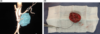
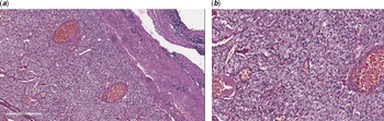
A 10-year-old girl weighing 25 kilos presented with severe cyanosis and squatting, with recent-onset episodes of paroxysmal headaches and giddiness. She was an unoperated case of Tetralogy of Fallot. She had previously undergone palliation with an emergency Blalock–Taussig shunt as an infant and was lost to follow-up. She was evaluated a year back for headaches with MRI brain which was normal. A detailed clinical evaluation done revealed persistently high blood pressures (200/110 mmHg) and severe desaturation (oxygen saturation of 55–60%) Cardiac examination revealed a single second heart sound with no pulmonary outflow or a shunt murmur. Chest X-ray showed cardiomegaly with oligemic lung fields. Echocardiogram revealed Tetralogy of Fallot with severe pulmonary stenosis, blocked shunt and biventricular dysfunction. Ventricular dysfunction, in the presence of systemic hypertension, was possibly due to catecholamine toxicity of the heart causing cardiomyopathy. She underwent investigations for hypertension. An ultrasound abdomen revealed normal renal artery Dopplers and a left-sided extra-renal mass. CT abdomen confirmed a well-defined, heterogeneous, contrast-enhancing lobulated mass in left para-aortic region, suggestive of a paraganglioma (Fig 1a). 24 hours urinary metanephrine level measured was significantly elevated (511 microgram/24 hours – normal range – 79–297 mics/24 hours), consistent with paraganglioma. She was started on Prasozin and Propranolol for control of her hypertension. Though her hypertension could be kept under control with some fluctuations, the alpha blockade effect and drop in systemic vascular resistance resulted in worsening of her cyanosis with severe hypoxia that her saturations dropped to 30–40%. It was hence decided to do a palliative procedure, an aortopulmonary shunt to improve the pulmonary blood flow and improve her oxygen saturations. Her blood pressures stabilised in the post-operative period, and she was discharged on two antihypertensives. She later underwent an isotope (metaiodobenzyl guanidine – MIBG) scan to localise the paraganglioma and to look for any other lesions in the body. Isotope scan revealed localised lesion with no isotope uptake in other parts of the body. Following this, 3 months later, she underwent excision of the paraganglioma. The lesion was large, measuring 7 × 6 cm in the left infra-renal para-aortic region. The tumour was highly vascular and densly adherent and it was excised completely (Fig 1b). There were gross fluctuations in blood pressure during tumour handling in the intraoperative period, which was effectively managed. She had a good clinical recovery and was off all antihypertensive medications in a week following removal of the tumour. Histopathological examination of the tumour confirmed the diagnosis of extra-renal paraganglioma with no evidence of capsular invasion, (Fig 2a and b) suggesting a benign nature of the tumour. Three months later, she underwent a repeat metaiodobenzyl guanidine (MIBG) scan which revealed no isotope uptake, suggesting the complete absence of tumour activity, including other sites of the body. She then underwent cardiac catheterisation to delineate the intracardiac anatomy and for presence of any aortopulmonary collaterals. Following this, she underwent complete repair of Tetralogy of Fallot, closure of ventricular septal defect and infundibular resection. She had an uneventful post-operative recovery. At 1-year-follow-up, she is asymptomatic with normal post-operative cardiac status and is normotensive.
( a ) 3D Reconstructed image from CT abdomen showing tumour in the para-aortic region. ( b ) Pathological specimen of excised tumour.

( a ) Haematoxylin ans Eosin section shows tumour cells surrounded by thick capsule. Tumour cells are present in tight nests in Zell-Ballen pattern (100 X). ( b ) Individual cell showing rond to fusiform nuclei, granular eosinophilic cytoplasm with no atypical mitotic figures (400 X).

Discussion
Concomitant occurrence of phaeochromocytoma/paraganglioma with unoperated Tetralogy of Fallot is rare. Considering the pathophysiology of both these conditions, the clinical picture is complicated and poses a challenge in management. The association between the occurrence of a neuroendocrine tumour with conditions causing chronic hypoxia-like cyanotic congenital heart disease has been described by Opotowsky et al. Reference Opotowsky, Moko and Ginns1 They have analysed the largest series of such cases and have speculated on the link between chronic hypoxia and development of neuroendocrine tumours. De la Monte et al. Reference de la Monte, Hutchins and Moore2 reported one of the earliest associations between peripheral neuroblastic tumours and chronic hypoxia.
Paragangliomas are catecholamine secreting tumours, which have persistent alpha and beta-agonistic actions, though alpha action predominates, and this causes systemic hypertension. The elevated systemic vascular resistance alters the haemodynamics in Tetralogy of Fallot and helps to maintain pulmonary blood flow. Such patients may have delayed cyanosis due to presence of adequate pulmonary blood flow. Reference Khoury, Kallile and May3 However in long-standing cases, when the pulmonary obstruction is also very severe, it leads to biventricular dysfunction and heart failure. Reference Kannan4 Our patient had significant biventricular dysfunction at presentation, which promptly reversed after treatment.
Though co-occurrence of paraganglioma and cyanotic heart disease has been repeatedly reported, management of both the conditions simultaneously has been rarely reported. Balakrishnan et al. Reference Balakrishnan, Ravikumar, Rao and Balakrishnan5 reported the management of both these conditions in the same hospitalisation. In most patients, removal of the endocrine tumour first is recommended, however, our patient needed a staged approach, considering the severity of cyanosis and hypoxia, presence of malignant hypertension, worsening of hypoxia with antihypertensive drugs and presence of significant biventricular dysfunction. An initial palliation with an aortopulmonary shunt improved the pulmonary blood flow, allowed better control of hypertension using alpha and beta-blocking agents and remarkable improvement in ventricular function. Perioperative management during tumour excision was easier after the palliative surgery for congenital heart disease. This was followed by intracardiac repair of Tetralogy of Fallot.
Paragangliomas are known to have a recurrence and malignant transformation depending on the genetic variability. Reference Welander, Söderkvist and Gimm6 In the case series associated with cyanotic heart disease, all tumours identified were benign with no metastasis. Our patient also showed benign nature of tumour on histopathology. Imaging with metaiodobenzyl guanidine (MIBG) revealed no evidence of metastasis. However, MIBG scan is less sensitive and it is important to follow-up this patient with preferably with newer somatostatin analogues like DOTA-NOC-PET can be used imaging for tumour recurrence. Genetic tests have not been performed on this patient due to lack of resources.
Conclusion
Paragangliomas occurring in the setting of chronic hypoxia in congenital cyanotic heart disease is a known entity. Understanding the pathophysiology of both these conditions and planning management is important while treating these children.
Acknowledgements
None.
Financial support
This research received no specific grant from any funding agency, commercial or not-for-profit sectors.
Conflicts of interest
None.
Ethical standards
The authors assert that all procedures contributing to this work comply with the ethical standards of the relevant national guidelines on human experimentation and with the Helsinki declaration of 1975, as revised in 2008 and has been approved by the Institutional committees.