Cor triatriatum sinistrum (subdivided left atrium) is a rare congenital cardiac anomaly whose embryologic basis has not been fully established Reference Nassar and Hamdan1,Reference Gheissari, Malm, Bowman and Bierman2 . It has been reported in 0.1–0.4% of patients with congenital heart disease Reference Hayes, Liu, Tam and Kass3–Reference Rudienė, Hjortshøj and Glaveckaitė6 . In this rare condition, the left atrium is abnormally divided into distinct compartments by fenestrated fibromuscular septum leading to obstruction in the blood flow Reference Kokotsakis, Anagnostakou and Almpanis7,Reference Zepeda, Morcos and Castellanos8 . The post-erosuperior chamber having pulmonary vein receives venous blood and an anteroinferior chamber in contact with the mitral valve contains left atrial appendage Reference Kokotsakis, Anagnostakou and Almpanis7–Reference Diestro, Regaldo, Gonzales, Dorotan, Espiritu and Pascual9 . Cor triatriatum sinister is generally diagnosed during infancy or early childhood; however, a few cases remain asymptomatic and hence diagnosed in adults Reference Nassar and Hamdan1 . The mortality rate in the untreated patients with cor triatriatum sinister is as high as 70% and surgical excision of the obstructing membrane is the treatment of choice Reference Gheissari, Malm, Bowman and Bierman2 . The cor triatriatum sinister anomaly can be easily diagnosed with echocardiography including measurement of the trans-membrane pressure gradient to evaluate the functional significance Reference Nassar and Hamdan1,Reference Saxena, Burkhart, Schaff, Daly, Joyce and Dearani10,Reference Stiermaier, Reil and Eitel11 . However, investigators have to be aware of this abnormality to ensure accurate diagnosis. This rare case report discusses the case of a 25-year-old patient with a cor triatriatum sinister and a mobile left atrial thrombus which was surgically excised leading to rapid recovery of the patient.
Case presentation
A 25-year-old man was presented to our institution with dyspnoea that gets aggravated during exertion. On physical examination, initial blood pressure was 103/71 mmHg and heart rate was 100 bpm. He had a history of moderate mitral and tricuspid regurgitation and atrial fibrillation but was not receiving any anticoagulation therapy as there was no history of systemic embolisation.
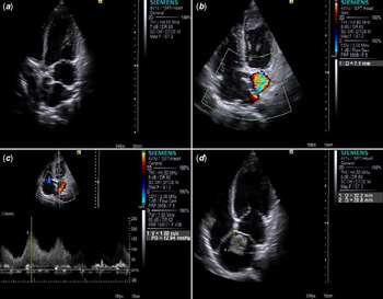
During the present admission, the patient was diagnosed with cor triatriatum sinister with left ventricle ejection fraction of 54% using transthoracic echocardiography. Echocardiography and transthoracic echocardiography showed a fibromuscular membrane-like structure traversing and dividing the left atrium, into two chambers: a post-erosuperior chamber that received blood from the pulmonary veins (PS-LA) and an anteroinferior (AI-LA) chamber that contained the left atrial appendage and mitral valve. (Fig 1a). Further examination with Doppler echocardiography confirmed the anomaly as Loeffler’s group II cor triatriatum sinister, as there was a small non-restrictive orifice of about 7.1 mm within the membrane (Fig 1b) and measured a mean pressure gradient of 12.94 mmHg across the orifice (Fig 1c). Transthoracic echocardiography also revealed a mobile mass probably a thrombus of approximately 32.2 × 28.8 mm in the post-erosuperior chamber of the left atrial (Fig 1d). We found the dilatated valvular ring in the mitral and tricuspid valve. Hence, the patient was scheduled for traditional mitral and tricuspid annuloplasty for rectification of mitral and tricuspid regurgitation. Regurgitation in the valve was rectified by DeVega tricuspid annuloplasty and Kay’s mitral annuloplasty. Following annuloplasty, the fibromuscular membrane and the mobile mass were excised by an open cardiac surgery. The patient had an uneventful recovery. The regurgitation of mitral and tricuspid valve was mild after annuloplasty as revealed by the transthoracic echocardiography. Further, post-operative transthoracic echocardiography and echocardiography showed complete excision of the fibromuscular membrane without any traces of the thrombus.
( a ) Transthoracic 2-dimensional echocardiography shows a membrane-like structure traversing the left atrium. ( b ) Colour Doppler echocardiography shows a small communicating orifice about 7.1 mm of the membrane. ( c ) Colour Doppler echocardiography reveals significant flow acceleration across the membrane (estimated mean pressure gradient, 12.94 mmHg). ( d ) Transthoracic 2-dimensional echocardiography shows a mass in the post-erosuperior chamber.

Discussion
Cor triatriatum sinister is a rare congenital cardiac anomaly first described by Church in 1868, and named by Borst in 1905 Reference Jha and Makhija12 . Since the first report of the cor triatriatum sinister in 1868, several case reports and case series have been reported in the literature Reference Gheissari, Malm, Bowman and Bierman2–Reference Rozema, Arruda and Snyder5,Reference Kokotsakis, Anagnostakou and Almpanis7–Reference Diestro, Regaldo, Gonzales, Dorotan, Espiritu and Pascual9,Reference Baris, Bogers, van den Bos and Kofflard13–Reference Shah, Mittal and Gupta15 . Cor triatriatum sinister is mostly diagnosed in neonates and it is a rare condition in adults Reference Hayes, Liu, Tam and Kass3 . It has been reported in equal proportion in both men and women Reference Nassar and Hamdan1,Reference Jha and Makhija12,Reference Troxclair, Ross and Newman16 . As per Loeffler’s classification, there are three types of cor triatriatum sinister based on the number and size of fenestrations in the fibro-muscular membrane Reference Nassar and Hamdan1,Reference Loeffler17 . In type 1, there will be no connection between two chambers; in type 2, there will be one to few small openings in the intra-atrial membrane; and in type 3, a large single opening exists between accessory chamber and true atrium. The first two types are mostly diagnosed in infants and children and are associated with 75% mortality if not diagnosed and treated at earlier stage Reference Troxclair, Ross and Newman16 . Whereas, the third type is frequently diagnosed in adults Reference Nassar and Hamdan1 . However, type 2 cor triatriatum sinister, which is a rare anomaly in adults, has been reported in the patient reported in our study. There are very limited case reports reporting this unusual cor triatriatum sinister in adults Reference Hayes, Liu, Tam and Kass3,Reference Khanra, Soni, Ola and Duggal18 . Cor triatriatum sinister is hypothesised to originate due to the mal-incorporation of the common pulmonary vein into the left atrial during embryological growth Reference Hayes, Liu, Tam and Kass3 .
The cor triatriatum sinister in adults is mostly asymptomatic, due to the presence of large foramen that decreases the intra-atrial pressure. However, symptoms will be visible later in life after calcification of the muscular fibers leading to cardiovascular comorbidities like mitral regurgitation or stenosis and calcification of the orifice or development of atrial fibrillation Reference Shah, Mittal and Gupta15 . Symptoms of cor triatriatum sinister can range from mild respiratory distress to severe congestive heart failure. We speculate that, in our case also, the patient was asymptomatic for last 25 years and cor triatriatum sinister was diagnosed along with mitral regurgitation and atrial fibrillation with symptoms of dyspnoea. The mobile mass of thrombus was probably due to the long-lasting haemodynamic change created by the additional septa. The aggravation of symptoms depends on the degree of obstruction Reference Rudienė, Hjortshøj and Glaveckaitė6 . In a recent systematic review involving 406 publications with 171 patients diagnosed with cor triatriatum sinister in adulthood, obstructive membrane physiology was reported in 41% of patients. Patients with obstructive membrane physiology experienced higher incidence of congestive heart failure, pulmonary hypertension, haemorrhage, and infections Reference Rudienė, Hjortshøj and Glaveckaitė6 . As reported in our case, dyspnoea on exertion was the frequent symptom observed among other patients with cor triatriatum sinister. In par with other studies, surgical excision was recommended for the patient in our study, as surgery is the treatment of choice in 91.6% of the patients diagnosed with cor triatriatum sinister. Previous studies have reported excellent clinical outcomes in cor triatriatum sinister patients undergoing adequate diagnosis and right treatment Reference Nassar and Hamdan1,Reference Rudienė, Hjortshøj and Glaveckaitė6 . The 5-year, 10-year, and 15-year survival rate was 96, 83, and 88% respectively in the two studies conducted by Saxena et al Reference Loeffler17 and Alphonso et al Reference Alphonso, Nørgaard, Newcomb, d’Udekem, Brizard and Cochrane19 . Hence, surgical correction of cor triatriatum sinister was safe and effective in many studies with very low risk of remission. In a previous study, a 39-year-old man diagnosed with cor triatriatum sinister refused the surgery and his condition critically deteriorated gradually and he died at 59 years due to severe pulmonary hypertension, severe mitral and right ventricular failure with moderate tricuspid regurgitation Reference Rudienė, Hjortshøj and Glaveckaitė6 . If the cor triatriatum sinister is not surgically rectified, the average survival is approximately 3 months if the defect in the separating membrane is <0.3 cm and 16 years if the defect is >0.3 cm Reference Troxclair, Ross and Newman16,Reference Khanra, Soni, Ola and Duggal18 . The patient in our case was asymptomatic for 25 years even with the orifice of >0.3 cm (0.71 cm).
Cardioembolic stroke has been reported in cor triatriatum sinister patients with or without concurrent atrial fibrillation Reference Hayes, Liu, Tam and Kass3,Reference Kokotsakis, Anagnostakou and Almpanis7,Reference Park, Park and Sir14,Reference Ridjab, Wittchen and Tschishow20,Reference Spengos, Gialafos and Vassilopoulou21 . Similarly, the presented case in our study was diagnosed with a large size mobile thrombus. We suspect that in our patient the mitral regurgitation and atrial fibrillation would have facilitated clot formation and a mobile thrombus in the left atrial. However, the smaller orifice in the membrane prevented the thrombus from migrating into the systemic circulation which otherwise would lead to cardioembolic stroke. The process of clot formation in cor triatriatum sinister resembles the pathophysiology of mitral stenosis Reference Hayes, Liu, Tam and Kass3,Reference Kokotsakis, Anagnostakou and Almpanis7,Reference Park, Park and Sir14 . Hence, adult patients diagnosed with cor triatriatum sinister have to undergo routine scan to avoid fatal embolic stroke. Additionally, investigators have to be aware of this rare abnormality to ensure timely diagnosis and surgical treatment in cor triatriatum sinister patients with significant degree of obstruction between the two left atrial chambers.
Conclusion
We report a rare case of Loeffler’s group 2 cor triatriatum sinister with a mobile thrombus diagnosed in a timely manner using transthoracic and Doppler echocardiography and treated with a right choice of surgical resection achieving complete removal of the thrombus that could have prevented stroke.
Acknowledgements
None.
Financial support
This research received no specific grant from any funding agency, commercial, or not-for-profit sectors.
Conflicts of Interest
None.
Ethical standards
The case report was exempted from ethical clearance and informed consent was provided by the next of kin.