Published online by Cambridge University Press: 21 September 2005
As recently as 35 years ago, children born with a functionally single ventricle were destined to a lifetime of multiple palliative operations, and disrupted growth and development. Physical signs of their cardiac disease were evidenced by chronic hypoxemia, polycythemia, clubbing, small stature, and severely reduced exercise ability. A normal lifestyle, with typical landmarks such as uninterrupted adolescence and potential childbearing, was unlikely. The successful application of right heart bypass by Fontan and Kreutzer in the early 1970s heralded an era of multiple medical and surgical breakthroughs that have systematically improved the quality of life and long-term outlook for these children.
Until the early 1980s, most forms of functionally single ventricle with duct-dependant pulmonary or systemic blood flow were uniformly fatal. The nearly simultaneous introduction of prostaglandin E1, echocardiography, and improvements in cardiopulmonary bypass, ushered in the exciting early experiences of neonatal open-heart surgery. These advances were quickly followed, in 1983, by successful management of hypoplastic left heart syndrome utilizing infant heart transplantation by Bailey et al.,1 and first stage reconstruction followed by a Fontan procedure by Norwood et al.2
As recently as 10 years ago, surgical mortality remained high, hospital stays were long and costly, and short-term morbidity was common. The present era has seen continued improvements in surgical techniques, such as the addition of an intermediate stage with construction of a bi-directional Glenn anastomosis prior to the Fontan palliation, and the “fenestrated” Fontan procedure. Overall, there is a better understanding of the sequels and future implications of the Fontan circulation. Similarly, cardiac transplantation has benefited from time and experience. Improved surgical techniques, better preservation of grafts, and improved regimes for immunosuppression have all provided better longevity and quality of life subsequent to transplantation. Significant advances in critical and perioperative care, such as modified ultrafiltration, and better management of pulmonary blood flow, have contributed to enhanced survival after both Fontan-type surgery and transplantation. The focus of management for children with the functionally single ventricle has now shifted from minimizing mortality to minimizing morbidity.
Though not yet an actuality, patients are now expected to live into their fourth decade or longer, to have minimal limitations in day-to-day activities, to have families, and to become productive members of society. It is important to emphasize that truly long-term results are still decades away, and are likely to change with every new medical or surgical modification of therapy. Available follow-up studies of adolescents and young adults reflect the medical and surgical management strategies used 15 to 25 years ago. The outcomes of those measures are likely different from the future outcomes of our present strategies. Some long-term limitations to good health and survival, however, such as post-Fontan arrhythmias and graft dysfunction after transplantation, may be greatly reduced with current strategies of management, but are unlikely to be completely eliminated. It is likely that with each newly introduced technique, different risks may become apparent, such as thrombosis of the extra-cardiac conduits that are part of the newer Fontan technique, or post-transplant lymphoproliferative disease with enhanced immunosuppression.
In a recent editorial on long-term follow-up of tetralogy of Fallot, Gutgesell wisely commented that “the incidence of long-term squeals of the treatment of congenital heart disease is directly related to how thoroughly they are sought. The challenge is to determine which of the laboratory abnormalities are of clinical relevance”.3 It is incumbent upon all involved in the care and follow-up of children and young adults with functionally single ventricles prospectively and compulsively to detail the potential morbidity of our surgical and medical strategies, to be critical of our findings, and accurately to inform our patients and their families of the current outcomes, so that we all may have reasonable expectations for “long-term outcomes”.
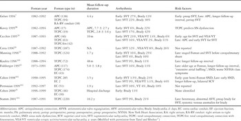
Brady- and tachy-arrhythmias are the most common adverse outcome following the Fontan procedure, and their recognition 10 to 20 years after surgery has become one of the driving forces behind recent surgical modifications, such as routine use of an extra-cardiac conduit, or atrial cryoablation at the time of the Fontan procedure or its revision.4 The most common type of tachyarrhythmia following the Fontan procedure is intra-atrial reentry tachycardia or atrial flutter. The incidence of postoperative atrial flutter ranges from 4 to 24%, and appears to increase with longer intervals of follow-up.5–19 In addition to intra-atrial reentry tachycardia, other types of supraventricular tachycardia occur, such as junctional ectopic tachycardia, ectopic atrial tachycardia, and atrioventricular reciprocating tachycardia.
All forms of tachycardia that occur after the Fontan operation are poorly tolerated compared to patients with structurally normal hearts. Recurrent and sustained intra-atrial reentry tachycardia is associated with increased risk of morbidity and mortality. Creation of the Fontan circulation makes some cardiac chambers less accessible for invasive electrophysiologic procedures, especially when an extra-cardiac conduit has been inserted supporting the concept of preoperative radiofrequency catheter ablation, or intra-operative cryoablation with or without atrial reduction at the time of the Fontan operation.
Although poor hemodynamics and atriomegaly may predispose individual patients to atrial flutter, the hemodynamic state in any given patient may have little predictive value for the risk of developing postoperative arrhythmias. It is not uncommon for a patient with favorable baseline hemodynamics, and a functional lifestyle, to be burdened by recurrent atrial flutter, prompting potentially high-risk surgical revision.
Furthermore, many of the variables found to be predictive of arrhythmia in a particular operative era may not be applicable to current surgical techniques. As a group, patients that had a Fontan operation performed in the 1970s had less complicated forms of functionally single ventricle, and were generally much older at the time of surgery. In the present era, patients undergoing the Fontan procedure have more complex anatomy, an increased number of preoperative risk factors, and are of younger age. The present Fontan palliation offers a wider variety of modifications compared to the surgical technique presented originally.
Medical therapy for tachyarrhythmia is generally difficult and unrewarding. Patients have a variable frequency of recurrence, but often develop a progressively more difficult substrate for medical treatment. As a result, patients often receive multiple anti-arrhythmic medications, sometimes in combination, in an attempt to control the tachycardia. Although one study20 reported a success rate of almost nine-tenths in controlling atrial arrhythmias with multiple medications at early follow-up, most have reported a disappointingly low rate of success, with significant side effects from medication. Interventional therapies include radiofrequency catheter ablation and surgical modifications of the Fontan circuit, with simultaneous atrial reduction by techniques such as the Cox Maze and/or cryoablation.21–24 The truly long-term results of these innovative strategies requires continued follow-up.
Bradyarrhythmias also occur with increasing frequency during follow-up. Sinus nodal dysfunction is the most common cause of late bradyarrhythmia. While the newer surgical modifications may reduce the long-term incidence of atrial flutter, there is some evidence that routine “staging” may result in a high incidence of late sinus nodal dysfunction,25 although this remains controversial.26 Sinus nodal dysfunction occurs with increased frequency after the total cavopulmonary anastomosis compared to the atriopulmonary connection, presumably due to direct trauma to the sinus node, or indirectly by injury to the sinus nodal artery. Chronotropic impairment, especially apparent during exercise, is essentially universal following Fontan palliation. The combination of the inappropriately slow atrial rate, coupled with the relatively fixed stroke volume following the Fontan procedure, conspire to limit the ability of these patients to exercise, especially as they approach adolescence and early adulthood.
While patients with isolated sinus nodal dysfunction are generally candidates for single chamber atrial pacing, and patients with isolated supraventricular tachycardia for routine anti-arrhythmic medications, the combination of supraventricular tachycardia and sinus nodal dysfunction as seen in the “tachycardia– bradycardia” syndrome is not uncommon in patients who have had a Fontan operation. Treatment of the supraventricular tachycardia not infrequently worsens the underlying slow atrial rhythm. A combination of medical therapy for the tachyarrhythmia, and back-up atrial pacing, is becoming more common as the population of patients with a Fontan operation ages. Table 1, modified from Tanel and Rhodes27 summarizes a number of the recent reviews of the problems occurring due to arrhythmias after the Fontan operation.
Table 1. Arrhythmias after the Fontan operation.
Protein losing enteropathy remains a puzzling and poorly understood disorder following the Fontan procedure. The syndrome comprises the triad of high filling pressures, extravasation of protein into the intestinal lumen, and diarrhea. Protein losing enteropathy may be mild or life threatening, and the clinical features include diarrhea, gastrointestinal pain, edema, ascites and effusions. The symptoms are due, in great measure, to chronic venous congestion and the loss of serum proteins and lymphocytes via dilated intestinal lacteals.28 The combination of lymphopenia and hypogammaglobulinemia due to loss of protein results in immunodeficiency. Serum proteins important in maintaining the normal coagulant cascade may also be lost, resulting in a hyper-coagulable state.29 Severe protein loss, in conjunction with food intolerance, may result in nitrogen imbalance, cachexia, and chronic debilitation. Virtually every published report on long-term outcome after the Fontan suggest an incidence of protein losing enteropathy, with proportions between 3% and 15%, depending upon the diagnostic methods and thresholds used to make the diagnosis and the duration of follow-up.30, 31 Protein losing enteropathy has serious consequences, and studies with large numbers of subjects report greater than mortality rates of more than 50% within 5 years from the time of initial diagnosis of the enteropathy.31 Because of its poor prognosis, some centers consider the enteropathy to be an indication for cardiac transplantation, despite the fact that resolution is not universal with this approach.31
In most patients with protein losing enteropathy, the hemodynamics in the Fontan circulation are fairly typical, and treatable anatomic and/or physiologic problems are, unfortunately, rarely identified. Medical and surgical management is limited, and frequently unsatisfying. It includes a high protein diet, intravenous hyperalimentation, diuretics, supplemental albumin infusions, corticosteriods, subcutaneous heparin, atrial pacing, fenestration or revision of the Fontan procedure, operative or interventional catheterization to relieve obstruction if present within the flow pathways, and cardiac transplantation.32–38 Post-transplant management of patients with “failed Fontan physiology” and protein losing enteropathy is extremely challenging and complicated. While return of pulsatile blood flow, relief of venous congestion, and better cardiac output should theoretically improve the situation, recovery after transplantation is often slow, and loss of protein may continue for many months. Aggressive management of these patients prior to transplantation is necessary to carry them through the difficult period subsequent to transplantation. Nutritional support should be instituted, and may include hyperalimentation and albumin infusions. Physical conditioning is necessary to maintain overall strength and endurance. Pulmonary consultation, or at least pulmonary function testing, is useful to determine the extent of limitations associated with prior surgeries, the Fontan physiology and pulmonary congestion related to protein losing enteropathy.
With increasing frequency, it is becoming recognized that thromboembolism, both clinically apparent and sub-clinical, may be a major long-term problem in survivors of the Fontan procedure. DuPlessis reported an incidence of stroke in nearly 3% in nearly 600 patients,39 and Rosenthal recently reported thromboembolic events in one-fifth of patients followed at the Yale-New Haven Medical Center over a five-year period of time.40 Other studies have suggested an even higher incidence of identified thrombus when highly sensitive tools such as transesophageal echocardiography are used. Balling et al.29 prospectively examined 52 asymptomatic subjects after the Fontan operation, and found thrombus in one-third. This suggests a significant underestimate of the problem and under detection rate. Coon41 recently reported a retrospective review of nearly 600 patients who underwent the Fontan operation at The Children's Hospital of Philadelphia. Thrombus was detected in one-tenth of patients, with freedom from thrombus of 92%, 90%, and 84% at one, three, and eight years after surgery, respectively.
A number of factors likely conspire to increase the risk of thrombosis after the Fontan operation, including venous stasis and low cardiac output, enteric loss of protein (see above), arrhythmia, and inherent abnormalities of the coagulation cascade, such as low levels of protein C, factors II, V, VII and X, and anti-thrombin III.42, 43 Mild hepatic dysfunction with synthetic impairment due to venous hypertension may contribute to these findings.44 Significant elevation in liver enzymes following the Fontan procedure, however, is rare.45
It is also important to emphasize the inter-relationship of many of these adverse outcomes following the Fontan procedure. Arrhythmia, sinus nodal dysfunction, formation of thrombus, embolism, and protein losing enteropathy may coexist in varying degrees, and may all be due to the same substrate: namely large atriums with multiple suture lines, low cardiac output, non-pulsatile flow and venous stasis, along with the long-term effects of the anatomic and physiologic abnormalities over time.
The number of infants receiving cardiac transplantation as primary therapy for functionally univentricular heart is small compared with those who enter the reconstructive pathway towards the Fontan procedure. Further, the number of children listed for transplantation as primary therapy for hypoplastic left heart syndrome has decreased over the last several years. A recent study from the Pediatric Heart Transplant Group shows that the number of infants with hypoplastic left heart syndrome listed for transplant in 1993 was 58 of 215, or just over one-quarter of the children listed. By 1998, the percentage of patients was 14%, specifically 33 of 229, representing a consistent decline over the six years of the study. The reasons for this decline likely include improved results with reconstructive surgery, and the severe, ongoing shortage of donors. In juxtaposition, the number of infants entering the staged reconstruction track most likely approaches 1500 to 2000 each year in the United States of America alone.
At the time of transplantation, the anatomy of the recipient will influence the intra-operative course. Additional donor tissue may be harvested for necessary anastomosis or vascular reconstructions.46 Subsequent to transplantation, these sites may narrow, and careful monitoring by echocardiography and/or catheterization is necessary. Interventional catheterization, such as stenting of a narrowed caval anastomosis, or balloon dilation of a narrowed pulmonary artery or re-coarctation, is typically sufficient to correct such issues.47
Aside from concerns imposed by the original anatomy, patients with functionally univentricular hearts have similar issues after transplantation to patients receiving hearts for other indications. In children care after transplantation is complicated by somatic growth, changes in metabolism, and personal development. In general, these normal developmental transformations influence all aspects of care, including regimes for immunosuppression, compliance with medication compliance, adverse events, and survival.
Calcineurin inhibitors, such as cyclosporine or FK 506, remain the mainstay of regimes for immunosuppression in children. At most transplant centers, cyclosporine is the drug of choice. The metabolism, bioavailability, and pharmacokinetics of cyclosporine are different in infants, children, adolescents and adults. Infants and young children with normal hepatic function metabolize cyclosporine faster than do adults. Further, there is some evidence to suggest that pre-hepatic metabolism of cyclosporine could contribute to the variable bioavailability of the drug.48 Because of these metabolic idiosyncrasies, the total daily dose of cyclosporine is titrated to a therapeutic blood level, and this titration becomes even more important in patients who have underlying hepatic dysfunction or protein losing enteropathy. Cyclosporine is a potent vasoconstrictor, and is nephrotoxic, so close monitoring of levels of the drug mandatory. Other side effects include hypertension, hyperlipidemia, and cosmetic side effects such as hirsuitism. Frequent surveillance of the levels of the drug is necessary in growing children to monitor the effects of somatic growth on doses and administration. Familiar interactions exist between the metabolism of cyclosporine and other common drugs.
While chemically different from cyclosporine, FK506, or tacrolimus, acts similarly to inhibit the immune response. Tacrolimus, however, has slightly different activity, and is considered a more potent immunosuppressive agent. It is often administered as a monotherapy49 and as “rescue” therapy for patients who have persistent rejection episodes despite therapeutic cyclosporine levels. While tacrolimus does not have cosmetic side effects, it can also cause nephrotoxicity, hepatotoxicity, abnormalities of glucose metabolism, and neurotoxicity.50 Drug interactions are common, and the same caveat applies to tacrolimus as to cyclosporine.
Until a few years ago, azathioprine was the usual adjunct immunosuppressive agent. The toxicities of this drug are well documented, and include generalized suppression of the bone marrow, pancytopenia, and hepatitis. Recently, mycophenolate mofetil has been used effectively as adjunct routine immunosuppression in children receiving cardiac transplants. Mycophenolate more specifically blocks production of lymphocytes, and has less marrow suppression and pancytopenia than does azathioprine. Its main side effects are abdominal pain, nausea, diarrhea, and vomiting.51, 52 Monitoring the level of the drug is recommended.
Two-thirds of children receive triple immunosuppression, a regimen inclusive of steroids, as maintenance during the first year after the transplant.53 Steroids are slowly discontinued in about half of these patients by 3 years after transplantation. The side effects associated with steroids are not inconsequential, especially to teenage patients, and include diabetes, hypertension, Cushingoid appearance, acne, appetite stimulation, abnormal fat distribution, water retention, osteoporosis, and growth retardation.
Notably all immunosuppressives increase the risks of infection and post-transplant lymphoproliferative disease. Post-transplant lymphoproliferative disease is a spectrum of diseases, ranging from B cell hyperplasia to true lymphoma. Risk factors for developing this process include exposure after transplantation to the Ebstein Barr virus, seroconversion to antibody production, and persistent elaboration of viral deoxyribonucleic acid.54 Testing for Ebstein Barr viral production has been useful in tracking the progress of the disease, but is not an absolute indicator of progression.55 Lymphoproliferative disease occurs in about one-tenth of transplanted children, and less commonly in the adult population. Treatment for post-transplant lymphoproliferative disease, as a first line, calls for diminishing immunosuppression to allow the body to clear the diseased cells. Conventional chemotherapy is, by and large, ineffective. New agents, such as rutuximab, have shown promising results in small numbers of patients.
Graft coronary vasculopathy56, 57 is also a significant limitation to long-term survival, and again occurs in about one-tenth of transplanted children. Graft coronary arterial disease may be considered vascular rejection. The hallmark of this process is intimal hyperplasia, which circumferentially narrows the vessel, unlike the eccentric lesions of typical atherosclerosis. Graft vasculopathy can occur proximally or distally along the length of the vessel, and can effect small and large coronary arteries. Most centers survey for this disease at yearly intervals, and because the risk of developing the complication increases with time after transplantation, continued surveillance is necessary. Coronary angiography is most commonly used, though in patients larger than about 15 kg intravascular ultrasound is safe and effective in determining early vasculopathy.58, 59 Once the disease is detected, retransplantation is the usual ultimate outcome. Typically the process is unremitting, though there are reports that enhanced immunosuppression may slow disease progression.60 Addonizio et al.56 reported that children receiving triple immunosuppression, including cyclosporine, azathioprine and steroids, had improved survival, and protection from graft vasculopathy.57
While most parents are optimistic about the immediate improvement in their child, long-range concerns prevail. An often-asked question concerns child-bearing potential after transplantation. Based on the published data, and anecdotal experience, there are no procreative restrictions or limitations due to chronic immunosuppression for young men or for young women subsequent to transplantation, albeit that pregnancy should be a planned and well-monitored event, and will likely require some modification in regime.61 Adolescent patients, and their parents, must understand that their reproductive capabilities are not attenuated by immunosuppression. Contraception, therefore, is strongly recommended.
“Fontan failure” is a commonly used term for a series of events that culminate in poor functional outcome and quality of life. Chronic arrhythmias, protein losing enteropathy, and ventricular failure may occur in isolation but frequently coexist. In virtually all published series of survivors of the Fontan procedure sudden death, presumably due to arrhythmia, has been reported in 1 to 2%. Longer duration of follow-up is accompanied by growing numbers of patients listed for cardiac transplantation, typically about 5% during the first 10 to 15 years after surgery. There has been speculation that late transplantation will be more common in patients with a functionally single morphologically right ventricle compared to a functionally single morphologically left ventricle, but at the current time, most published data does not support this concern. However, a recent study shows that in a cohort of 188 adult patients with functionally single or systemic right ventricles who received various surgical palliations, 17.6% were in classes III or IV of the New York System. Systemic ventricular ejection fraction in symptomatic versus asymptomatic patients at rest was 34.8 ± 15.7% versus 46.7 ± 13.4% (p = 0.00001). Mortality was 47.1% in symptomatic patients, and 5% in asymptomatic patients, at 15.7 years of post-operative follow-up.62 Regardless of the ventricular morphology, it is impossible to determine whether the current risk of late transplantation will increase as patients live into their third and fourth decades of life, implying an eventual certainty of transplantation for all patients, with the Fontan circulation, or whether current modifications, such as early volume unloading by a staged interim Glenn procedure, improvements in myocardial protection, long-term medical therapies, and so on will improve the long-term outcome and decrease the “inevitability” of transplantation.
Following primary transplantation, there is a continued risk of late mortality, as well as limitations to survival, including graft vasculopathy, late rejection, graft failure, and post-transplant lymphoproliferative disease. Primary graft failure and rejection are still significant causes of death in children at 3 to 5 years after the transplant. Lymphoma accounts for a small percentage of such deaths, and the incidence of post-transplant lymphoproliferative disease remains relatively low. The vast majority of patients are free of malignancy at 5 years after transplantation. The cumulative incidence of transplant coronary disease or graft vasculopathy is 2.5% by 1 year, and 11.4% by 5 years after the transplant.63 For all children receiving cardiac transplantation, from infancy up to 18 years of age, 5% or less have retransplantation as their main indication with a primary diagnosis of graft failure, rejection, or graft vasculopathy.63, 64 As with patients who have had a Fontan procedure, recent modifications of medical therapy may reduce morbidities, and the incidence of late graft loss and retransplantation.
Although more has been written about the long-term neurological outcomes following the Fontan operation compared with transplantation, there are increasing numbers of reports of formal developmental studies in survivors of both strategies.65–75 Despite the marked differences in the physiology and surgery of the two strategies, the developmental outcomes are surprisingly similar for both.
Children with functionally single ventricle may have additional congenital anomalies of the central nervous system, adding to their risk for adverse neurological outcome.76, 77 Hypoxic-ischemic injury may occur with prolonged distress at presentation.78 Prolonged hypoxemia alone, either prior to transplant while awaiting surgery, or during staged reconstruction until the Fontan procedure is completed, has been implicated in causing subsequent cognitive and developmental limitations.74, 79 The effects of cardiopulmonary bypass and deep hypothermic circulatory arrest may be similarly implicated, and the effects of these processes may be cumulative. Further, the additional burden of chronic disease contributes significant psychosocial stress on these patients and their families. In a recent study by Gentles, as yet unpublished, where questionnaires were mailed to 363 patients and their families following the Fontan procedure, nearly half of the patients had received or were receiving special services, and one-fifth of the families had undergone psychotherapy44 (from Gentles TL, unpublished data, personal communication).
Psychosocial readiness for transplantation is an essential part of the success of this complicated and end-stage therapy. In the present era, therefore, most transplant teams dealing with children include a pediatric psychologist or psychiatrist. Psychosocial evaluation before transplantation has become a standard part of the process. “Stress management” and short-term therapy is welcomed by most patients and their families before and after the procedure. During the process of evaluation, and while awaiting transplantation, patients and their families must confront mortality, acceptance of a donated organ, life-long medications, and potential limitations to long-term survival. Skilled psychologists and psychiatrists will typically avail patients of definitive coping skills, such as creative imagery, meditation, and other techniques and tools to reduce stress. Infrequently, pharmacotherapy is required to treat more severe depression or anxiety. Recipients of school age, and adolescents, have additional concerns to deal with that directly result from chronic disease, including absences from school for illness or visits to their doctors, physical deconditioning, and delayed sexual maturation. These issues may not quickly resolve after transplantation. After transplantation, cosmetic side effects, such as hirsuitism and gingival hyperplasia due to cyclosporine, or steroid-induced weight gain and strias, can be particularly distressing, especially to adolescent patients. In this age group, non-compliance to medication is a well-documented entity, and is associated with late mortality. Though the challenges faced by recipients and their families are on-going, consistent psychosocial support is beneficial in decreasing morbidity and mortality. A retrospective review of a large center dealing with children showed that patients were less likely to have early episodes of rejections, from 3 to 12 months after transplantation, if they were children with “maximal personal support,” that is, with two parents, aid from relatives, friends and community, when compared to children without such support (6.7% vs. 36%, p < 0.05). Additionally, 17% of children who had at least one parent employed experienced a rejection, versus 70% of children with unemployed parents (p < 0.01). The marital status, education, and type of medical coverage of the parent did not affect the incidence of rejection (unpublished observations).
In children with a functionally single ventricle, perioperative neurological events may complicate the initial surgery irrespective of the strategy for management, and may be the presenting sign of neurological injury due to thromboembolic events, low cardiac output, or both. Kurth et al.80 recently reported on non-invasive assessment of brain oximetry by near-infrared spectroscopy in a large cohort of heterogeneous patients with congenital heart disease. Patients with a functionally single ventricle had the lowest cerebral oxygenation of any subgroup, most likely due to the combination of hypoxemia and borderline systemic blood flow conspiring to reduce delivery of oxygen to the brain. The fragile nervous system may be further damaged by the effects of cardiopulmonary bypass, circulatory arrest, or additional pre-operative insults.
The initial surgical procedure, whether first stage palliation or cardiac transplantation, is necessarily done utilizing cardiopulmonary bypass, and in most institutions, with variable periods of deep hypothermic circulatory arrest. Post-operative short-term equals include seizures, stroke, periventricular leukomalacia and intracranial hemorrhage in varying degrees. Specific to recipients of cardiac transplantation, cyclosporine, the mainstay of most immunosuppression regimens, lowers the threshold for seizure, and also causes magnesium wasting. The combination of a supratherapeutic level of cyclosporine and hypomagnesemia may cause seizures that typically resolve with correction of the electrolytic imbalance and level of cyclosporine.81 Cyclosporine neurotoxicity may present as more subtle though no less troublesome findings. Tremor, dysesthesias such as tingling, burning or numbness of the palms and soles, headaches, transient blindness, tunnel vision, and generalized skeletal muscle weakness occur not infrequently. Factors that enhance the neurotoxic effects include previous injury to the central nervous system, prior history of headache or migraine, concomitant use of high-dose steroids which elevate levels of cyclosporine and hypomagnesemia.82 Most of these side effects tend to be temporary or transient, and are commonly, but not exclusively, related to an elevated level of cyclosporine. Intolerance to cyclosporine is also a possible etiology, though it is relatively rare that, for this reason, the drug must be discontinued and exchanged for another immunosuppressant. The incidence of cyclosporine-related neurotoxicity ranges from 10 to 25% of patients receiving the drug. Diagnostic imaging in patients receiving cyclosporine has shown symmetric, low density areas on computerized tomographic scanning and high-resolution images on resonance imaging. White matter seems to be affected more often than gray matter. These findings are consistent with cerebral edema, which may be due to an inflammatory response to accumulation of cyclosporine.83, 84 Despite its inherent neurotoxicity, neurological symptoms in cardiac transplant recipients must always be seriously considered, and usually warrant a comprehensive evaluation. For example, seizure or headache may be the presenting sign of cerebral infection or post-transplant malignancy, and meningitis, encephalitis, abscesses and tumors must be ruled out.
In our most recent two years experience with the first stage Norwood reconstruction at the Children's Hospital of Philadelphia, routine perioperative electroencephalographic monitoring during the first 48 h after surgery has identified seizures in 17% of patients (unpublished data). Following either staged reconstruction or transplantation, feeding difficulties are prominent. These are likely a combination of peripheral and central oral-motor dysfunction.66, 74, 85
Irrespective of their management, patients with functionally single ventricle are at risk for thromboembolic events. Stroke has been reported in up to 5% of survivors of the Fontan procedure.39 The incidence of stroke following cardiac transplantation is a bit more difficult to ascertain. Risks must be considered before and after the procedure, and the aggregate risk is high. The course prior to transplantation can typically include low cardiac output, intracardiac thrombus, mechanical ventilatory and circulatory support. After transplantation, the effects of cardiopulmonary bypass, circulatory arrest, and labile post-operative blood pressure potentially enhance the risk. Even at therapeutic levels, cyclosporine causes vasoconstriction, nephrotoxicity, and resultant systemic hypertension, all effectively enhancing the possibility of intracerebral hemorrhage. Despite all this, the actual occurrence of stroke post-transplant is relatively low, at about one-tenth in one series, and long-term disability is rare.86 The most significant neurological complications before or after transplantation seem to be related to mechanical circulatory support and the necessary anticoagulation. In the majority of children who require extreme care and survive, those who suffer neurological insults usually go on to heal remarkably well, with little residual neurological deficit.86, 87 Still, these patients and their families are significantly challenged. Uzark et al.88 showed in 1992 that there is strong need for systematic assessment of psychosocial adaptation in children undergoing cardiac transplantation and their families. Other authors have supported this stand, adding that early psychological evaluation prior to the procedure, and at regular intervals after transplantation, with appropriate tailoring of the school curriculum and psychotherapeutic regime, may improve overall cognitive and behavioral outcome.89, 90
The combined pre-operative, intra-operative, and post-operative factors conspire in most cases to adversely impact long-term neurodevelopmental outcome. Eke et al. reported data up to 2.5 years following infant cardiac transplantation using deep hypothermia and profound hemodilution. They found that neither the rate of cooling, the duration of circulatory arrest, nor low ionized levels of calcium had any measurable effect on neurological outcome measured by the mean scores on the psychomotor development index of the Bayley Scales of Infant Development.69
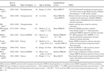
With both strategies, the percentage of patients with abnormal scores on a variety of standardized neurological tests are abnormally high, up to one-quarter, especially in the area of fine and gross motor functioning.66, 74 While in general, most patients who have had either a Fontan operation or transplantation have outcomes within the normal range,75 outcomes for groups are significantly below normal.75 Table 2 summarizes a number of recent reports on short- and long-term neuro-developmental outcomes. As discussed previously, strategies for management have changed considerably since the early 1970s, and given the improvements in cardiopulmonary bypass, and the reduction of postoperative morbidity in recent years, outcomes over the next 10 to 20 years are likely to be significantly improved for patients born in the current era.
Table 2. Central nervous system outcomes in patients with functionally single ventricle.
Adequate ability to exercise requires a complex interaction between the neuromuscular, pulmonary, and cardiovascular systems. Children with Fontan physiology, as well as those following transplantation, may have diminished exercise performance based upon dysfunction of any or all of these systems. Following transplantation, the pulmonary and systemic circulations act as separate circuits arranged in series. Although they are interdependent, and changes in the hemodynamics of one circuit effect the other, at any point in time they have independent preload and afterload. Changes in the pulmonary and systemic vascular resistance, therefore, may occur that impact on one ventricle with minimal impact on the other. In the Fontan circulation, the ventricle(s) must pump the blood across both the systemic and the pulmonary vascular beds. The total afterload seen by the ventricle is equal to the combined resistance of the systemic and pulmonary beds.91 The implication of this physiology is that, during exercise, the ventricle will see an increased afterload when compared to biventricular physiology at any given cardiac output, even assuming a normal physiologic drop in the resistances in the pulmonary and systemic beds. The impact of the Fontan pulmonary vascular physiology on exercise performance remains poorly understood. While it appears that a very low resistance across the pulmonary bed is needed to permit cardiac output to increase during exercise, it is not clear what factors are responsible for the maintenance of this low resistance. Any obstruction of the large arteries appears to be poorly tolerated. Small vessel disease that restricts vasodilation with exercise also impairs exercise performance. It does appear that the primary ability of the pulmonary vascular bed to dilate in a normal fashion in response to exercise is critical to exercise performance in those with the Fontan physiology.
Both groups of patients have a blunted response of heart rate to exercise. This is most likely due to denervation in those undergoing transplantation, and local surgical issues in those with the Fontan physiology. The end result is to reduce overall capacity to exercise. Most published series of survivors of the Fontan procedure have maximal exercise capacity of up to three-quarters of age and sex matched controls. Longer duration of follow-up, and longer duration of volume loading prior to the Fontan procedure, are associated with worse exercise performance.92–94 Again, it is not yet clear whether current modifications to the Fontan circulation will improve the overall exercise performance as these younger patients approach adolescence.
After cardiac transplantation, aerobic exercise is encouraged, and most patients have few limitations regarding the types of activity. Aggressive, competitive contact sports, such as football, are frequently discouraged because of the potential for direct chest trauma. Similarly, sports such as wrestling, which may result in prolonged Valsalva maneuvers, and impeded cerebral blood flow because of awkward body position, are discouraged or should be undertaken with the patient and parental knowledge that syncope may occur. All patients receiving prolonged maintenance with steroids should be aware that osteoporosis is a long-term risk, and with this, bony fragility, easy fractures, and prolonged healing. Patients at risk for osteoporosis should have a bone density assessment, especially prior to beginning an exercise regimen. Outside of these few specific limitations, recipients may freely engage in aerobic exercise and moderate weight-lifting. More typically, patients are deconditioned subsequent to transplantation as a result of long illness, and frequently require strong encouragement to resume activities appropriate to their age. Two studies have shown that children have diminished exercise capacity and tolerance after transplantation, similar in degree to studies following the Fontan procedure. In one study of 31 children after transplantation, only 6 had normal exercise capacity with maximum work load, as measured by progressive cycle ergometry testing. Compared with the other 25 patients, these 6 also had higher maximal consumptions of oxygen. As with patients undergoing a Fontan procedure, patients undergoing transplantation at a younger age were more likely to have normal exercise capacity.95, 96

More than nine-tenths of school-age children fare well after both staged reconstruction or transplantation, with a normal day-to-day lifestyle, and minimal to no restrictions in activity.44, 97 The majority of children develop appropriately, meeting typical milestones, but a minor proportion have delayed development because of chronic illness and hospitalization. There is no doubt that both of these fundamentally different surgical strategies will continue to play a role in the management of children with complex congenital cardiac malformations. Research should now focus on stratification of risk, and identification of factors affecting the outcomes for each strategy of management. In this way, it may be possible to determine at presentation which option for treatment should be considered for each infant with functionally univentricular anatomy.
Table 1.
Table 2.