Implications
This transversal study shows the role of oxidative stress as a link between animal behavior and the postmortem muscle metabolism, which may have an effect on the ultimate meat quality. Based on the results of this study, variations in animal behavior, as assessed by a novel cognitive bias test, correlate with significant differences in the oxidative stress levels of the muscle after slaughter. In turn, these changes modulate the postmortem autophagic pathway and the antioxidant defences of muscle cells, which are likely involved in the first steps of meat tenderization. This discovery improves the understanding of animal’s decision making and its influence on the postmortem muscle metabolism, and allows the improvement of knowledge regarding animal welfare biomarkers.
Introduction
Concern for farm animal welfare is based on the belief that animals can suffer physically and psychologically during their rearing, transport, lairage at the abattoir and obviously at slaughter. This concern is clearly an important issue for ordinary people across Europe who demand that animals are treated as humanely as possible (European Union, 2007).
It is known that psychological stress due to environmental conditions (heat and cold) and novel experiences, such as weaning and road transportation, induce the release of neurotransmitters (NTs), particularly catecholamines and their metabolites, which may be mediated by the cognitive bias of each animal (Berridge and Waterhouse, Reference Berridge and Waterhouse2003; Logue and Gould, Reference Logue and Gould2013). Recent research has focused on the development of non-invasive tests that allow the characterization of animals based on their cognitive bias (Carreras et al., Reference Carreras, Mainau, Rodríguez, Dalmau, Manteca and Velarde2015) and refer to the influence of the emotional state on cognitive processes and information processing including attention, learning, memory and decision making. Our hypothesis is that this characterization could be linked to the animal’s susceptibility to stress at slaughter, which may have important effects on its redox status and therefore on the postmortem muscle metabolism and the resulting meat quality.
It has been shown that psychological stress is usually related to increased levels of reactive oxygen species (ROS), which cause an accumulation of oxidative damage when antioxidant defences are unbalanced (Jorgensen, Reference Jorgensen2013; Nakhaee et al., Reference Nakhaee, Shahabizadeh and Erfani2013). Furthermore, at slaughter the process of exsanguination deprives cells and tissues of nutrients and oxygen, leading to massive accumulation of ROS, halting ATP production and causing cytoplasmic acidification and calcium dysregulation (Rubio-Gonzalez et al., Reference Rubio-Gonzalez, Potes, Illan-Rodriguez, Vega-Naredo, Sierra, Caballero, Fabrega, Velarde, Dalmau, Olivan and Coto-Montes2015). This massive production of ROS is the pivotal event of the muscle-to-meat conversion, and stimulates muscle cells to react in order to cope with the oxidative stress (Lana and Zolla, Reference Lana and Zolla2015).
These stimuli may trigger different muscle cell responses, including changes in autophagy (Garcia-Macia et al., Reference Garcia-Macia, Sierra, Palanca, Vega-Naredo, de Gonzalo-Calvo, Rodriguez-Gonzalez, Olivan and Coto-Montes2014; Lana and Zolla, Reference Lana and Zolla2015), which is a cell survival mechanism in which eukaryotic cells self-digest part of their cytosolic components to degrade long-lived proteins and organelles in response to starvation and other stressors (Coto-Montes et al., Reference Coto-Montes, Boga, Rosales-Corral, Fuentes-Broto, Tan and Reiter2012). Previous studies from our group have shown that some rearing practices, like mixing unfamiliar animals at the farm and/or before slaughtering, produce increased oxidative stress in the muscle tissue and triggers autophagy in the first hours after slaughter (Rubio-Gonzalez et al., Reference Rubio-Gonzalez, Potes, Illan-Rodriguez, Vega-Naredo, Sierra, Caballero, Fabrega, Velarde, Dalmau, Olivan and Coto-Montes2015).
The objective of this work was to study the effect of the cognitive bias (positive bias (PB) or negative bias (NB)) on the animal’s susceptibility to stress at slaughter and the resulting muscle postmortem metabolism, with a special examination to the muscle oxidative status and cell autophagic processes. This study is part of a broader investigation focused on the development of novel animal-based approaches to animal welfare, including methods to assess the animal’s emotional state (Carreras et al., Reference Carreras, Mainau, Rodríguez, Dalmau, Manteca and Velarde2015) and physiological biomarkers of fear and stress susceptibility (Arroyo et al., Reference Arroyo, Carreras, Valent, Pena, Mainau, Velarde, Sabria and Bassols2016).
Material and methods
This study was approved by the Institutional Animal Care and Use Committee of Institut de Recerca i Tecnologia Agroalimentàries (IRTA) (Monells, Spain). The care and use of animals were performed in accordance with the European Union Directive 2010/63 on the protection of animals used for experimental and other scientific purposes (European Union, 2010).
Animal management and the cognitive bias test
A total of 36 entire male pigs (crosses of Large White×Landrace halothane gene-RYR1-free (NN) sows with Pietrain heterozygous (Nn) boars) were raised at the experimental farm of IRTA from weaning to slaughter. Animals were housed in slatted pens (5×2.7 m) under natural light conditions at a constant environmental temperature of 22°C±3°C. Each pen was provided with one steel drinker bowl (15×16 cm) connected to a nipple and a concrete feeder (58×34 cm) with four feeding places. Pigs had water and feed ad libitum. The pigs were inspected daily and no health problems were observed during the experimental period.
A period of 1 month before slaughter, the pigs were subjected to a cognitive bias test that allows the classification of the animals into ‘PB’ or ‘NB’, with non-classifiable animals being considered as ‘neutral’ (Carreras et al., Reference Carreras, Mainau, Rodríguez, Dalmau, Manteca and Velarde2015). In brief, pigs were individually trained during 16 sessions to discriminate between a bucket with or without access to chopped apples according to its position (left or right) in a 34-m2 test pen. Then, each animal was subjected individually to the test session, where the bucket was placed at a central situation (ambiguous cue). To classify the animals by their cognitive bias, the time to contact the bucket in the test session was compared with the mean time of the last four training sessions. If the time taken in the test session was between the mean±SEM of the last two sessions with access to feed, then the animal was classified as ‘PB’ for cognitive bias. If the time taken in the test session was between the mean±SEM of the last two sessions with no access to food, then the animal was classified as ‘NB’ for cognitive bias. As a result of this test, 19 animals were classified as PB, six as NB and 11 were neutral, being this proportion homogeneously distributed between the groups.
Slaughtering conditions
When pigs reached 100 to 110 kg weight, they were fasted 8 h before being transported to the experimental abattoir of IRTA (1.2 km of distance), where they were handled calmly and slaughtered after a lairage time ranging from 30 min to 1 h, maintaining the housing groups during lairage. Animals were stunned in groups of two by exposure to 90% CO2 at atmospheric air for 3 min and exsanguinated by vertical system afterwards.
There were two slaughtering batches performed in two different dates. For this experiment, animals of the same slaughtering batch (n=18) were selected to avoid any possible difference of handling that could influence on the animal’s susceptibility to stress. Within the 18 pigs, nine were classified as PB, three were NB and six were non-classifiable, thus reflecting the proportion 3 : 1 (PB:NB) of the original population. Then, a complete analysis of the brain NTs and the biomarkers of muscle postmortem metabolism was performed on the 12 classified animals (nine PB and three NB) and non-classifiable pigs were discarded.
Tissue collection and sampling procedure
Immediately after slaughter, the skull was opened carefully, and the selected brain structures (amygdala, hippocampus and pre-frontal cortex) were dissected and samples collected, dipped into liquid nitrogen as quickly as possible and kept frozen at −80°C until analysis. In addition, muscle samples (20 g) were taken from the Longissimus dorsi (LD) muscle of each animal, starting at the last rib and following the cranial direction, at 0, 4, 8 and 24 h postmortem. These samples were immediately snap-frozen in liquid nitrogen and stored at −80°C until the analyses were performed.
Brain neurotransmitter analysis
Frozen brain samples from specific areas were weighed and homogenized rapidly using a steel mortar in the continued presence of liquid nitrogen in ice-cold 0.25 M perchloric acid containing 0.1 M Na2S2O5 and 0.25 M ethylenediamine tetra-acetic acid (EDTA) (1 : 10 w/w). Dihydroxybenzylamine and Nω (Nω-methyl-5-hydroxytryptamine oxalate salt) were added as internal standards for catecholamines and indoleamines, respectively. The mixture was homogenized by sonication, followed by centrifugation at 3000×g for 10 min at 4 °C, and the supernatants were kept frozen at −80°C. After centrifugation at 12 000×g for 10 min at 4°C, concentrations of NA, DA, serotonin (5HT) and its metabolite 5-hydroxyindoleacetic acid (5-HIAA) were determined in 20-µl aliquots using HPLC (Elite LaChrom; Merck, Hitachi, Japan) equipped with a Cromolith Rp-18e 100×4.6 mm column (Merck KGaA, Darmstadt, Germany) with electrochemical detection (ESA Coulochem II 5200; ESA, Bedford, MA, USA). The mobile phase consisted of 0.5 M citrate buffer (pH 2.8), 0.05 mM EDTA, 1.2 mM sodium octyl sulphate and 1% acetonitrile (v/v). Electrochemical detection was achieved using a dual-electrode analytical cell with porous graphite electrodes set at potentials of 0.05 and 0.4 V, respectively, and the flow rate was 1 ml/min (Sabria et al., Reference Sabria, Torres, Pasto, Peralba, Allali-Hassani and Pares2003). Linearity was verified in triplicate with six different standard concentrations over the concentration range of the samples, resulting in correlation coefficients >0.999. For NA, DA, 5HT and 5-HIAA, the intra-assay coefficients of variation values were 3.76%, 2.08%, 4.07% and 2.43%, respectively, and the inter-assay coefficients of variation values were 3.37%, 2.83%, 3.63% and 2.40%, respectively. The concentrations of monoamines and metabolites were expressed as ng/g weight tissue.
Muscle biochemical measurements
Small muscle pieces (0.5 g) were homogenized using a Ultra-turrax homogenizer (Ultra-Turrax T25 digital; IKA, Staufen, Germany) at 4°C in 4.5 ml of homogenization buffer (10 mM potassium phosphate buffer (pH 7.4), 50 mM sodium chloride and 0.1% Triton-X 100 (BDH Prolabo Chemicals, VWR, Radnor, PA, USA) (1 : 10 w/v)). Then, the tissue homogenates were centrifuged at 1500×g for 6 min at 4°C, and the supernatants were collected. The amount of protein in the supernatants was measured using the method described by Bradford (Reference Bradford1976).
The concentrations of the end products of the lipid peroxidation (LPO) cascade, reactive aldehyde malondialdehyde (MDA) and 4-hydroxy-2-(E)-nonenal (4-HNE), were determined using an LPO Assay Kit (No. 437634; Calbiochem, San Diego, CA, USA) based on the condensation of the chromogene 1-methyl-2-phenylindole with either MDA or 4-HNE. The results are expressed as nmol (MDA+4-HNE)/g protein. Total antioxidant activity (TAA) was determined using the ABTS/H2O2/HRP method (Arnao et al., Reference Arnao, Cano and Acosta2001; de Gonzalo-Calvo et al., Reference de Gonzalo-Calvo, Neitzert, Fernandez, Vega-Naredo, Caballero, Garcia-Macia, Suarez, Rodriguez-Colunga, Solano and Coto-Montes2010). The results are expressed as equivalents of mg Trolox/mg protein.
Muscle western blot analysis
The muscle tissue homogenates (100 μg of protein per sample) were mixed with Laemmli sample buffer (Bio-Rad Laboratories Inc., Hercules, CA, USA) and denatured by boiling at 100°C for 5 min. The samples were fractionated using SDS-PAGE at 100 V per two gels and subsequently transferred onto polyvinylidene fluoride membranes at 100 V (Immobilon TM-P; Millipore Corp., Bedford, MA, USA). The membranes were blocked for 1 h at room temperature with 10% (w/v) skim milk dissolved in Tris-buffered saline (TBS) (50 mM Tris/HCl (pH 7.5) and 150 mM NaCl). Subsequently, the membranes were incubated with the respective primary antibodies anti-Beclin-1 (4445; Cell Signaling, Danvers, MA, USA), anti-light chain 3 (LC3) (PD014; Medical & Biological Laboratories Co. Ltd, Naka-ku Nagoya, Japan) and anti-glyceraldehyde-3-phosphate dehydrogenase (anti-GAPDH) (sc-20356; Santa Cruz Biotechnology, Santa Cruz, CA, USA), which were diluted previously in TBS buffer containing 1% (w/v) skim milk. After three 10 min washes (for Beclin-1 and GAPDH) or 20 min washes (for LC3) in TBS-T (50 mM Tris/HCl (pH 7.5), 150 mM NaCl and 0.05% Tween-20), the membranes were incubated with the corresponding horseradish peroxidase-conjugated secondary antibody (Sigma-Aldrich, St Louis, MO, USA), which was diluted in TBS buffer containing 1% (w/v) skim milk, for 2 h at room temperature, followed by three 10 or 20 min washes in TBS-T. The membranes were developed using a chemiluminescent horseradish peroxidase substrate (WBKLS0500; Millipore Corp., Darmstadt, Germany) according to the manufacturer’s instructions. The levels of proteins were analyzed quantitatively using Quantity One 5.5.1 software (Bio-Rad Laboratories Inc.). The results were normalized to GAPDH as a loading control.
Proteasome activity assay
Proteasome activity was assessed in homogenized muscle tissue using a 20 S proteasome activity assay kit (Chemicon International Inc., Temecula, CA, USA). This assay is based on the detection of the fluorophore 7-amino-4-methylcoumarin (AMC) after its cleavage from the labeled substrate llVY-AMC by proteasome chymotrypsin-like activity. Free AMC is detected by fluorimetric quantification (380/460 nm). The data are presented as μM AMC/mg protein.
Statistical analysis
The individual animal was the experimental unit. The effect of cognitive bias on NT brain levels was determined by ANOVA using the GLM procedure of SPSS version 15.0 (SPSS Inc., Chicago, IL, USA). For variables measured at different postmortem times (TAA, LPO, proteasome activity, Beclin-1, LC3-II/LC3-I ratio), the model included the effects of cognitive bias, aging time and their interaction as main effects. Once the interaction between the main factors was established, the effect of cognitive bias or the effect of aging time (with animal as the random factor) was tested. When significant, differences between individual means were analyzed with the Bonferroni post hoc test. It is worth to notice that the experimental design was unbalanced, due to the unequal sample size that reflected the reality of the population. For this reason, the general lineal model procedure that applies sums of squares type III was used as a correction method, although its estimates may be slightly conservative (1% to 5% lower rejection rates of H 0 in comparison with analyses of balanced datasets, as shown by Landsheer and van den Wittenboer, Reference Landsheer and van den Wittenboer2015).
Results
Neurotransmitter profile study
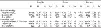
The analysis of NT levels in the selected brain structures showed that DA level in the pre-frontal cortex was significantly lower (P<0.001) in NB pigs than in PB pigs. Also, a significant increase in 5HT (P<0.01) and higher but not significant values of NA and 5-HIAA in the hippocampus of NB individuals were observed compared with those in the PB individuals (Table 1).
Effect of cognitive bias (positive bias (PB) or negative bias (NB)) on the concentrations of catecholamines and indoleamines in different brain regions

SE=pooled standard error; ns=not significant.
**P<0.01, ***P<0.001.
Oxidative stress status
Table 2 shows the results of biomarkers of oxidative stress, including muscle LPO (indicator of oxidative damage) and TAA (antioxidant defence). There was a significant effect (P<0.001) of cognitive bias on the muscle TAA, which showed higher values in NB pigs compared with PB ones at any postmortem time. This difference was significant even at 0 h postmortem, probably as a response of the muscle cells to an increased free radical generation at slaughter, and was maintained throughout the whole postmortem period. However, this antioxidant response was not fast enough to prevent higher levels of LPO in the muscle tissue of NB pigs at 0 h, but contributed to a significant decrease in oxidative damage during meat maturation (P<0.001).
Effect of cognitive bias (CB) (positive bias (PB) or negative bias (NB)), postmortem time (T) (0, 4, 8, 24 h) and their interaction (CB×T) on total antioxidant activity (TAA, mg Trolox/mg protein) and lipid peroxidation (LPO, µmol malondialdehyde +4-hydroxy-2-(E)-nonenal/g protein) in Longissimus dorsi muscle

SE=pooled standard error; ns=not significant.
a,bFor a given CB, means in the same row followed by different letters are significantly different at P<0.05.
***P<0.001.
This decrease of LPO at early postmortem time (0 to 4 h) was greater (P<0.001) in NB animals, thus showing a significant interaction of cognitive bias and postmortem time on meat oxidative damage.
Proteasome activity
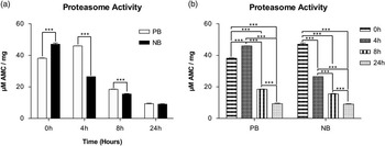
Consistent with the oxidative stress status, the results indicate significantly higher (P<0.001) 20S proteasome activity in the muscle tissues of NB pigs at 0 h postmortem (Figure 1a), whereas PB pigs showed significantly increased 20 S proteasome activity at 4 h postmortem (P<0.001) (Figure 1b), thus showing an evident lag in the system response of the muscle cells. These results seem to indicate higher free radical production in the muscle tissues of NB animals due to higher peri-slaughter stress, whereas this effect was retarded in PB pigs. The gradual decay in proteasome activity over time (P<0.001) was equivalent in both cases (Figure 1b).
Proteasome activity in the muscle (mean±SEM), expressed as μM 7-amino-4-methylcoumarin (AMC)/mg protein; (a) effect of cognitive bias (positive bias (PB) or negative bias (NB)) on the proteasome activity at different postmortem time; (b) effect of postmortem time (0, 4, 8 and 24 h) on the muscle proteasome activity within the different animal groups for the cognitive bias test (PB and NB). ***P<0.001.

Characterization of autophagic processes
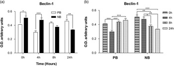
The levels of the autophagy biomarkers Beclin-1 and microtubule-associated protein I LC3 were analyzed in the muscle extracts. Beclin-1 is a class III phosphatidylinositol 3-kinase-interacting protein (60 kDa) that plays an important role in promoting autophagy. The immunoblot analysis in both animal groups (NB and PB) showed active Beclin-1 and therefore autophagic activities in the postmortem muscle tissue (Supplementary Figure S1), although the evolution of these activities differed depending on the animal’s cognitive bias. Beclin-1 evolution in NB animals showed a similar trend to that found for the proteasome activity, with higher levels than in PB pigs at 0 (P<0.05) and 4 h postmortem (P<0.001, Figure 2a), followed by a gradual decrease during meat conditioning (from 4 to 24 h postmortem). However, an opposite trend was observed in the muscle of PB pigs, with significant increased Beclin-1 expression over the final hours of the study, that is, from 4 to 24 h postmortem (Figure 2b).
(a) Effect of cognitive bias (positive bias (PB) or negative bias (NB)) on Beclin-1 expression at different postmortem time; (b) effect of postmortem time (0, 4, 8 and 24 h) on Beclin-1 expression within the different animal groups for the cognitive bias test (PB and NB). *P<0.05, **P<0.01, ***P<0.001. Data expressed as semi-quantitative optical density (arbitrary units) of blot bands normalized to GAPDH.

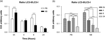
The conversion of LC3-I (soluble unlipidated form) to LC3-II (membrane-bound, phospholipid conjugate form) correlates with completed autophagosomes and autolysosomes and with inevitable autophagy; thus, the immunoblot analysis of the autophagy flux (i.e. the ratio LC3-II/LC3-I) is a useful marker of autophagic activity. Western blot analysis showed two bands corresponding to LC3-I and LC3-II (18 and 16 kDa, respectively, Supplementary Figure S2). Significant differences were observed between both animal groups in the autophagy flux (Figure 3a), with higher level of the ratio LC3-II/LC3-I in the LD muscle of NB pigs at early postmortem time (0 h), followed by a significant decrease during meat conditioning (Figure 3b). However, the muscle of PB pigs showed a delay in the presence of these autophagy markers, thus showing higher level (P<0.001) than in NB pigs at later postmortem times (8 h).
(a) Effect of cognitive bias (positive bias (PB) or negative bias (NB)) on the autophagy flux (LC3-II/LC3-I ratio) at different postmortem time; (b) effect of postmortem time (0, 4, 8 and 24 h) on LC3-II/LC3-I ratio within the different animal groups for the cognitive bias test (PB and NB). *P<0.05, **P<0.01, ***P<0.001. Data expressed as semi-quantitative optical density (arbitrary units) of blot bands normalized to GAPDH.

Discussion
The results of this study show that biochemical and cellular mechanisms involved in the postmortem conversion of muscle into meat may vary as a function of the psychological condition of animals at slaughter, which, in turn, depends on their cognitive bias, that reflects their emotional state and its influence on cognitive processes and information processing including attention, learning, memory and decision making (Mendl et al., Reference Mendl, Burman, Parker and Paul2009).
In the present study, pigs were classified using a novel cognitive test that allows individual animals to be classified into ‘PB’ or ‘NB’, thus reflecting a positive or negative emotional state. Our hypothesis was that the pig’s emotional state would affect its interpretation of the different stimuli related to slaughter and consequently its susceptibility to stress at slaughter and this should be associated with a specific pattern of NTs, that have been proposed as physiological markers of stress (Yeates and Main, Reference Yeates and Main2008). In the literature, changes in brain NT profile in genetically stress-susceptible pigs and their involvement in aggressiveness and dominance have been reported (Adeola et al., Reference Adeola, Ball, House and O’Brien1993; Poletto et al., Reference Poletto, Cheng, Meisel, Garner, Richert and Marchant-Forde2010, Reference Poletto, Cheng, Meisel, Richert and Marchant-Forde2011). Also, the relationship between brain NTs and fear-related behavior in pigs have recently been demonstrated by our group (Arroyo et al., Reference Arroyo, Carreras, Valent, Pena, Mainau, Velarde, Sabria and Bassols2016) in a study in which fearful animals subjected to stressful handling showed higher concentration of catecholamines and indoleamines, specially serotonin, in the hippocampus.
Our findings are in accordance with these previous publications, although it is worthwide to mention that the low number of animals classified as NB limits a general interpretation of our results. In our study NB pigs, that were presumably more prone to regard with suspicion and fear any strange and threatening stimuli related to slaughter, showed higher serotonin levels in the hippocampus at slaughter, despite having a calm and smooth pre-slaughter handling, which indicate higher susceptibility to slaughtering stress. Furthermore, higher serotonin concentration found in the hippocampus of NB individuals agree with several studies that have described that under different situations of stress there is an increased release of noradrenaline and serotonin in the hippocampus in rats (Mora et al., Reference Mora, Segovia, Del Arco, de Blas and Garrido2012) and pigs (Piekarzewska et al., Reference Piekarzewska, Sadowski and Rosochacki1999). Our results also agree with the report of Bauer (Reference Bauer2015) who associated higher levels of serotonin with fear or anxiety. This could be due to the fact that the hippocampus is the brain structure that was first recognized as the target for stress hormones (Mora et al., Reference Mora, Segovia, Del Arco, de Blas and Garrido2012). All these results support the idea that animals in a negative psychological state may have poorer management of stress situations.
Dopamine has also been linked to a number of important psychological processes including reward-motivated behavior, hedonic reactions to positive reward, provision of an error detection signal during the acquisition of new learning, response to novel stimuli, provision of reinforcement signals essential for acquisition of new action patterns and incentive motivation (Phillips et al., Reference Phillips, Vacca and Ahn2008). All these arguments reinforce the suggestion that the higher levels of DA in the pre-frontal cortex of PB animals could be linked to a positive reaction to new stimuli, even those related to pre-slaughter management.
It is widely accepted that the pre-slaughter management is likely the most stressful situation for the animal destined to meat production, whose effects are clearly related to each animal’s ability to cope with it. A recent review indicated the relationship between mood disorders, including anxiety and stress, and several potential contributors to cellular oxidative stress and proposed LPO as an important damage marker for anxiety disorders (Salim, Reference Salim2014). Also, it has been shown that psychological stress results in oxidative damage in the muscle (Li et al., Reference Li, Zhang, Chen, Wang, Huang and Liu2011; Rubio-Gonzalez et al., Reference Rubio-Gonzalez, Potes, Illan-Rodriguez, Vega-Naredo, Sierra, Caballero, Fabrega, Velarde, Dalmau, Olivan and Coto-Montes2015). Our results are in agreement with these authors in showing higher oxidative damage in the LD muscle of NB pigs at slaughter (0 h postmortem), which demonstrate a relationship between psychological stress at slaughter and muscle cell damage. Increased enzymatic antioxidant systems have also been observed as possible adaptive response to stress under different situations (Caballero et al., Reference Caballero, Sierra, Oliván, Vega-Naredo, Tomás-Zapico, Álvarez-García, Tolivia, Hardeland, Rodríguez-Colunga and Coto-Montes2006; Rubio-Gonzalez et al., Reference Rubio-Gonzalez, Potes, Illan-Rodriguez, Vega-Naredo, Sierra, Caballero, Fabrega, Velarde, Dalmau, Olivan and Coto-Montes2015). In the present study, this adaptive response to stress was higher in the muscle of NB pigs compared with PB ones. However, this antioxidant response was not fast enough to prevent higher levels of LPO in the muscle tissue of NB pigs at 0 h postmortem, but contributed to a significant decrease in oxidative damage during meat maturation.
All these factors, together with the animal’s cognitive bias and its susceptibility to stress may affect to the weakening of muscle cells under postmortem conditions, which depends on the proteolysis extent of key target proteins that are regulated by the synergic and combined action of different proteolytic systems, such as lysosome and proteasome (Houbak et al., Reference Houbak, Ertbjerg and Therkildsen2008; Ouali et al., Reference Ouali, Gagaoua, Boudida, Becila, Boudjellal, Herrera-Mendez and Sentandreu2013). It has been demonstrated the role of lysosomal cathepsins in meat maturation (Maribo et al., Reference Maribo, EWrtbjerg, Andersson, Barton-Gade and Mùller1999; Caballero et al., Reference Caballero, Tomas-Zapico, Vega-Naredo, Sierra, Tolivia, Hardeland, Rodriguez-Colunga, Joel, Nevo, Avivi and Coto-Montes2007) and also that lysosomal permeabilization and cathepsins release is often an early event in the cell response to oxidative stress (Conus and Simon, Reference Conus and Simon2008). Also, It is known that the proteasome degrade most intracellular proteins, including both short- and long-lived proteins, being the Ub/20S proteasome system responsible for this protein degradation (Ouali et al., Reference Ouali, Gagaoua, Boudida, Becila, Boudjellal, Herrera-Mendez and Sentandreu2013). However, the role of the proteasome system in early meat tenderization is still unknown, but its activation is considered one of the first responses to cellular stress. In this study, the higher proteasome activity in the LD muscle of NB at 0 h postmortem compared with PB pigs and its faster postmortem decline could have been induced by the psychological and oxidative stress suffered by these animals at slaughter. Based on these results, NB pigs seem to be more susceptible to slaughter stress than PB pigs, that showed a delay in the postmortem evolution of the proteasome activity in the muscle.
Oxidative stress produced by pre-slaughter stress can induce in the muscle another proteolytic mechanism known as autophagy (Rubio-Gonzalez et al., Reference Rubio-Gonzalez, Potes, Illan-Rodriguez, Vega-Naredo, Sierra, Caballero, Fabrega, Velarde, Dalmau, Olivan and Coto-Montes2015). This can be related to an increase in the tissue ROS level, which can activate autophagy as a defence mechanism to prevent cellular damage. Based on our previous findings that demonstrated the occurrence of autophagy processes in the pre-rigor phase of beef conditioning (Garcia-Macia et al., Reference Garcia-Macia, Sierra, Palanca, Vega-Naredo, de Gonzalo-Calvo, Rodriguez-Gonzalez, Olivan and Coto-Montes2014), and that certain degree of psychological stress at slaughter may influence the time-scale evolution of autophagy biomarkers in the postmortem muscle of pigs (Rubio-Gonzalez et al., Reference Rubio-Gonzalez, Potes, Illan-Rodriguez, Vega-Naredo, Sierra, Caballero, Fabrega, Velarde, Dalmau, Olivan and Coto-Montes2015), the present study aimed to extend the knowledge regarding the relationship between autophagy markers and the animal’s cognitive bias and its subsequent susceptibility to peri-slaughter stress. Our results show a significant effect of the animal’s emotional state on the postmortem evolution of autophagy in the muscle tissue, with NB pigs showing increasing values of Beclin-1 at early postmortem time (0 to 4 h postmortem), most likely resulting from the attempt of the muscle cell to counteract oxidative stress at slaughter, followed by a significant decrease during meat conditioning. However, PB pigs showed a delay in the muscle autophagy onset, with lower level of Beclin-1 at early postmortem times.
A similar pattern of variation showed the ratio LC3-II/LC3-I (considered marker of autophagy) in the LD muscle of NB pigs, then showing that the autophagy onset occurred earlier in the muscle of NB animals, that had higher susceptibility to stress, and it appeared as a mechanism of cell survival in order to counteract the increased oxidative stress.
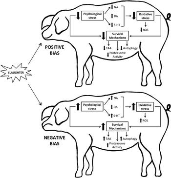
In conclusion, the present study reveals the effect of the animal’s cognitive bias and its related response to pre-slaughter stress on the postmortem muscle metabolism, and its high influence on the muscle oxidative stress, the proteasome activity and the autophagy onset during the first phases of postmortem tenderization (Figure 4). Furthermore, our results highlight the potential contribution of biomarkers of proteolysis, autophagy and oxidative stress in the cell muscle for the assessment of pre-slaughter stress. Our findings expand the current knowledge regarding biomarkers of animal welfare and we propose the use of the proteasome activity (20 S), the main biomarkers of autophagy (Beclin-1, LC3-II/LC3-I ratio) and the muscle antioxidant defence (TAA, LPO) as potential biomarkers of peri-slaughter stress. However, our study was limited due to the low number of NB animals detected in the population, so further studies will be necessary in order to confirm these results.
Summary of primary factors implicated in this study and the relation established between them. An animal’s susceptibility to stress at slaughter depends on its cognitive bias and shows some neurotransmitter variations, which induce physiological responses and changes in the muscle response to oxidative stress, triggering autophagy as a muscle cell survival mechanism. NA=noradrenaline; DA=dopamine; 5HT=serotonin; ROS=reactive oxygen species; TAA=total antioxidant activity.

Acknowledgment
The authors would like to thank to the staff of IRTA involved in animal handling and sample collection. This work was supported by FISS-13-RD12/0043/0030 and FISS-14-PI13/02741 (Instituto de Salud Carlos III), GRUPIN-071 (PCTI), AGL2010-21578-C03 and AGL2011-30598-C03 (Ministerio de Economía y Competitividad) and FEDER funds. Y. P. thanks a pre-doctoral fellowship from the Vicerrectorado de Investigación y Campus de Excelencia Internacional (Universidad de Oviedo) and FISS pre-doctoral fellowship (FI14/00405) from the Ministerio de Economía y Competitividad (Instituto de Salud Carlos III). B. d. L.-D. is a FICYT pre-doctoral fellowship (BP11-135) from PCTI of Gobierno del Principado de Asturias. Some of the authors are members of the INPROTEOLYS and INEUROPA networks.
Supplementary materials
To view supplementary material for this article, please visit https://doi.org/10.1017/S1751731117000714