INTRODUCTION
Large animals require a circulatory system for the supply of O2, nutrients, hormones and growth factors etc., to all parts of the body, as well as the removal of waste products, such as, CO2, lactic acid and urea. This is the function of the blood vascular system. A secondary, lymphatic vascular system has evolved in parallel to return capillary-derived, extravasated fluid back to the blood. Tumours, with the same basic requirements, co-opt directly and indirectly the body's circulatory system(s). As relatively large, metazoan organisms, tissue-dwelling parasitic helminths, have the same constraints for the successful colonization of the parasitized host.
The circulatory system is laid down during early embryonic development by the formation de novo and in situ of a primitive capillary plexus from angioblasts (vasculogenesis). Physiological angiogenesis (formation of new blood vessels from pre-existing capillaries and post-capillary venules) in later, embryonic development, is the transformation of this structure by endothelial cell (EC) proliferation-migration-sprouting-pruning into the fully formed and mature, blood-lymphatic vascular systems. Neoangiogenesis, by both physiological angiogenesis in the adult (wound healing, female reproductive cycle) and pathological angiogenesis (tumours, metastases, various vascular diseases), recapitulate these cellular and molecular pathways of vasculature development.
In this review, we wish to submit and discuss the working hypothesis as to how parasitic helminths achieve the formation of a parasite-defined vasculature as one of the prerequisites for successful parasitism i.e. growth, maturity, reproduction and reinfection. We envisage at least 2 possible, or a combination of, pathways for parasitic helminth-associated angiogenesis: in tissue-dwellers, the angiogenic switch for parasitic vasculature formation occurs, either by upregulating a host- or parasite-derived, pro-angiogenic trigger of the host's neovascularization machinery; in vasculature-dwellers, through the co-option of the host's vascular system as, either active or passive, intra-vascular and/or –lymphatic, obligate ‘inquilines’.
CIRCULATORY SYSTEM
General properties and characteristics
All the cells, tissues and organs of the body are dependent on an efficient supply of O2 and nutrients, as well as the removal of CO2 and metabolic waste products (Alitalo et al. Reference Alitalo, Tammela and Petrova2005). These tasks are performed by the circulatory/vascular system. The circulatory system consists of 2 highly branched, tubular networks formed from endothelial cells (ECs), one of which is closed, the primary blood vascular system, and one of which is open, the secondary lymphatic vascular system. Blood circulates through the blood vessels in the direction of arteries-arterioles-capillary beds-venules-veins. The exchange of gases, nutrients and metabolic waste products occurs in the capillaries, and extravasation of macromolecules and cells in the post-capillary venules (Jain, Reference Jain2003; Adams and Alitalo, Reference Adams and Alitalo2007). The lymphatic vascular system functions to maintain fluid balance, in that the blood pressure causes a continuous leakage of plasma into the interstitial space. This extravasated, protein-rich fluid (lymph), along with extravasated leucocytes and activated antigen-presenting cells, is returned via lymphatic capillaries-collecting lymphatic vessels-lymph nodes-lymphatic trunks and ducts to the blood circulation (Alitalo et al. Reference Alitalo, Tammela and Petrova2005).
Development
The development of a functional vascular system is of vital importance in organogenesis as signified by it being one of the earliest events in embryogenesis (Coultas et al. Reference Coultas, Chawengsaksophak and Rossant2005). The initial phase of vasculogenesis is the differentiation and proliferation of ECs in situ and in vivo (i.e., haemangioblasts, haematopoietic and EC precursors; the latter angioblasts migrate and assemble as the outer cells of blood islands). Fusion of these blood islands leads to the appearance of the primary vasculature comprising major vessels, such as dorsal aorta and cardinal veins, and a connecting, primitive capillary plexus. Whereby the capillaries are already specified as, either arterial or venous. The vasculogenic system is characterized as being immature, active and unstable in producing leaky-permeable-haemorrhagic blood vessels (Yancopoulos et al. Reference Yancopoulos, Davis, Gale, Rudge, Wiegand and Holash2000; Karamysheva, Reference Karamysheva2008).
The subsequent phase of angiogenesis, to increase blood flow to the growing tissues, requires the expansion of the pre-existing arteries and veins by circumferential growth and remodelling (arteriogenesis), as well as the sprouting-branching-pruning-remodelling from pre-existing capillaries and post-capillary venules to more complex networks (Carmeliet, Reference Carmeliet2005). Angiogenic sprouting is a complex, morphogenetic event typified by EC sprouting-sprout outgrowth and guidance-sprout fusion and lumen formation-perfusion and maturation (Carmeliet, Reference Carmeliet2003; Adams and Alitalo, Reference Adams and Alitalo2007). The resultant secondary vasculature is a highly organized, hierarchical and stereotyped vascular network, with nascent arteries and veins clad by the differentiated mural cells of vascular smooth muscle cells (VSMCs) and capillaries enveloped by pericytes (Carmeliet, Reference Carmeliet2005; Ferrara and Kerbel, Reference Ferrara and Kerbel2005). This leads to the phenotype of a mature, stable, quiescent and functional blood vascular system.
However, in physiological angiogenesis this adult blood vascular system can still exhibit plasticity, in that, under specific circumstances it can recapitulate developmental angiogenesis’ patterns of growth-branching-pruning and remodelling-repression with ECs displaying cell proliferation-migration-survival-morphogenesis or apoptosis (Jain et al. Reference Jain, Munn and Fukumura2002; Jain, Reference Jain2003). This can occur during wound healing, physiological organ growth and the cyclicity of the female reproductive tract. Pathological angiogenesis, as exemplified by tumours and metastases, shows continual remodelling due to inappropriate patterns of growth and regression to yield an unstable, continually evolving network of abnormal structure and function (Jain, Reference Jain2003).
During embryonic development, lymphangiogenesis occurs by the sprouting of lymphatic endothelial cells (LECs) from embryonic veins, that migrate to lymphatic sacs before growth by sprouting-branching-proliferation-remodelling (Adams and Alitalo, Reference Adams and Alitalo2007). Lymphatic capillaries have a discontinuous basement membrane without mural cells. Collecting lymphatic vessels possess VSMCs, basement membrane and valves (Alitalo et al. Reference Alitalo, Tammela and Petrova2005).
Molecular angiogenic factors
Developmental and physiological angiogeneses are of fundamental importance to the ontogeny and homeostasis of vertebrate physiology. Of necessity, therefore, neovascularization and concomitant, vascular vessel maturation are highly regulated, complex and co-ordinated processes, that require the sequential and balanced activation of a series of specific stimulatory and inhibitory, ligand-receptor signalling pathways (Ferrara et al. Reference Ferrara, Gerber and LeCouter2003). Besides a range of anti-angiogenic factors, the key pro-angiogenic, regulatory system of angiogenesis is the vascular endothelial growth factor (VEGF)-VEGFR (receptor) family-signalling pathway (Ho and Kuo, Reference Ho and Kuo2007; Ferrara, Reference Ferrara2009). However, a number of other ligand-receptor regulators are of major importance: the angiopoietin (Ang)-Tie family; the ephrin B2-EphB4 system; the Delta-like-4 ligand (Dll4)-Notch family signalling cascade; platelet-derived growth factor (PDGFB)-PDGFRβ signal transduction; and transforming growth factor (TGF)β-receptor signalling pathway (Adams and Alitalo, Reference Adams and Alitalo2007; Shibuya, Reference Shibuya2008).
The critical molecule regulating blood vessel-morphogenesis is VEGF-A (synonym, vascular permeability factor [VPF]) and belongs to a family of potent, angiogenic regulators, including, VEGF-B, VEGF-C, VEGF-D and placental growth factor (PlGF). Alternative splicing produces various VEGF-A isoforms, the most important and dominant of which is VEGF-A165 (containing 165 amino acids). The heparin binding-induced VEGF-A165 concentration gradient promotes EC proliferation-migration-survival-differentiation-sprouting and direct assembly of vascular structures that are necessary for the morphogenetic processes of blood vessel vasculogenesis and angiogenesis (Adams and Alitalo, Reference Adams and Alitalo2007; Karamysheva, Reference Karamysheva2008). This includes the angiogenic capillary sprouting-steps of: vasodilation and plasma protein-extravasation formation of a provisional matrix, containing fibronectin and fibrin etc., for activated EC migration; extra-cellular matrix (ECM) remodelling through EC-mediated, pericellular proteolysis by surface-bound and secreted proteases of the matrix metalloproteinase (MMP2, MMP9 and MT1-MMP) and plasminogen activator-plasmin systems (Pepper, Reference Pepper2001; van Hinsbergh and Koolwijk, Reference Van Hinsbergh and Koolwijk2008); differential expression of tip-cell sprouts; and induction of the angiogenic switch i.e. of EC invasiveness and motility (Bergers and Benjamin, Reference Bergers and Benjamin2003). Signalling is mainly through the EC-located receptor tyrosine kinase, VEGFR2. Under specific circumstances, such as embryonic/developmental angiogenesis, this signal is negatively modulated by VEGFR1 binding of the EC-located receptor. PlGF, a specified VEGFR1-specific ligand, only shows an enhancement of neovascularization during pathological angiogenesis i.e. tumour growth (Carmeliet, Reference Carmeliet2003) and primary tumour metastasis (Hiratsuka et al. Reference Hiratsuka, Nakamura, Iwai, Murakami, Itoh, Kijima, Shipley, Senior and Shibuya2002; Shibuya, Reference Shibuya2006). The key molecule regulating lymphatic vessel morphogenesis (lymphangiogenesis) is VEGF-C, signalling through the EC-located VEGFR3 (Adams and Alitalo, Reference Adams and Alitalo2007). Signal amplification is dependent on co-receptor neuropilins (NRP), which are differentially distributed on arteries (NRP1), and veins and lymphatics (NRP2). The upregulation of many angiogenic genes, primarily the induction of VEGF-A, is triggered and driven by hypoxia. The tissue- and organ growth-generated, hypoxic conditions are signalled through hypoxia-inducible transcription factors (Ferrara et al. Reference Ferrara, Gerber and LeCouter2003).
The vascular endothelium-located, Dll4 (transmembrane ligand)-Notch receptor signalling system is responsible for the complex cascade involved in selection of the non-proliferating tip cell-phenotype from a subpopulation of ECs engaged in angiogenic capillary sprouting. The Notch pathway is also required for formation of the arterial branch during arteriovenous differentiation (Adams and Alitalo, Reference Adams and Alitalo2007; Karamysheva, Reference Karamysheva2008).
The ephrin B2 transmembrane ligand serves as an arterial EC marker, whilst the cognate tyrosine kinase receptor EphB4 is a venous EC marker. The ephrin B2-EphB4 pathway is essential for angiogenesis, in that it is also concerned with the establishment of arterial and venous identity (Carmeliet, Reference Carmeliet2003; Adams and Alitalo, Reference Adams and Alitalo2007). However, the ultimate regulator of these arterial differentiation pathways is VEGF-A. In addition, EC specialization can be organ-specific with the finding that endocrine gland EC-capillary, pore-like fenestrations are induced by the tissue-specific, angiogenic regulator, endocrine gland (EG)-VEGF (LeCouter et al. Reference LeCouter, Kowalski, Foster, Hass, Zhang, Dillard-Telm, Frantz, Rangell, DeGuzman, Keller, Peale, Gurney, Hillan and Ferrara2001).
These nascent, immature and unstable blood vessels are stabilized by mural cell recruitment and ECM formation (Jain, Reference Jain2003). This process is organized by the Ang1-Tie2, PDGFB-PDGFRβ and TGFβ signalling systems. PDFGB plays the key role in pericyte recruitment. The EC-secreted PDFGB facilitates both mural cell proliferation, differentiation and migration-recruitment via interaction with the latter cell type's PDGFRβ-receptor tyrosine kinase (Karamysheva, Reference Karamysheva2008). Pericyte- and VSMC-secreted Ang1 interacts with the EC-located receptor tyrosine kinase-Tie2, to promote pericyte-endothelium association and to lower vascular permeability (Karamysheva, Reference Karamysheva2008). TGFβ1, a pericyte- and EC-derived, multifunctional cytokine, contributes to blood vessel maturation with ECM production and mural cell differentiation (Jain, Reference Jain2003). Less is known on lymphatic vessel maturation, but it is acknowledged to involve the angiopoietins and ephrin B2 (Alitalo et al. Reference Alitalo, Tammela and Petrova2005).
The autocrine, EC-expressed Ang2-Tie2 system has different roles in developmental as opposed to physiological and pathological angiogeneses. In embryonic angiogenesis, Ang2 is a competitive inhibitor of Ang1, i.e. a Tie2 antagonist, to block its vascular, stabilizing action. In physiological and pathological angiogeneses, Ang2 has a key role in destabilizing the vasculature as a necessary prerequisite to subsequent remodelling. The destabilized blood vessels have 2 fates depending on the presence or absence of VEGF. If VEGF is absent vascular regression and EC apoptosis ensue, whilst in the presence of VEGF Ang2 supports renewed angiogenesis (Yancopoulos et al. Reference Yancopoulos, Davis, Gale, Rudge, Wiegand and Holash2000; Karamysheva, Reference Karamysheva2008).
A number of endogenous, anti-angiogenic factors have been reported, for example, thrombospondin-1 (TSP-1; Rodriguez-Manzaneque et al. Reference Rodriguez-Manzaneque, Lane, Ortega, Hynes, Lawler and Iruela-Arispe2001), angiostatin (O'Reilly et al. Reference O'Reilly, Holmgren, Shing, Chen, Rosenthal, Moses, Lane, Cao, Sage and Folkman1994, Reference O'Reilly, Holmgren, Chen and Folkman1996) and endostatin (O'Reilly et al. Reference O'Reilly, Boehm, Shing, Fukai, Vasios, Lane, Flynn, Birkhead, Olsen and Folkman1997). TSP-1 is anti-angiogenic, in that it inhibits EC proliferation and migration, and induces EC apoptosis. Angiostatin and endostatin belong to the ‘statins’, inhibitory molecules derived by specific cleavage from larger proteins, which themselves are inactive. Thus, angiostatin is a plasminogen fragment and endostatin is a C-terminal fragment of collagen type XVIII.
Tumours are also dependent on a blood supply thereby neovascularization is usually considered a prerequisite for tumour growth, progression and metastasis. It is, therefore, imperative for the tumour to gain access to the host's vascular system and generate a tumour vasculature. The induction of the tumour vasculature is termed the angiogenic switch (Bergers and Benjamin, Reference Bergers and Benjamin2003) and is characterized, among other things, by an imbalance of pro- and anti-angiogenic factors. The loss of tight regulation results in tumour vessels that are unstable (due to altered EC-pericyte interactions)-immature-non-quiescent-haemorrhagic-leaky, in part, due to VEGF-A overproduction (Carmeliet, Reference Carmeliet2000, Reference Carmeliet2003, Reference Carmeliet2005). In the following review, we wish to develop and reinforce our working hypothesis as being analogous to that of tumour angiogenesis; that successful, metazoan parasitism of growth, maturation, egg production and reinfection are reliant on parasitic helminth-associated angiogenesis, which expresses, at the least, some of the features of neovascularization-dependent tumorigenesis.
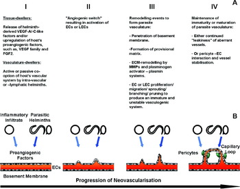
At least 2 pro-angiogenic components of the angiogenic switch have been found to be VEGF-A (Inoue et al. Reference Inoue, Hager, Ferrara, Gerber and Hanahan2002) and MMP9 (Bergers et al. Reference Bergers, Brekken, McMahon, Vu, Itoh, Tamaki, Tanzawa, Thorpe, Itohara, Werb and Hanahan2000; Hiratsuka et al. Reference Hiratsuka, Nakamura, Iwai, Murakami, Itoh, Kijima, Shipley, Senior and Shibuya2002). Induced and upregulated MMP9 is a functional constituent of the angiogenic switch in both carcinogenesis and primary tumour metastasis, either by increasing the bioavailability of ECM-sequestered VEGF or preparing distant, premetastatic niches for specific, neoplastic cell invasion, respectively. An anti-angiogenic component of the angiogenic switch in tumour progression has been found to be the endogenous angiogenesis inhibitor TSP-1 (Rodriguez-Manzaneque et al. Reference Rodriguez-Manzaneque, Lane, Ortega, Hynes, Lawler and Iruela-Arispe2001). TSP-1 upregulation was correlated with the suppression of spontaneous tumour growth and the inhibition of MMP9 activation, with the consequent prevention of VEGF mobilization from ECM sequestration, for example, perlecan (Jiang and Couchman, Reference Jiang and Couchman2003) for VEGFR2-mediated transduction (Fig. 1).
Hypothesized pathway(s) of parasitic helminth-associated (lymph)angiogenesis. (A) Partition of (lymph)angiogenesis into four developmental stages: I, release of helminth- and/or upregulation of host-derived, proangiogenic factors; II, angiogenic switch of endothelial cell activation; III, remodelling events of parasite vasculature-formation; IV, maintenance of immaturity or maturation of parasite vasculature. (B) Cartoon of the four phases of (lymph)angiogenesis: I, priming of vascular or lymphatic, endothelial cells by parasitic helminth- and/or inflammatory infiltrate-generated, proangiogenic factors; II, angiogenic switch and focal breakdown of vessel-basement membrane; III, remodelling and formation of parasite-determined vasculature; IV, maintenance of immaturity or pericyte-identified maturation. ECs, vascular endothelial cells; ECM, extracellular matrix; FGF, fibroblast growth factor; LECs, lymphatic endothelial cells; MMPs, matrix metalloproteinases; VEGF, vascular endothelial growth factor.

PARASITIC HELMINTHS
During the development of numerous tissue- and/or vasculature-dwelling helminths, it is apparent that there are direct and indirect interactions with the host's blood- and/or lymphatic-vascular systems. Potentially, such parasitic helminths would or could have the capability to initiate pro-angiogenic events. Our intention is to present and discuss all these ‘impacts’ that have been investigated in relation to parasitic helminth-associated angiogenesis. The emphasis on the selection of reviewed helminths has been guided by PubMed-literature searches using the basic keywords of parasitic helminths/angiogenesis/neovascularization.
Cestoda
The only previous systematic studies of the Cestoda have been carried out on the metacestode larval stage in intermediate hosts of Mesocestoides corti, present in mice and the ‘pork’ tapeworm, Taenia solium, in humans and pigs. This cyst stage can develop in the central nervous system (CNS) as a causative agent of neurocysticercosis. The resultant, protective, granulomatous immune response surrounding the lesion(s) is characterized by an inflammatory blood immune cell-infiltrate, fibrosis, a loss of blood-brain barrier (BBB) function, i.e. permeability, and wound healing (Restrepo et al. Reference Restrepo, Alvarez, Castano, Arias, Restrepo, Trujillo, Colegial and Teale2001). The wound-healing response involved astrocyte-dependent, glial scar formation, fibroblast-derived collagen type I deposition and angiogenesis (Alvarez et al. Reference Alvarez, Colegial, Castano, Trujillo, Teale and Restrepo2002). The rapid neovascularization of astrocyte endfeet-deficient blood vessels and enhanced BBB permeability is associated with the upregulation of astrocytes’, parenchymal vessels’ and inflammatory blood immune cells’ VEGF expression (Alvarez and Teale, Reference Alvarez and Teale2006; Sikasunge et al. Reference Sikasunge, Johansen, Phiri, Willingham and Leifsson2009). This was accompanied by the neovascular and perivascular localization of mast cell-juxtaposed, basic fibroblast growth factor (FGF2; Alvarez et al. Reference Alvarez, Colegial, Castano, Trujillo, Teale and Restrepo2002), a known vasoactive and pro-fibrotic cytokine. The FGF family of heparin-binding, pleiotropic growth factors, among other properties, participate in both adult physiological and pathological angiogeneses, through cross-talk and synergism with VEGF-VEGFR system activation (Presta et al. Reference Presta, Dell'Era, Mitola, Moroni, Ronca and Rusnati2005). Even cases of intraocular, neovascular glaucoma (=neovascularization), with surgical and anti-VEGF MAb treatments, have been demonstrated to be due to T. solium metacestode cysts (Ratra et al. Reference Ratra, Phogat, Singh and Choudhari2010).
These findings have been interpreted as supporting the proposed, working hypothesis of a parasitic helminth-provoked, host-elicited angiogenic response. The larval stage of the fox tapeworm, Echinococcus multilocularis (human alveolar echinococcosis), in the intermediate host's liver, exhibits a tumour-like, infiltrative growth of the multivesicular metacestodes through asexual reproduction and is surrounded by newly formed blood vessels in the granulation tissue (Guerret et al. Reference Guerret, Vuitton, Liance, Pater and Carbillet1998; Weiss et al. Reference Weiss, Bauer and Kohler2010). From a molecular and functional comparison with the multifunctional, mammalian protein phosphoglucose isomerase (PGI), both parasitic growth and parasite-associated angiogenesis involve the highly conserved E. multilocularis analogue (EmPGI; (Stadelmann et al. Reference Stadelmann, Spiliotis, Muller, Scholl, Muller, Gottstein and Hemphill2010). It can be speculated that the pro-angiogenic potential of EmPGI is not only required for neovascularization, but is responsible, at least in part, for the tumorigenic- and metastatic-like growth of E. multilocularis metacestodes.
Nematoda
Caenorhabditis elegans
The sequenced genome of the free-living nematode, Caenorhabditis elegans, has been successfully used to trace the structure and supplementary functions of highly conserved, evolutionarily ‘favourable’ factors during the evolution from simple to more complex organisms (vertebrates/mammals). The basement membrane, muscle tissue-located nematode perlecan/UNC-52 has become crucial for mammalian vasculogenesis and chondrogenesis (Jiang and Couchman, Reference Jiang and Couchman2003). The conserved role of the collagen type XVIII NC/endostatin domain in regulating neural cell migration and axonal guidance of nematode neurogenesis, now functionally overlaps with vertebrate angiogenesis (Ackley et al. Reference Ackley, Crew, Elamaa, Pihlajaniemi, Kuo and Kramer2001). The original FGF-FGFR signalling system may resemble that of the single FGFR and 2 FGFs (related to subfamilies FGF8 and FGF9) of C. elegans, with developmental and physiological properties, respectively (Coulier et al. Reference Coulier, Pontarotti, Roubin, Hartung, Goldfarb and Birnbaum1997; Birnbaum et al. Reference Birnbaum, Popovici and Roubin2005). The nematode homologues of VEGFRs were primarily and solely involved in cell migration and adhesion of neurogenesis and morphogenesis, later subverted by vertebrates/mammals to include angiogenesis (Popovici et al. Reference Popovici, Isnardon, Birnbaum and Roubin2002). In fact, C. elegans generates a PDGF/VEGF-like ligand (PVF1) that is biologically active, in that it binds mammalian VEGFRs and induces angiogenesis (Tarsitano et al. Reference Tarsitano, De Falco, Colonna, McGhee and Persico2006). It is, therefore, hard to escape the conclusion that nematodes have the capacity to produce and secrete biologically active, VEGF-like molecules. However, whether only host- and/or parasitic nematode-derived angiogenic factors are alone responsible for neovascularization is still an open question. For example, one component of ocular onchocerciasis (river blindness), caused by L1 microfilariae of the filarial worm, Onchocerca volvulus, is the parasite-associated angiogenesis (Pearlman et al. Reference Pearlman, Diaconu, Hazlett, Merriweather and Unnasch1997; Pearlman and Hall, Reference Pearlman and Hall2000; Tawe et al. Reference Tawe, Pearlman, Unnasch and Lustigman2000). The angiogenic response to an O. volvulus homologue of the Ancylostoma-secreted protein family (OvASP2) proved to be indirect, with the recombinant protein not interacting directly with ECs to stimulate proliferation or VEGF generation (Higazi et al. Reference Higazi, Pearlman, Whikehart and Unnasch2003).
Filarial nematodes
These two theses as to whether the parasite vasculature is due to parasitic nematode-elicited, (lymph)angiogenic-like factors and/or with/without inflammation-involved, host-upregulated angiogenesis has proved particularly difficult to resolve, in no small part because their pathogeneses are multifactorial. Taking this into account, therefore, it would be pertinent to review 4 filarial nematodes of different predilection sites: the intra-lymphatic, vascular Wuchereria bancrofti and Brugia malayi of lymphatic filariasis, including the lower trunk and limbs (Noroes et al. Reference Noroes, Addiss, Amaral, Coutinho, Medeiros and Dreyer1996); the tissue-dwelling O. volvulus of nodular and ocular onchocerciasis (Collins et al. Reference Collins, Lujan, Figueroa and Campbell1982); and the intra-blood, vascular Dirofilaria immitis of canine cardiopulmonary dirofilariasis in pulmonary arteries (Manfredi et al. Reference Manfredi, Di Cerbo, Genchi, Genchi, Rinaldi and Cringoli2007).
The development and progression of lymphatic filariasis is characterized by an initial adult worm-dependent, but inflammation- and obstruction-independent, lymphatic vessel dilation (lymphangiectasia). Adult worm death can lead to inflammatory- and fibrotic-dependent, immune reactions of lymph vessel inflammation (lymphangitis), followed by chronic syndromes of lymphatic dysfunction, beginning with serous fluid accumulation of hydrocele and, on recurrent bacterial infection, results in the interstitial fluid accumulation of lymphoedema and eventual elephantiasis (Dreyer et al. Reference Dreyer, Noroes, Figueredo-Silva and Piessens2000; Peixoto and Figueiredo-Silva, Reference Peixoto and Figueiredo-Silva2001; Lammie et al. Reference Lammie, Cuenco and Punkosdy2002). Target LECs are differentially stimulated by B. malayi-produced antigens (Rao et al. Reference Rao, Sutton, Zometa and Vickery1996a,Reference Rao, Zometa, Vickery, Kwa, Nayar and Suttonb) that regulate lymphangiogenesis and lymphatic vessel differentiation via the MMPs/TIMPs balance and not, as expected, by prototypic lymphangiogenic factors (Bennuru and Nutman, Reference Bennuru and Nutman2009). More typically, W. bancrofti adult worm- and/or host-derived responses identified lymphatic dilation and lymphoedema with overexpressed VEGF-C/soluble(s) VEGFR3 (Debrah et al. Reference Debrah, Mand, Specht, Marfo-Debrekyei, Batsa, Pfarr, Larbi, Lawson, Taylor, Adjei and Hoerauf2006) and equated hydrocele with VEGF-A overexpression (Debrah et al. Reference Debrah, Mand, Toliat, Marfo-Debrekyei, Batsa, Nurnberg, Lawson, Adjei, Hoerauf and Pfarr2007, Reference Debrah, Mand, Marfo-Debrekyei, Batsa, Pfarr, Lawson, Taylor, Adjei and Hoerauf2009).
However, a further complication is the obligate presence of the endosymbiont Wolbachia, whereby its depletion by doxycycline-rifampicin antibiotic therapy (Volkmann et al. Reference Volkmann, Fischer, Taylor and Hoerauf2003; Fainaru et al. Reference Fainaru, Hornstein and Folkman2009) ameliorates the chronic lymphatic filariasis symptoms. This raises the possibility of bacteria-induced, pro-inflammatory cytokines being involved in the nematode-related lymphangiogenesis (Debrah et al. Reference Debrah, Mand, Specht, Marfo-Debrekyei, Batsa, Pfarr, Larbi, Lawson, Taylor, Adjei and Hoerauf2006; Pfarr et al. Reference Pfarr, Debrah, Specht and Hoerauf2009). As regards the lymphatic filariasis-correlated fibrosis, the elevated endothelin-1 (ET-1) levels on W. bancrofti infection observed in elephantiasis-associated fibrosis were in agreement with its known pathophysiological properties (Esterre et al. Reference Esterre, Plichart, Huin-Blondey and Nguyen2005).
Vascular perfusion has revealed an intimate relationship between the adult O. volvulus and proliferated, vascular system capillaries within subcutaneous nodules (George et al. Reference George, Palmieri and Connor1985; Smith et al. Reference Smith, Cotter, Williams and Guderian1988), but with no extravasation of the perfusate. Ocular onchocerciasis was confirmed to involve neovascularization and neutrophil infiltration-exacerbated corneal opacification (Hall et al. Reference Hall, Berger, Diaconu and Pearlman2002). The induced, blood and lymphatic vessel neovascularization, either by parasitic worm-dependent (lymph)angiogenic factors or host-generated, inflammatory immune responses, revealed a particular pattern of the chemokine CXCL12-CXCR4, and of Ang1, Ang2 and VEGF-C (Attout et al. Reference Attout, Hoerauf, Denece, Debrah, Marfo-Debrekyei, Boussinesq, Wanji, Martinez, Mand, Adjei, Bain, Specht and Martin2009).
Pulmonary artery-located, D. immitis-mediated, endovascular lesions are conspicuous for their intimal hyperplasia and hypertrophy along with obstructing thrombi, which can lead to chronic hypertension (Grandi et al. Reference Grandi, Zivicnjak, Beck, Genchi, Rinaldi and Cringoli2007; Venco, Reference Venco, Genchi, Rinaldi and Cringoli2007). The pathogenesis of intimal thickening and vessel lumen-stenosis due to medial VSMC proliferation and migration, has been linked to platelet-generated PDGF (Ross, Reference Ross1986) and, more especially, the pathophysiological properties of overexpressed ET-1 (Uchide and Saida, Reference Uchide and Saida2005). A word of caution is necessary here, in that, concerning the antigens of D. immitis and the Wolbachia endosymbiont implicated in this inflammatory pathology, only the latter were able to upregulate EC-derived VEGF in vitro (Simon et al. Reference Simon, Morchon, Rodriguez-Barbero, Lopez-Belmonte, Grandi and Genchi2008).
Non-filarial nematodes
The origin of Trichinella spiralis (trichinosis), parasitic nematode-associated angiogenesis has been well defined. The unique ‘intramulticellular’ L1 larva-stage is responsible for the transformation of invaded myocytes into nurse cell-parasite complexes; a long-term survival strategy to guarantee a constant nutrient supply and waste disposal. The nurse cell-parasite complex is surrounded by a collagenous capsule, that is a circulatory rete of enlarged, permeable, sinusoid-type capillaries (Humes and Akers, Reference Humes and Akers1952; Despommier, Reference Despommier1990; Baruch and Despommier, Reference Baruch and Despommier1991) and an inflammatory cell infiltrate. The coincident processes of angiogenesis, fibrogenesis and nurse cell formation are envisaged as a secreted, L1 larva antigen-activated and -driven reprogramming of the host's genomic pattern of gene expression (Despommier, Reference Despommier1993). The parasitic nematode-triggered, host's angiogenic factor-derived angiogenesis is contributed to by nurse cell VEGF (=VPF, vascular permeability factor; correlated with sinusoid-type permeability) and activated macrophage VEGF and FGF2 (Capo et al. Reference Capo, Despommier and Polvere1998; Despommier, Reference Despommier1998; Shariati et al. Reference Shariati, Perez-Arellano, Lopez-Aban, Arefi, Martinez-Fernandez and Muro2009). The initial angiogenic switch is presumed to be hypoxia-activated in that nurse cell-anaerobiosis is maintained by the uncoupling of mitochondrial ATP generation (Capo et al. Reference Capo, Despommier and Polvere1998). Both the encapsulating species, T. spiralis, and the non-encapsulating species, Trichinella pseudospiralis, had been implicated in the participation of angiogenesis during nurse cell-parasite complex formation (Ko et al. Reference Ko, Fan, Lee and Compton1994). The tissue-dweller, Capillaria hepatica (syn. Calodium hepaticum; rodent hepatic capillariasis), a relative of T. spiralis, is the causative agent of septal fibrosis pathogenesis as an adaptive response to chronic liver injury. Along with an inflammatory infiltrate, nematode-triggered angiogenesis precedes fibrogenesis in generating a vascular shunt of accessory vessels to maintain the flow of portal blood to hepatic sinusoids (De Souza et al. Reference De Souza, Tolentino, Assis, Cristina De Oliveira Gonzalez, Maria Correia Silva and Andrade2006). Whereby the actin-containing pericytes of proliferated capillaries exhibit phenotypic transformation into ECM-synthesizing myofibroblasts to anticipate fibrogenesis (Lemos and Andrade, Reference Lemos and Andrade2010).
There is an ever-increasing number of examples of parasitic nematode-associated angiogenesis, that ranges from the observation itself to its systematic investigation. These nematodes are characterized by life cycles that involve, either an intravascular, migratory, larval phase development and/or a peri- to intra-vascular, stationary phase of the adult at its predilection site. Pathogenesis most frequently appears in the elaboration of parasitic helminth-triggered, host immune system-mediated, inflammatory and granulomatous lesions as a prerequisite for the associated neovascularization. As a start, adults of the canine lungworm, Filaroides hirthi, result in little more than the perivasculitis of pulmonary arterioles, venules and veins, the marked dilation of lymphatic vessels and perilymphangitis, and fibrosis (Hirth and Hottendorf, Reference Hirth and Hottendorf1973; Bahnemann and Bauer, Reference Bahnemann and Bauer1994). In contrast, larvae, adults and eggs of the feline lungworm, Aelurostrongylus abstrusus, initiate pulmonary granulomas of altered arterioles and arteries: hyperplasia of the tunica intima; mitoses of the tunica media; and tunica adventitia surrounded by eosinophilic cuffs (Stockdale, Reference Stockdale1970). Further, the intravascular and migratory larvae of Spirocerca lupi (canine spirocerciasis) that travel along arteries, mature in the thoracic aorta and eventually reside in oesophageal nodules, generate an initial, highly vascularized, loose connective or granulation tissue (Van der Merwe et al. Reference Van der Merwe, Kirberger, Clift, Williams, Keller and Naidoo2008). It may be considered a carcinogenic helminth, on account of S. lupi-associated, oesophageal sarcomas. The perivascular lungworm Stenurus ovatus of the bottlenose dolphin is accompanied by an inflammatory reaction and a pulmonary, bronchiolar, vascular proliferation to yield capillary- and artery-type vessels (Kuwamura et al. Reference Kuwamura, Sawamoto, Yamate, Aoki, Ohnishi and Kotani2007). The intravascular, migratory L4 larvae of Strongylus vulgaris (equine verminous endarteritis), moving from the large intestine to the anterior mesenteric artery-predilection site, are responsible for the pathogenesis of inflammatory reaction, thrombosis and thickening of vessel walls (Ogbourne and Duncan, Reference Ogbourne and Duncan1977). The intimal thickening with accompanying intimal and medial fibrosis are due to the accumulation and proliferation of VSMCs, together with the inflammatory cell infiltrate (Morgan and Van Houten, Reference Morgan and Van Houten1990). One possible source of the VSMC mitogen was found to be the L4 larvae themselves (Morgan et al. Reference Morgan, Storts, Stromberg, Sowa and Lay1989). In fact, the L3 infective larva-triggered, chronic, inflammatory granuloma of ocular toxocariasis (Toxocara canis) has been considered to be the cause of vasoproliferative tumour of the retina and choroidal neovascular membrane formation in the paratenic human host (Monshizadeh et al. Reference Monshizadeh, Ashrafzadeh and Rumelt2000; Mori et al. Reference Mori, Ohta and Murata2007).
The intravascular rat lungworm, Angiostrongylus cantonensis (human angiostrongyliasis), as larvae are situated in intracerebral vessels and the subarachnoid space of the brain, but as mature adults are located in pulmonary arteries along with the symptomatic inflammatory cell infiltrate and intimal proliferation of the vascular wall (Yoshimura et al. Reference Yoshimura, Aiba, Oya and Fukuda1980; Wang et al. Reference Wang, Lai, Zhu, Chen and Lun2008). However, in the non-permissive, human host the brain-arrested, immature adults are the cause of BBB dysfunction and the pathophysiology of eosinophilic meningitis (Lee et al. Reference Lee, Tsai, Chen, Wang, Hsiao and Yen2006). The parasitic nematode-induced, inflammatory reaction and infection-compromised BBB permeability result in leucocyte transmigration, plasma protein exudation and vasogenic brain oedema. The upregulated, angiogenic growth factor family, of VEGF and hepatocyte growth factor (HGF), and MMP9 have been considered to play roles in the pathogenesis i.e. VEGF as a vascular permeability factor (Tsai et al. Reference Tsai, Liu, Lee, Chen and Yen2007), HGF in protecting against EC injury and alleviating BBB dysfunction (Tsai et al. Reference Tsai, Huang, Liu, Wann, Lee, Chen, Yen, Tai, Shi and Chen2009), and the leucocyte infiltrate-derived MMP9 as correlated with BBB impairment (Tsai et al. Reference Tsai, Chung, Chen, Liu, Lee, Chen, Sy, Wann and Yen2008). The overlap of various pathogenetic criteria from S. vulgaris and A. cantonensis with the French heartworm, Angiostrongylus vasorum (canine pulmonary angiostrongyliasis), would suggest that such aspects of parasitic nematode-associated angiogenesis to be more widespread. Thus, A. vasorum at the permissive host's pulmonary arteries-target site exhibits the following: a thickened tunica media of VSMC hypertrophy and hyperplasia in juxtaposition with fibrosis; lumen occlusion by organized thrombi; recanalization of organized thrombi and resultant, pulmonary hypertension (Bolt et al. Reference Bolt, Monrad, Koch and Jensen1994; Bourque et al. Reference Bourque, Conboy, Miller and Whitney2008).
The migratory L3 infective larvae of Strongyloides venezuelensis (rodent strongyloidiasis) are intravascular until reaching the provisional target organ of the lungs, thereafter, via tracheal passage disseminating to the small intestine mucosa. Angiogenic factors VEGF, FGF2 and endostatin play a role in pathogenesis, albeit by indirect mechanisms that involve the inflammatory mediator nitric oxide (NO) (Shariati et al. Reference Shariati, Perez-Arellano, Lopez-Aban, El Behairy and Muro2010). The numbers of L3 larvae and adults in the lungs and intestine, respectively, were VEGF and FGF2 levels-dependent, whilst somatic antigens of L3 infective larvae upregulated the macrophage-expression of VEGF and FGF2 mRNA.
Ancylostoma caninum (canine ancylostomiasis) has even yielded an anti-angiogenic factor of biomedical significance. The recombinant, small protein anti-coagulant rNAPc2 is found to block angiogenesis, as well as primary and metastatic tumour growth in mice; this via a mechanism including the inhibition of tissue factor/factor VIIa complex-proteolytic activity in the coagulation cascade of thrombosis (Cappello et al. Reference Cappello, Vlasuk, Bergum, Huang and Hotez1995; Hembrough et al. Reference Hembrough, Swartz, Papathanassiu, Vlasuk, Rote, Green and Pribluda2003).
Trematoda
Most investigations of parasitic helminth-associated neovascularization have been carried out on the definitive hosts of intravascular members of the Trematoda. For the adult Unitubulotestis sardae in the teleost Sarda sarda, an inflammatory infiltrate was accompanied by a gill arch-originating network of anastomosing, variably sized blood vessels (Marino et al. Reference Marino, Giannetto, Cavallaro, Paradiso, Bottari and De Vico2003). Avian schistosomiasis in the swan Cygnus cygnus, a probable infection by Trichobilharzia spp., revealed vascular lesions of venous hypertrophy distinguished by an intense medial VSMC proliferation with eventual occlusion (Akagami et al. Reference Akagami, Nakamura, Nishino, Seki, Shimizu and Yamamoto2010).
Cat and Syrian hamster models of the American lung fluke, Paragonimus kellicotti (American paragonimiasis), displayed an early, pulmonary pathogenesis of intense, inflammatory granulomas and fibrosis, which was accompanied by significant alterations in vascularity. This remodelling comprised pleural neovascularization, tunica media hyperplasia and hypertrophy of pulmonary arterioles, arteries and veins, and increased vascular permeability (Weina and England, Reference Weina and England1990; Weina and Burns, Reference Weina and Burns1992). Histopathology of late, not early, bovine fascioliasis due to Fasciola hepatica liver infection is characterized by vascular modification in portal and hepatic veins and, in particular, hepatic arteries. However, due to the temporal separation from infection does this transformation represent indirect and secondary, rather than direct and primary, effects of parasitism (Shirai et al. Reference Shirai, Sato, Shibuya, Naito and Tsukise2006)?
Because of their pathobiology, we have decided to include in this review the oriental liver fluke, Opisthorchis viverrini (opisthorchiasis), as well as the closely related Chinese liver fluke, Clonorchis sinensis (clonorchiasis). The diseases are distinguished by chronic inflammation, cholangiocyte proliferation and fibrosis of the target organ, intra- and extrahepatic bile ducts. Opisthorchiasis and, to a lesser extent, clonorchiasis are associated with cholangiocarcinogenesis e.g. cholangiocarcinoma, a cancer of the bile duct (Sripa et al., Reference Sripa, Kaewkes, Sithithaworn, Mairiang, Laha, Smout, Pairojkul, Bhudhisawasdi, Tesana, Thinkamrop, Bethony, Loukas and Brindley2007, Reference Sripa, Bethony, Sithithaworn, Kaewkes, Mairiang, Loukas, Mulvenna, Laha, Hotez and Brindley2010). They are representatives of the so-called group of carcinogenic, metazoan helminths (Mayer and Fried, Reference Mayer and Fried2007). For O. viverrini, a putative, secreted, pathogenic growth factor has been identified by proteomic analysis as an orthologue of human granulin (Smout et al. Reference Smout, Laha, Mulvenna, Sripa, Suttiprapa, Jones, Brindley and Loukas2009), and one of the properties of granulin believed to promote carcinoma progression is the angiogenesis potential! We consider these data, along with investigation of this liver fluke's transcriptome (Laha et al. Reference Laha, Pinlaor, Mulvenna, Sripa, Sripa, Smout, Gasser, Brindley and Loukas2007; Young et al. Reference Young, Campbell, Hall, Jex, Cantacessi, Laha, Sohn, Sripa, Loukas, Brindley and Gasser2010), warrant the inclusion of O. viverrini among the ‘hot-spot species’ group with significance and relevance to the topic parasitic helminth-associated neovascularization.
Urinary schistosomiasis (Schistosoma haematobium) is a major disease of humans, because infection is strongly correlated with the prevalence of bladder-adenocarcinoma malignancy. Along with lymph node metastasis, tumour angiogenesis as quantified by microvessel density is being used as a prognostic marker of carcinogenesis (El-Sobky et al. Reference El-Sobky, Gomha, El-Baz, Abol-Enein and Shaaban2002; Elsobky et al. Reference Elsobky, El-Baz, Gomha, Abol-Enein and Shaaban2002). The disease itself is delimited by inflammatory infiltrates, granulomas surrounding eggs and periovular angiogenesis within the portal tract of the liver, as well as the urinary bladder (Botros et al. Reference Botros, Hammam, El-Lakkany, El-Din and Ebeid2008). The EC mitogenic activity of the egg-generated, soluble schistosome egg antigen (SEA) has been proposed to be due to the presence of proangiogenic growth factors (El-Awady et al. Reference El-Awady, Gad, Wen, Eassawi, Effat, Amr, Ismail and Christ2001).
Most of the work on SEA-induced neovascularization, however, has been performed on intestinal schistosomiasis (Schistosoma mansoni). The characteristic trait of both S. mansoni and S. haematobium infections is the periovular granuloma in the intestine, liver and/or bladder. The resultant, inflammatory response culminates in the activation of blood leucocytes, fibroblasts and ECs, whereby the wound-healing reaction is co-ordinated with an earlier angiogenesis and a later fibrogenesis (Lenzi et al. Reference Lenzi, Sobral and Lenzi1988; Baptista and Andrade, Reference Baptista and Andrade2005; Van de Vijver et al. Reference Van de Vijver, Colpaert, Jacobs, Kuypers, Hokke, Deelder and Van Marck2006), or concomitant geneses as in the rodent, Calomys callosus (Lenzi et al. Reference Lenzi, Mota, Pelajo-Machado, Vale, Vale, Andrade and Lenzi2002). This is a probable example of EC-inflammatory immune cell cross-talk evident in the physiology and pathophysiology of inflammation-associated angiogenesis i.e., of angiogenesis-inflammation and angiogenesis-tumour-inflammation cross-talk (Mor et al. Reference Mor, Quintana and Cohen2004; Noonan et al. Reference Noonan, De Lerma Barbaro, Vannini, Mortara and Albini2008). The nascent blood vessels bridging the periovular granuloma-occluded, pre-sinusoidal capillaries in the interlobular spaces of the liver are mainly of arterial, as well as of venous origin, but are aberrant in being tortuous, thin-walled, dilated, congested and varicose (Bogliolo, Reference Bogliolo1957; Andrade and Cheever, Reference Andrade and Cheever1971; Bloch et al. Reference Bloch, Wahab and Warren1972). Pathogenesis, pathophysiology and vascular remodelling are all more severe in hepatic lesions of periportal-pipestem fibrosis in hepatosplenic, as opposed to hepato-intestinal schistosomiasis (Silva et al. Reference Silva, Ribeiro-Dos-Santos, Soares and Andrade2006).
Earlier findings had shown ECs to be responsive to, in particular, pro-inflammatory cytokine-and parasite contact-mediated activation, involving the generation of and NO-dependent killing of schistosomula (Oswald et al. Reference Oswald, Eltoum, Wynn, Schwartz, Caspar, Paulin, Sher and James1994a,Reference Oswald, Wynn, Sher and Jamesb), migration and attachment during the active extravasation of eggs (Ngaiza et al. Reference Ngaiza, Doenhoff and Jaffe1993; File, Reference File1995), upregulation of the cell-adhesion molecules, intercellular adhesion molecule (ICAM)-1, E-selectin and vascular cell adhesion molecule (VCAM)-1 during egg attachment (Ritter and McKerrow, Reference Ritter and McKerrow1996; Lejoly-Boisseau et al. Reference Lejoly-Boisseau, Appriou, Seigneur, Pruvost, Tribouley-Duret and Tribouley1999), and presentation of an anti-inflammatory phenotype in cerebral capillary ECs by lung-stage schistosomula (Trottein et al. Reference Trottein, Descamps, Nutten, Dehouck, Angeli, Capron, Cecchelli and Capron1999). In addition, the angiogenic phenotype during periovular granuloma morphogenesis requires the full-spectrum of activated EC activities, including, proliferation (Andrade, Reference Andrade2009), upregulation of cell-adhesion molecules (Esterre et al. Reference Esterre, Plichart, Huin-Blondey and Nguyen2005) and previous, focal remodelling of the ECM (Singh et al. Reference Singh, Gerard, Hudson and Boros2004).
Specifically, egg granuloma-derived and -secreted factors were found to stimulate fibroblast and VSMC proliferation (Dunn et al. Reference Dunn, Mansour, Guindy and Rojkind1986; Wyler et al. Reference Wyler, Prakash and Libby1987), as well as material that promoted EC growth (Wyler et al. Reference Wyler, Prakash and Libby1987). These inflammatory immune cell-elicited activities were assumed to be involved in the pathogenesis of hepatic fibrosis and compensatory neovascularization of fibrous scar tissue, respectively. Thus, fibrosin is the CD4+ T-lymphocyte- and fibroblast-generated, novel, fibrogenic cytokine modulating tissue fibrosis as a complication of schistosomiasis mansoni-induced, chronic inflammation (Wyler, Reference Wyler1996; Prakash et al. Reference Prakash, Paul and Robbins2007). In the absence of inflammatory immune cells, SEA still mediated direct effects on fibroblast (Wyler and Tracy, Reference Wyler and Tracy1982) and EC (Freedman and Ottesen, Reference Freedman and Ottesen1988) proliferation. Such EC-stimulating activity in vitro (mitogenic) frequently translates into a pro-angiogenic growth factor(s) in vivo. The SEA-mediated activator of vascular ECs supported this conclusion in being anti-apoptotic, and capable of both proliferation and in vitro angiogenesis i.e., differentiation as defined by capillary-tube formation (Loeffler et al. Reference Loeffler, Lundy, Singh, Gerard, Hudson and Boros2002). The angiogenic switch was elicited by upregulation of EC VEGF and FGFR2, but not of FGF2, FGFR1, VEGFR1 or VEGFR2. Our own work has specified a second, direct-acting, egg-derived, pro-angiogenic factor, whose biological properties were concluded as not being homologous to VEGF and whose biochemical properties were characteristic, in part, of a highly glycosylated moiety (Kanse et al. Reference Kanse, Liang, Schubert, Haas, Preissner, Doenhoff and Dennis2005; Dennis et al. Reference Dennis, Geyer, Geyer and Kamerling2007). Whatever the geneses of the egg-induced, pro-angiogenic factor-mediated neovascularization of periportal fibrotic tissue, the angiogenic phenotype encompasses MMPs and TIMPs, in particular MMP9, in the pathophysiology of acute granulomatous and fibrotic responses at 9 weeks post-infection (Vaillant et al. Reference Vaillant, Chiaramonte, Cheever, Soloway and Wynn2001; Singh et al. Reference Singh, Gerard, Hudson and Boros2004). As a biomedical application, circulating levels of VEGF and anti-SEA IgG4 have been assessed as markers of disease progression and development of periportal fibrosis, respectively (Tawfeek et al. Reference Tawfeek, Alafifi and Azmy2003).
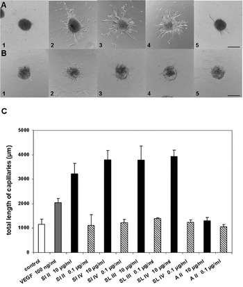
As a prelude to N-terminal tag sequence-characterization, as well as the standardized protocols of tryptic in-gel digestion, peptide mass fingerprinting, mass spectrometric fragmentation analysis and database searches (Galuska et al. Reference Galuska, Rollenhagen, Kaup, Eggers, Oltmann-Norden, Schiff, Hartmann, Weinhold, Hildebrandt, Geyer, Muhlenhoff and Geyer2010) etc., the isolation of the pro-angiogenic factor has proved particularly intractable. In this case, firstly, samples have been re-tested with a second in vitro angiogenesis system, the EC spheroid assay (Fig. 2; Korff and Augustin, Reference Korff and Augustin1998, Reference Korff and Augustin1999). Secondly, repetitive fractionation by multiple runs on C18 reverse-phase HPLC and screening with VSMCs demonstrated the loss of biological activity. Preliminary data with a re-pooled, fractionated aliquot have been interpreted as being a result of synergism (unpublished results), whereby biological activity consists of more than one component, for example, the male mating and dauer pheromones of C. elegans (Edison, Reference Edison2009). Thirdly, the future use of polymyxin B-bound beads to avoid any LPS contamination and the liability of upregulated VEGF expression (Marx et al. Reference Marx, Mulder, Van Den Bergh, Overbeeke, Haanen and Vermes1999).
Response of endothelial cells to SEA as verified by the spheroid assay. (A) Response of BRECs to high levels of SEA (10 μg/ml): 1, negative control; 2, positive control FGF2 at 5 ng/ml; 3, intestinal SEA; 4, hepatic SEA; 5, adult worm extract. Calibration bar=500 μm. (B) Response of HUVECs to high levels of SEA (10 μg/ml): 1, negative control; 2, positive control FGF2 at 5 ng/ml; 3, intestinal SEA; 4, hepatic SEA; 5, adult worm extract. Calibration bar=500 μm. (C) Analysis in vitro of capillary total-length formation by spheroid assay at high and low levels of SEA (10 and 0·1 μg/ml). A II, adult worm extract of batch II; BRECs, bovine retinal endothelial cells; FGF, fibroblast growth factor; HUVECs, human umbilical vein endothelial cells; SEA, soluble schistosome egg antigen; SI II/IV, intestinal SEA of batches II/IV; SL III/IV, hepatic SEA of batches III/IV; VEGF, vascular endothelial growth factor as positive control at 100 ng/ml.

CONCLUDING REMARKS
This ‘selectively’ comprehensive survey of parasitic helminth-associated neovascularization had the ‘hidden’ agenda of revealing ‘hot-spot species’ of relevant significance to this topic. From the literature, these would include E. multilocularis (Cestoda); C. elegans, B. malayi, W. bancrofti, A. caninum and T. spiralis (Nematoda); and S. mansoni and O. viverrini (Trematoda). Of particular importance, in this regard, are those helminths where the partial or complete genome is manifest for the screening of databanks in genomic and proteomic profiling: T. solium (Cestoda; Aguilar-Diaz et al. Reference Aguilar-Diaz, Bobes, Carrero, Camacho-Carranza, Cervantes, Cevallos, Davila, Rodriguez-Dorantes, Escobedo, Fernandez, Fragoso, Gaytan, Garciarubio, Gonzalez, Gonzalez, Jose, Jimenez, Laclette, Landa, Larralde, Morales-Montor, Morett, Ostoa-Saloma, Sciutto, Santamaria, Soberon, de la Torre, Valdes and Yanez2006); C. elegans and B. malayi (Nematoda; C. elegans, 1998; Ghedin et al. Reference Ghedin, Wang, Spiro, Caler, Zhao, Crabtree, Allen, Delcher, Guiliano, Miranda-Saavedra, Angiuoli, Creasy, Amedeo, Haas, El-Sayed, Wortman, Feldblyum, Tallon, Schatz, Shumway, Koo, Salzberg, Schobel, Pertea, Pop, White, Barton, Carlow, Crawford, Daub, Dimmic, Estes, Foster, Ganatra, Gregory, Johnson, Jin, Komuniecki, Korf, Kumar, Laney, Li, Li, Lindblom, Lustigman, Ma, Maina, Martin, McCarter, McReynolds, Mitreva, Nutman, Parkinson, Peregrin-Alvarez, Poole, Ren, Saunders, Sluder, Smith, Stanke, Unnasch, Ware, Wei, Weil, Williams, Zhang, Williams, Fraser-Liggett, Slatko, Blaxter and Scott2007); and S. mansoni and O. viverrini (Trematoda; Laha et al. Reference Laha, Pinlaor, Mulvenna, Sripa, Sripa, Smout, Gasser, Brindley and Loukas2007; Berriman et al. Reference Berriman, Haas, LoVerde, Wilson, Dillon, Cerqueira, Mashiyama, Al-Lazikani, Andrade, Ashton, Aslett, Bartholomeu, Blandin, Caffrey, Coghlan, Coulson, Day, Delcher, DeMarco, Djikeng, Eyre, Gamble, Ghedin, Gu, Hertz-Fowler, Hirai, Hirai, Houston, Ivens, Johnston, Lacerda, Macedo, McVeigh, Ning, Oliveira, Overington, Parkhill, Pertea, Pierce, Protasio, Quail, Rajandream, Rogers, Sajid, Salzberg, Stanke, Tivey, White, Williams, Wortman, Wu, Zamanian, Zerlotini, Fraser-Liggett, Barrell and El-Sayed2009; Young et al. Reference Young, Campbell, Hall, Jex, Cantacessi, Laha, Sohn, Sripa, Loukas, Brindley and Gasser2010). This information combined with the corresponding, experimental data can then be used to analyse the ‘burning question’, as to whether the parasitic vasculature is a consequence of host- and/or helminth-derived angiogenic growth factors. As archetypes are the C. elegans-encoded PDGF/VEGF-like factor, which is able to bind mammalian VEGF receptors and induce angiogenesis (Tarsitano et al. Reference Tarsitano, De Falco, Colonna, McGhee and Persico2006) and the multifunctional, potentially pro-angiogenic EmPG1 of E. multilocularis (Stadelmann et al. Reference Stadelmann, Spiliotis, Muller, Scholl, Muller, Gottstein and Hemphill2010). The extrapolation of such data carries the present provisos that, nematodes may simply possess the synthetic machinery for the potential synthesis of certain vascular growth factors i.e., C. elegans, and the EmPG1 factor of E. multilocularis has yet to be tested in vivo for angiogenic activity.
ACKNOWLEDGEMENTS
The authors wish to thank Dr Kai Maass for excellent, professional computer work.