Alkaline phosphatase (ALP) is a naturally occurring enzyme in milk. Because thermal resistance of the enzyme is greater than that of non-spore forming pathogenic microorganisms commonly found in milk, the detection of ALP is used as a legal test to determine whether milk has been adequately pasteurized or recontaminated with raw milk (Aschaffenburg & Mullen, Reference Aschaffenburg and Mullen1949; Murthy & Cox, Reference Murthy and Cox1988). However, the presence of ALP activity in properly pasteurized milk as a consequence of contamination with bacterial ALPs has been reported (Hammer & Olson, Reference Hammer and Olson1941). To assist in identifying this microbially produced heat-resistant phosphatase activity, Knight & Fryer (Reference Knight and Fryer1989) proposed to test again after laboratory re-pasteurization any sample failing the ALP activity test. However, Murthy & Kaylor (Reference Murthy and Kaylor1990) demonstrated that it was not possible to differentiate between bovine milk and microbial ALP by the proposed re-pasteurization test, because both heat-labile and heat-stable ALP could be produced by microorganisms such as Bacillus anthracis, B. cereus, B. megaterium (Dobozy & Hammer, Reference Dobozy and Hammer1969), Micrococcus sadonesis (Glew & Heath, Reference Glew and Heath1971), Saccharomyces cerevisiae (Gorman & Hu, Reference Gorman and Hu1969). Moreover, when testing ALP by phenol release, false positive results have been reported with pesticides, such as baygon (2-isopropoxy-phenyl-n-methyl carbamate) and sevin (1-naphtyl-n-methyl carbamate) (Kumar et al. Reference Kumar, Sud and Gupta1973), or antibiotics such as penicillin and oxytetracyclin (Manolkidis & Alichanidis, Reference Manolkidis and Alichanidis1971). Conversely, false negative results can be due to the presence of pesticides such as phosphamidon and 0,0-dimethyl-2,2-dichlorovinyl phosphate (Kumar et al. Reference Kumar, Sud and Gupta1973), or antibiotics such as streptomycin, erythromycin and neomycin (Manolkidis & Alichanidis, Reference Manolkidis and Alichanidis1971).
To determine milk ALP without interference from microbial ALP, pesticides or antibiotics, an immunological approach based on polyclonal antibodies (PAbs) produced against bovine milk ALP has been proposed by Vega-Warner et al. (Reference Vega-Warner, Wang, Smith and Ustunol1999). The antibodies have been used to develop a competitive indirect ELISA specific for the bovine milk ALP (Vega-Warner et al. Reference Vega-Warner, Gandhi, Smith and Ustunol2000). However, the PAbs cross-reacted with heat-denatured ALP and the ELISA was thus not specific enough to be used to verify adequate heat treatment of milk. Chen et al. (Reference Chen, Tai, Shen, Tu, Wu and Chang2006) used the same approach with PAbs developed in hens. The antibodies cross-reacted with Escherichia coli ALP and their reactivity against bovine ALP in raw milk was not established.
The aim of the present study was to produce a monoclonal antibody highly specific for the bovine milk ALP and then to optimize a simple capture immunoassay that only detects bovine milk ALP and does not react with bacterial or fungal ALPs.
Materials and Methods
Materials and samples
ALP (type XXI, EC 3.1.3.1) from bovine skim milk (raw, P5395), from bovine intestinal mucosa (P3681, P4502, P6774) and from Esch. coli (P4377), Freund's complete and incomplete adjuvants and bovine serum albumin (BSA) were obtained from Sigma-Aldrich Chimie (St-Quentin-Fallavier, France). ALP from calf intestinal mucosa (79390) was purchased from Fluka (Sigma-Aldrich). Clarifying Reagent (R) and iso-butylalcohol (2-butanol) were from VWR-Prolabo (France). Ready to use p-nitrophenyl phosphate (p-NPP) substrate were purchased from Sigma (N-7653), BioFx and Uptima (Interchim, Montluçon, France). A sample of highly purified bovine milk ALP was kindly provided by the STLO unit (INRA, Rennes, France).
Fresh bulk raw milk was obtained from the INRA Research Centre dairy herd.
Monoclonal antibodies (MAbs)
Female BALB/c mice (8–10 weeks old) were immunized with 50 μg commercial bovine milk ALP in a volume of 100 μl of a 1:1 mixture of Freund's complete adjuvant and 0·01 m-phosphate buffered saline pH 7·2 (PBS), equally injected in each hind footpad (d 0) (Rachetti et al. Reference Rachetti, Fossati, Comitti, Putignano and Galante1987). A secondary immunization was administered on d 13 with the same amount of antigen in Freund's incomplete adjuvant. On d 16, popliteal lymph nodes were removed. A cell suspension was prepared by teasing with forceps, the cells were forced twice through a 70 μm mesh nylon sheet with a sterile plunger and washed three times with Dulbecco's Modified Eagle's Medium (Gibco). Cells were mixed with exponentially growing murine myelome cells X63-Ag.8-6.5.3 in a 1:5 (v/v) ratio. The mixture was centrifuged at 300 g for 5 min, and the medium was removed. Fusion was then conducted as previously described (Levieux & Venien, Reference Levieux and Venien1994). After 12 to 14 d, the supernatants of the hybridoma population were screened for specific antibody production by indirect capture ELISA. Positive hybrids were cloned by limiting dilution and then grafted into mice for ascites production.
MAbs were purified from ascitic fluids by anion-exchange chromatography on Q-Sepharose Fast-Flow (Amersham) using a linear gradient of NaCl (0–0·5 m) in 0·02 m-Tris buffer pH 8·2.
Butanol extraction of milk samples
Three ml butanol were added to 3 ml milk in glass tubes. The tubes were capped, vigorously inverted several times, vortexed for 15 s and centrifuged at 2500 g for 30 min. The lower aqueous phase was recovered and stored at 4°C until analysis within the day or stored at −20±5°C.
Indirect capture ELISA
The wells of flat-bottomed polystyrene microtitre plates (Maxisorp; Nunc, Kamstrup, Denmark) were coated with 100 μl affinity purified goat anti-mouse IgG antibodies at a concentration of 10 μg/ml in PBS for 60 min at room temperature. Four washes with PBS containing 0·1% Tween-20 (PBS-Tween) were followed by addition of the hybridoma supernatant (100 μl/well) diluted 1:3 in PBS-Tween. After 60 min incubation at room temperature, unbound antibodies were removed by washing as described above. Bovine whey (100 μl/well) diluted 1:5 in PBS-tween was then added as a source of ALP and incubated 60 min at room temperature. Then the plates were washed and ready to use p-NPP substrate (100 μl/well) was added and incubated in the dark for 30–60 min at 37±1°C. The absorbance was recorded at 405 nm using an IEMS microplate reader (Labsystem, Helsinky, Finland).
ALP immunoassay
Flat-bottomed polystyrene microtitre plates were coated by incubating 100 μl/well of chromatographically purified MAbs at a concentration of 10 μg/ml in PBS at room temperature for 60 min. After four washes with PBS-Tween, samples (100 μl/well) of butanol treated milks adequately diluted in PBS Tween containing 5 g BSA/l (PBS-Tw-BSA) were added and incubated at room temperature for 60 min. Then the plates were washed 4 times and the procedure for incubating p-NPP substrate and recording the absorbance was carried out as described for the indirect assay. Standard curve was mostly raw bulk milk diluted in heated milk (80±2°C, 5 min) or in PBS-Tw-BSA. ALP activity can alternatively be expressed in μmol of p-nitrophenol liberated per ml milk and per min by using a standard curve of p-nitrophenol. Results were expressed as mean of duplicate analysis.
Results and Discussion
Producing specific antibodies against bovine milk ALP is challenging because the enzyme purification is difficult. The crude bovine milk ALP available from Sigma as a starting material contained denatured ALP and other milk proteins. This could explain the 2·7% cross-reactivity of the PAbs obtained by Vega-Warner et al. (Reference Vega-Warner, Gandhi, Smith and Ustunol2000) with heat-denatured ALP. As a direct consequence of this cross-reactivity, the competitive indirect ELISA developed by these authors could not be used to verify adequate pasteurization of milk and milk products. Similar difficulties in obtaining specific antibodies were encountered by Chen et al. (Reference Chen, Tai, Shen, Tu, Wu and Chang2006) since their PAbs strongly cross-reacted against Esch. coli. Thus we turned to the MAb technology which allows selection of single antibody-secreting clones. The specificity of the clones was confirmed by the screening method since only clones able to capture the bovine milk ALP were evidenced by the ALP enzyme activity and thus selected. Moreover, in contrast with PAbs, the selected MAbs can be produced indefinitely and therefore used in commercial kits.
From the mice immunized with crude bovine milk ALP we obtained 11 hybrids positive for ALP activity in the indirect capture immunoassay. Titration curves of the culture supernatants allowed the selection, for cloning and further characterization, of 3 hybrids secreting MAbs with high binding activity. Mouse ascites were produced for the 3 hybrids and the MAbs were purified from the ascitic fluids by anion-exchange chromatography.
The purified MAbs were coated in microtitre plates to specifically capture the native ALP from bovine milk. After washing to eliminate uncaptured proteins, the ALP activity was revealed by adding the specific enzyme substrate. In contrast to the competitive ELISAs proposed by Vega-Warner et al. (Reference Vega-Warner, Gandhi, Smith and Ustunol2000) and Chen et al. (Reference Chen, Tai, Shen, Tu, Wu and Chang2006), this capture immunoassay does not require microtitre plates coated with bovine milk ALP which is no longer commercially available and hard to purify.
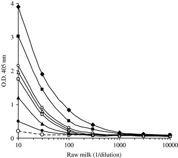
Each step of the capture immunoassay needed to be optimized for successful results. Optimal concentration of the coated MAbs (10 μg/ml) was defined by checkerboard titration of crude bovine milk ALP dilutions. However, when applied to raw milk sample the sensitivity of the immunoassay was very low, suggesting a non-specific inhibition of the antigen-antibody interaction. As ALP purification has been frequently performed using butanol extraction of dairy fluids or tissues, we tried to extract ALP from raw milk with different concentrations of butanol. Results showed that ALP activity in the immunoassay increases with the butanol concentration (Fig. 1). Optimal results were obtained with 50% butanol (final concentration) since at higher concentration the volume of the recovered aqueous phase decreases by partial dissolution in the butanol phase. No effect of temperature (4°C versus 20°C) or vortexing 15 to 60 s was observed.
Effect of milk ALP extraction with butanol or detergents on the immunoassay reactivity. Serial dilutions of raw milk in boiled milk were vortexed with different concentrations (v/v) of butanol or detergents before incubation for 60 min in MAb GD5 coated microplates. After washing, p-NPP substrate was incubated for 30 min and the O.D. was read at 405 nm. Dotted line: untreated milk. Closed symbols: butanol 10% (circles), 30% (triangles), 50% (squares) 75% (diamonds). Open symbols: Tween 20 5% (circles), deoxycholate 20 mm (triangles), Triton X100 5% (squares). All concentrations are given as a final concentration.

Detergents such as tween 20 (50 g/l, final concentration), triton X100 50 g/l, final concentration) and deoxycholate (20 mm, final concentration) were also tested as an alternative to butanol. An increase of the ALP reactivity in the immunoassay was obtained with the three detergents, while they remained less effective than 50% butanol (Fig. 1).
The necessity for butanol extraction of ALP from raw milk in order to be detected by antibodies has not been reported by Vega-Warner et al. (Reference Vega-Warner, Gandhi, Smith and Ustunol2000) or Chen et al. (Reference Chen, Tai, Shen, Tu, Wu and Chang2006) since they have not applied their competitive indirect ELISA to dilutions of raw milk in heated milk. The butanol effect can be explained by the fact that ALP activity is associated with the bovine milk fat globule membrane (MFGM) which is a derivative of the mammary gland plasma membrane (Morton, Reference Morton1953). The MFGMs are formed when secretory cell plasma membrane adhering to milk fat droplets are fragmented during extrusion of milk fat (Snow, Reference Snow1994). ALP being anchored on the mammalian cell surface by a phosphatidylinositol residue, the antibody interaction is probably inhibited by steric hindrance. Extraction with mild detergents such as tween, triton or deoxycholate could be used instead of butanol if the maximum sensitivity of ALP detection is not required.
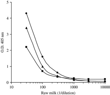
Three ready to use p-NPP substrates (Sigma, BioFx and KPL Uptima) were considered and the best signal to noise ratio was consistently obtained with the Sigma reagent (Fig. 2). An increase of sensitivity ×10 or 100 could be obtained using fluorometric substrates such as 4-methylumbelliferyl phosphate.
Comparison of three ready to use p-NPP substrates for the detection of milk ALP by capture-ELISA. Serial dilutions of raw milk in boiled milk were incubated for 1 h at room temperature in MAb GD5 coated microplates. After washing, Sigma (circles), Uptima (triangles) and BioFx (squares) p-NPP substrates were incubated 40 min at 37°C and the O.D. was read at 405 nm.

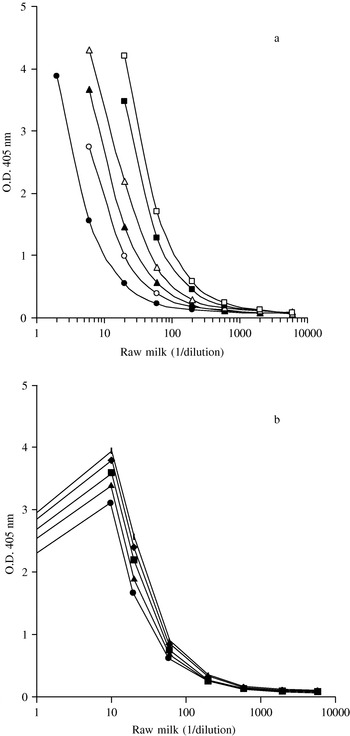
Sensitivity of milk ALP immunodetection increased with incubation time and agitation (Fig. 3a). Increasing the incubation time was mostly effective for the substrate step while agitation essentially increased the PAL capture by the MAbs (not shown). Incubation temperature also had pronounced effect on the immunodetection sensitivity, particularly for the substrate incubation step (Fig. 3b) and thus must be controlled.
Effect of incubation time, agitation and temperature on the sensitivity of milk ALP detection by the immunoassay. (a) Serial dilutions of raw milk in boiled milk were incubated in MAb GD5 coated microplates for 15 (circles), 30 (triangles) or 60 (squares) min at room temperature, with (open symbols) or without (closed symbols) agitation with Thermomixer Eppendorf. After washing, p-NPP substrate was added and incubated in the same conditions. O.D. was read at 405 nm. (b) Serial dilutions of raw milk in boiled milk were incubated in MAb GD5 coated microplates for 60 min at room temperature. After washing, p-NPP substrate was added and incubated at 25°C (circles), 30°C (triangles), 37°C (squares), 40°C (diamonds) or 45°C (bars) for 30 min. O.D. was read at 405 nm.

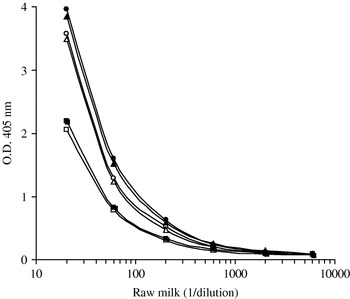
Effect of the diluant used for the serial dilutions of raw milk on the sensitivity of milk ALP detection by immunoassay using three different MAbs. Serial dilutions of raw milk in boiled milk (closed symbols) or BSA 0·5% (open symbols) were incubated in MAb GD5 (circles), MAb AD8 (triangles) or MAb EE7 (squares) coated microplates at room temperature for 60 min. After washing, p-NPP substrate was added and incubated at 37°C for 60 min. O.D. was read at 405 nm.

In order to simplify the test, BSA was tested at a 5 g/l concentration in PBS Tween as an alternative to the boiled milk used for the serial dilutions of raw milk. No significant difference in immunoreactivity was observed between the two diluents for MAb EE7 while a small decrease in optical density was observed for MAbs GD5 and AD8 (Fig. 4).
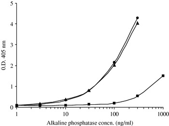
Reactivity of MAbs GD5, AD8 and EE7 against purified bovine milk ALP. Serial dilutions of purified ALP in boiled milk were incubated in MAb GD5 (circles), MAb AD8 (triangles) or MAb EE7 (squares) coated microplates at room temperature for 60 min. After washing, p-NPP substrate was added and incubated at 37°C for 60 min. O.D. was read at 405 nm.

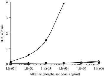
To control the specificity of the MAbs-based immunoassay, its reactivity was tested against highly purified bovine milk ALP. The purified ALP was detected at a concentration as low as 5 ng/ml with MAbs GD5 or AD8 (Fig. 5). Such a sensitivity can only be obtained with high affinity antibodies in the conditions of the test. Moreover, the immunoassay was not reactive with the commercially available bovine intestinal and microbial (Esch. coli) ALPs (Fig. 6). The cross-reactivity with non-milk ALPs was less than 0·001% since the optical density obtained for the raw milk ALP at 10 ng/ml was higher than that obtained for the non-milk ALP at 1 mg/ml (O.D. values: 0·174 and 0·150 respectively).
Specificity of the immunoassay against ALPs from bovine milk, bovine intestine or Esch. coli. Serial dilutions of commercially available ALPs in boiled milk were incubated in MAb GD5 coated microplates at room temperature for one hour. After washing, the p-NPP substrate was incubated 30 min at 37°C and the O.D. was read at 405 nm. Closed circles: raw bovine milk ALP (Sigma P5395). Open symbols: ALPs from bovine intestinal mucosa (Sigma P6774) (circles), calf intestinal mucosa (Fluka 79390) (triangles) and Esch. coli (Sigma P4377) (squares).

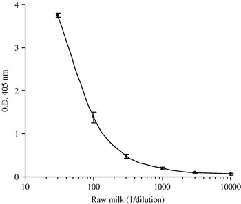
Typical immunoassay curve obtained for quantification of bovine milk ALP. Serial dilutions of raw milk in boiled milk were incubated in MAb GD5 coated microplates at room temperature for 60 min. After washing, p-NPP substrate was added and incubated at 37°C for 60 min. O.D. was read at 405 nm. Results are mean±sd of quadruplicate analyses.

Finally, the repeatability of the immunoassay was found to be 3·5 and 7·5% at 3 and 1% raw milk in boiled milk respectively, and the detection limit (mean±3 SD) was found at 0·02% raw milk in boiled milk for 60 min substrate incubation (Fig. 7).
In conclusion, a simple capture immunoassay for the specific detection of bovine milk ALP has been developed using monoclonal antibodies. It allows the detection of 0·02% raw milk in boiled milk, which is adequate in practice (Black et al. Reference Black, Kuzyk and Duggan1992) and does not react with ALPs of intestinal or bacterial origins. The usefulness of the capture immunoassay for kinetic studies of ALP heat denaturation and for determining mild time/temperature treatment of milk is presented in Levieux et al. (Reference Levieux, Geneix and Levieux2007). Moreover, work in progress indicates that the capture immunoassay can be successfully applied to solid dairy products such as cheese obtained from raw, thermized or pasteurized milk.
The authors wish to thank J Leonil, J Fauquant and MA Pilard (INRA, STLO, Rennes, France) for providing purified bovine milk ALP, and ARILAIT (France) for helpful discussions and constant interest during completion of this work. This project was partly funded by the French Ministry for Research (AQS n° 2002 T08).