


Introduction
First described in 1977, pneumorrhachis (PR) remains a rarely encountered radiological finding characterized by the presence of air within the spinal canal. Reference Oertel, Korinth, Reinges, Krings, Terbeck and Gilsbach1 PR can be classified by anatomical location as either extradural (epidural) or intradural (subdural or subarachnoid) and by etiology into traumatic, spontaneous or iatrogenic categories. Reference Goh and Yeo2 Extradural PR, often detected incidentally, is generally considered benign. Reference Kazufumi, Masahiro, Yasushi, Nobuhisa, Yoshihiro, Shogo, Kazuhiro, Kazuhide, Masatoki, Hideki, Koichi, Atsushi and Mitsuaki3 In contrast, intradural PR is frequently associated with severe trauma, dural disruption and an increased risk of neurological complications. Reference Place and Pezzuti4 Although often discovered on imaging performed for other reasons, the presence of PR can serve as an important indicator of severe underlying pathology, particularly in trauma cases involving high-energy mechanisms or penetrating injuries. Reference Kazuki and Ken5 Despite growing recognition, the literature on PR remains fragmented, largely comprising isolated case reports or small case series. Reference Tannouri, Noori, Alabboudi, Abdulhadi and Alobeid6 The purpose of this review is to synthesize current evidence regarding PR’s classification, pathophysiology, clinical presentation, imaging findings and management approaches. In addition, we present a representative case of traumatic PR to contextualize the literature and highlight its clinical and radiological significance.
Methods
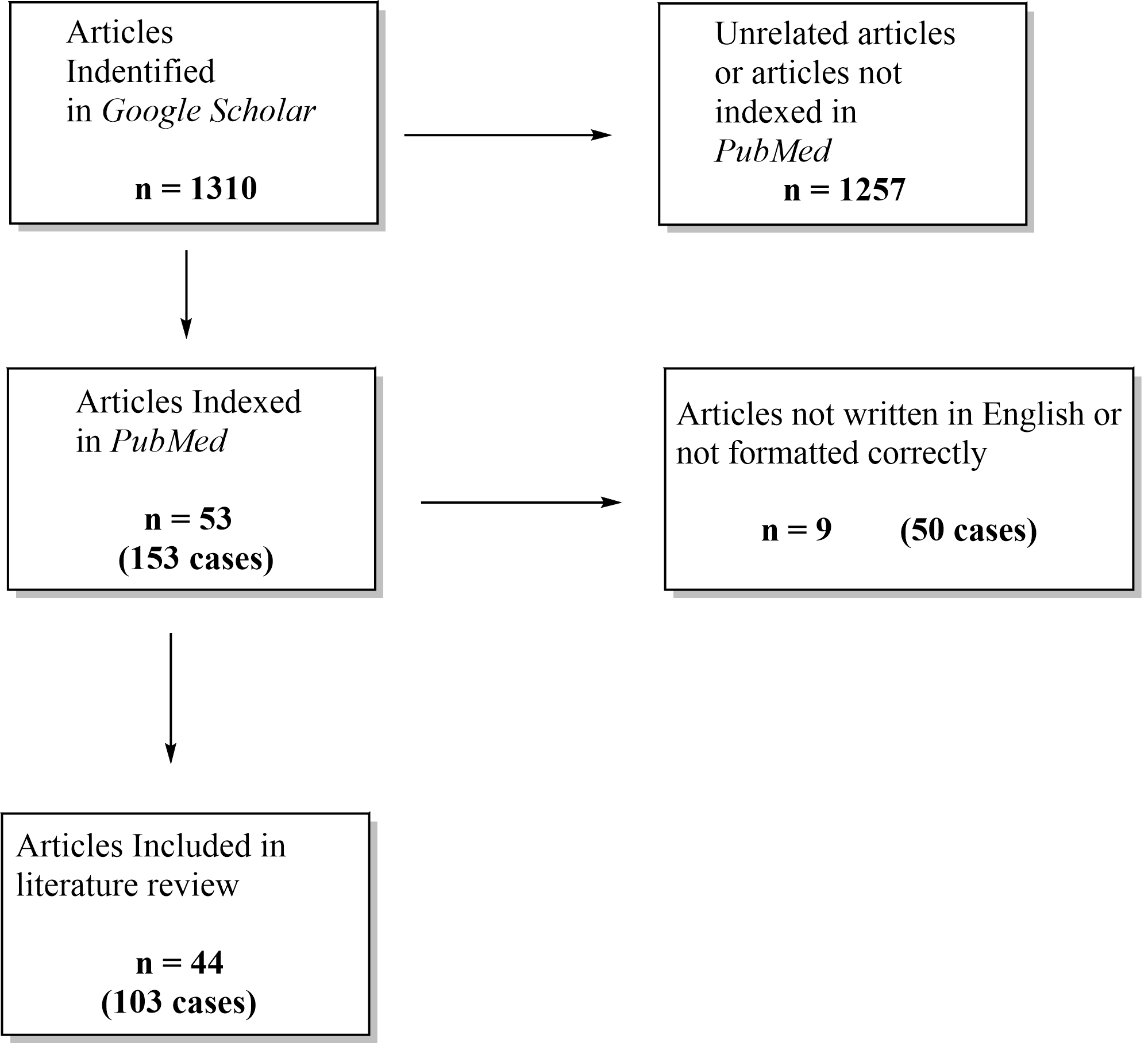
We performed a comprehensive review of the literature using Google Scholar with filters applied to identify only PubMed-indexed, English-language articles. The initial search yielded 1310 articles. After screening for relevance and adherence to our inclusion criteria – namely, English-language, PubMed-indexed articles reporting clinical cases of PR – 53 articles were selected. Subsequently, nine articles were excluded due to non-conforming formats or incomplete clinical data, resulting in a final pool of 44 articles for detailed review, collectively encompassing 103 individual cases. Data extracted from these reports included etiology, anatomical location, compartmental classification, associated injuries and management strategies, alongside reported clinical outcomes (Figure 1).
PRISMA-style flow diagram illustrating the selection process for studies included in the literature review. A total of 1310 articles were initially identified through Google Scholar. After removing 1257 unrelated or non-PubMed-indexed articles, 53 PubMed-indexed articles (153 cases) remained. Of these, 9 articles (50 cases) were excluded for being non-English or improperly formatted, leaving 44 articles (103 cases) for final inclusion.

Figure 1. Long description
A three-level flowchart depicting the article selection process. At the top left, a box reads ‘Articles identified in Google Scholar, n=1310.’ A rightward arrow leads to a box reading ‘Unrelated articles or articles not indexed in PubMed, n=1257.’ A downward arrow leads to a second-row box reading ‘Articles indexed in PubMed, n=53 (153 cases),’ which connects rightward to ‘Articles not written in English or not formatted correctly, n=9 (50 cases).’ A final downward arrow leads to the bottom box reading ‘Articles included in literature review, n=44 (103 cases).’
Illustrative Case Summary
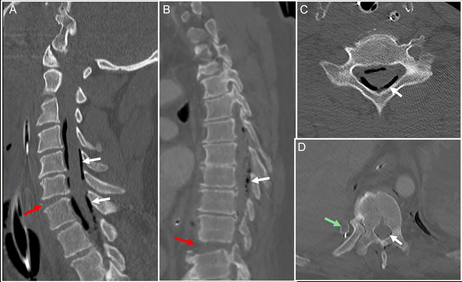
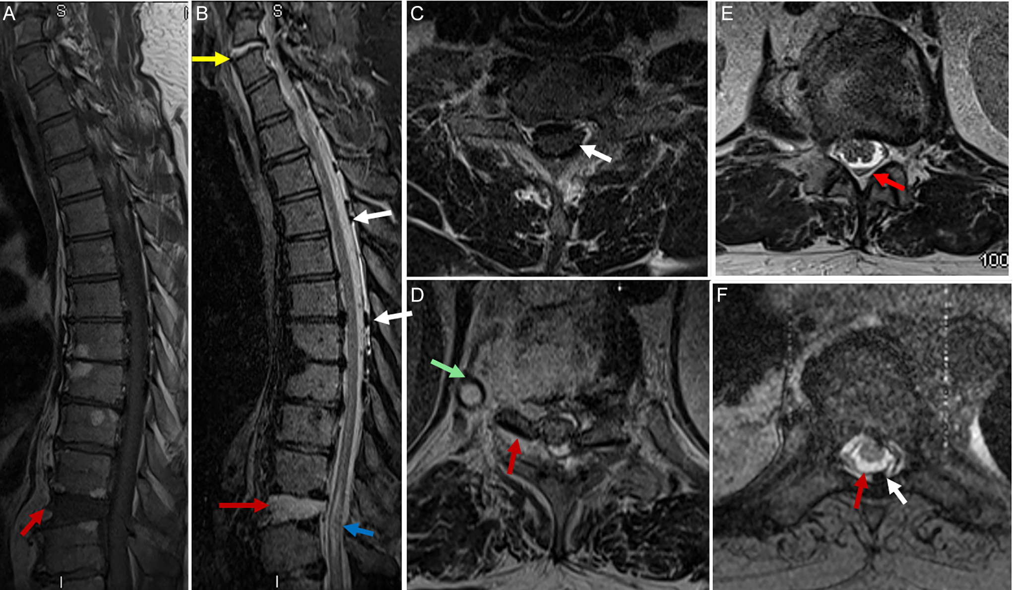
A 56-year-old man was brought to the emergency department as a CODE 1 trauma after a high-speed motorcycle crash, where he was ejected approximately 25 feet at an estimated speed of 80 km/h. The patient was found to be minimally responsive by paramedics with a Glasgow Coma Scale of 14. On arrival, he was hemodynamically stable but complained of severe thoracic back pain and mild compromise of respiration. Initial trauma imaging revealed complex spinal trauma. Cervical spine CT and MRI (Figures 2 and 3) revealed a three-column fracture at C6–C7 with mild retrolisthesis, widening of intervertebral disc space, right C6–C7 facet subluxation and anterior and posterior longitudinal ligament injuries. Prevertebral soft tissue edema and epidural air were evident, along with a small anterior epidural hematoma extending to T1–T2 (Figures 2A,C and 3B,C). Thoracic imaging was consistent with a Chance-type fracture-dislocation at T10–T11 with 8 mm retrolisthesis, retropulsion of bone into the canal, bilateral facet subluxation with intra-articular fluid and ligament disruption (Figures 2B,D and 3A,D–F). MRI showed a T2 hyperintense cord signal as a sign of contusion, focus of blooming artifact as a sign of intramedullary hemorrhage and a focal anterior epidural hematoma at T11–T12. Also, fluid-fluid levels within the thecal sac at T12–L1 consistent with subarachnoid hemorrhage (Figure 3B–F).
CT cervical spine sagittal (A) shows widening of the intervertebral disc space at C6–C7 (red arrow). Multiple foci of pneumorrhachis (white arrows). CT thoracic spine sagittal (B) shows translation injury at T10 and T11 (red arrow) with bilateral facet joint subluxation and foci of pneumorrhachis along the spinal canal (white arrow). CT cervical spine axial (C) shows foci of pneumorrhachis extending into the intradural space (white arrow). CT axial thoracic spine (D) shows tip of the chest tube adjacent to the area of fracture (green arrow), with foci of pneumorrhachis (white arrow) likely the site of pleurospinal fistula.

Figure 2. Long description
A composite of four CT images labeled A through D. Panel A (sagittal cervical spine): shows widening of the C6–C7 intervertebral disc space marked by a red arrow, with multiple hypodense foci of air within the spinal canal indicated by white arrows, consistent with pneumorrhachis. Panel B (sagittal thoracic spine): demonstrates translation injury at T10–T11 indicated by a red arrow, with foci of intraspinal air visible along the canal marked by a white arrow. Panel C (axial cervical spine): cross-sectional view demonstrating a hypodense air focus extending into the intradural space, marked by a white arrow. Panel D (axial thoracic spine): shows the tip of a chest tube adjacent to a vertebral fracture site, indicated by a green arrow, with an adjacent hypodense air focus (white arrow) consistent with the suspected site of the pleurospinal fistula.
Sagittal T1-weighted MRI of the thoracic spine (A) shows translation injury at T10–T11 (red arrow). Sagittal T2-weighted MRI of the thoracic spine including cervicothoracic junction (B) shows dislocation at C6–C7 (yellow arrow) and translation at T10–T11 (red arrow) with cord signal abnormality at this level (blue arrow). Axial T2-weighted MRI of the cervical spine (C) shows air within the thecal sac (white arrow). Axial T2-weighted MRI of the thoracic spine (D) shows a chest tube (green) and bone fragments at the level of fracture (red arrow), likely the site of the pleurospinal fistula and causing spinal cord compression. Axial T2-weighted MRI of the thoracic spine (E) shows a fluid-fluid level consistent with subarachnoid hemorrhage (red arrow) and (F) disruption of the dura (red arrow) and foci of pneumorrhachis (white arrow).

Figure 3. Long description
A composite of six MRI images labeled A through F. Panel A (sagittal T1 thoracic): demonstrates translation injury at the thoracic spine level, indicated by a red arrow, with visible vertebral misalignment. Panel B (sagittal T2 thoracic including cervicothoracic junction): shows dislocation at the C6–C7 level marked by a yellow arrow, with T2 signal changes and translation at T10–T11 (red arrow) with cord signal abnormality at this level (blue arrow). Panel C (axial T2 cervical): cross-sectional image showing a hypointense focus of air within the spinal canal, indicated by a white arrow. Panel D (axial T2 thoracic): depicts the chest tube as a signal void (green marker) with adjacent bone fragments at the fracture level, consistent with the pleurospinal fistula site and causing spinal cord compression; a red arrow marks associated injury. Panel E (axial T2 thoracic): demonstrates a fluid-fluid level within the thecal sac, indicated by a red arrow, consistent with subarachnoid hemorrhage. Panel F (axial T2 thoracic): shows disruption of the dura indicated by a red arrow, with adjacent hypointense foci of pneumorrhachis marked by a white arrow.
CT scans demonstrated PR with epidural and suspected intradural air from C2 to T12 (Figure 2A–D), with subcutaneous emphysema at T11–T12 and right fracture of the T11 rib. A small right pneumothorax and right hemothorax were managed by chest tube insertion adjacent to the fractures (Figure 2D). These were consistent with a combination of traumatic tears in the dura and retrograde air migration from the lungs in keeping with pleurospinal fistula. CT head scan was negative for intracranial trauma or pneumocephalus, with facial bone CT showing minimally displaced nasal bone fractures and nasal septal fracture, both of which were treated conservatively.
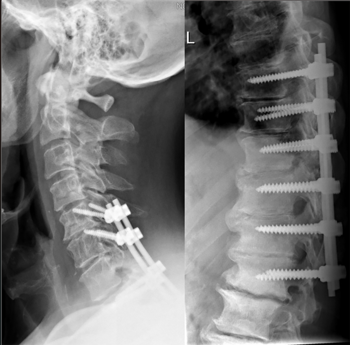
The patient underwent C5–T2 spinal fusion with decompression, C5–C6 decompression and T8–L1 decompression with fusion to stabilize cervical and thoracic injuries (Figure 4). Due to intradural air and risk of infection, the patient received empiric meningitic antibiotics. Despite aggressive surgical and medical management, the patient developed complete paraplegia. After stabilization of his spinal injuries and resolution of his thoracic pathology, he was discharged for outpatient rehabilitation.
X-ray cervical spine (A) and lateral view thoracic spine (B) show C5–T2 spinal fusion with decompression, C5–C6 decompression and T8–L1 decompression with fusion to stabilize cervical and thoracic injuries.

Figure 4. Long description
Two plain radiographs shown side by side. Panel A (lateral cervical spine): demonstrates posterior cervical instrumentation with pedicle screws and rods spanning C5 to T2, with hardware visible in lateral projection against the cervical vertebral bodies. Panel B (lateral thoracic spine): shows long-segment posterior spinal instrumentation extending from T8 to L1, with multiple pedicle screws and connecting rods visible in lateral view, consistent with thoracolumbar decompression and fusion.
Literature Review
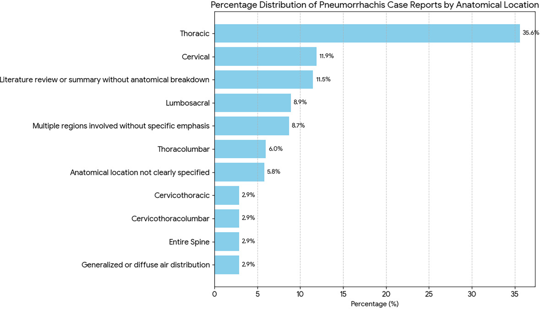
Literature review analysis revealed that traumatic PR was responsible for approximately 73.9% of the total cases. These cases were primarily due to high-energy blunt force trauma such as motor vehicle crashes, falls or penetrating trauma. Spontaneous cases represented about 20.3% and involved transient intrathoracic pressure elevation from conditions such as violent coughing, vomiting, Valsalva maneuvers or drug abuse. Iatrogenic cases accounted for approximately 5.8% and typically resulted from spinal surgeries such as epidural anesthesia, spinal surgeries or other invasive procedures. Anatomical distribution of PR showed that thoracic spine was most involved and appeared in approximately 35.6% of the cases. This can be explained by the anatomical proximity of the thoracic spine to the mediastinum and pleural spaces, which would provide a pathway for air migration to the spinal canal. The cervical region was indicated in 11.9% of the cases and then 8.8% in the lumbosacral region (Figure 5). The remaining cases were either found in more than one spinal region or were not specified as to location.
Percentage distribution of pneumorrhachis case reports categorized by anatomical location. Thoracic involvement accounted for the largest proportion (35.6%), followed by cervical (11.9%), literature reviews or summaries without anatomical breakdown (11.5%), lumbosacral (8.9%), multiple regions without specific emphasis (8.7%) and other less frequent distributions including thoracolumbar (6.0%), anatomical location not clearly specified (5.8%) and generalized or diffuse air distribution (2.9%). Rare combinations such as cervicothoracic, cervicothoracolumbar and entire spine involvement each represented 2.9% of reported cases.

Figure 5. Long description
A horizontal bar chart titled ‘Percentage Distribution of Pneumorrhachis Case Reports by Anatomical Location.’ The x-axis shows percentage (0–35%) and the y-axis lists twelve anatomical categories. From highest to lowest: Thoracic (35.3%), Cervical (11.8%), Literature review or summary without anatomical breakdown (11.4%), Lumbosacral (8.8%), Multiple regions without specific emphasis (8.6%), Thoracolumbar (5.9%), Anatomical location not clearly specified (5.7%), and Cervicothoracic, Cervicothoracolumbar, Entire Spine, and Generalized or diffuse air distribution each at 2.9%. All bars are rendered in light blue.
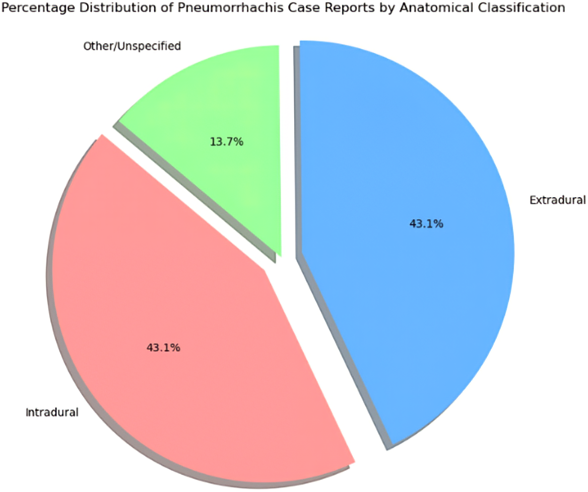
Regarding compartmental classification, intradural and extradural PR occurred with nearly equal frequency, each being about 43.1% of the cases. Extradural PR is typically incidental, spontaneous in course and not requiring treatment. Intradural PR carried a higher risk of complications, particularly when it was in association with CSF leaks, pneumocephalus or injury to the spinal cord. There existed a subset of cases (13.7%) involving mixed or unspecified compartments (Figure 6).
Compartmental distribution of pneumorrhachis cases by anatomical classification. Intradural and extradural pneumorrhachis occurred with equal frequency (43.1% each), while 13.7% of cases involved mixed or unspecified compartments.

Figure 6. Long description
A three-segment pie chart titled ‘Percentage Distribution of Pneumorrhachis Case Reports by Anatomical Classification.’ The blue segment represents extradural pneumorrhachis at 43.1%. The pink or salmon segment represents intradural pneumorrhachis at 43.1%. The green segment represents other or unspecified compartments at 13.7%. Each segment is labeled with its percentage and a legend identifies the three categories.
Discussion
The pathogenesis of PR varies depending on its underlying cause. Traumatic PR most commonly results from direct disruption of the dura mater, spinal fractures that breach the spinal canal or the retrograde migration of air from an associated pneumothorax or subcutaneous emphysema. Reference Hadjigeorgiou, Singh, Stefanopoulos, Petsanas, Hadjigeorgiou and Fountas7 Spontaneous PR typically occurs when air dissects along fascial planes during episodes of increased intrathoracic pressure, potentially entering the epidural space via neural foramina. Reference Charpentier and Cardos8 Iatrogenic PR generally arises from the introduction of air during spinal interventions, such as epidural injections or surgical procedures. Reference El-Halabi, Alkandari and Yaktien9 The anatomical distribution of PR reflects both the mechanism of injury and the available anatomical pathways for air migration. The thoracic spine appears susceptible due to its proximity to air-containing structures such as the mediastinum and pleura. Reference Haldane10
Although often asymptomatic, the presence of PR – particularly when intradural – can indicate significant underlying pathology. Reference Place and Pezzuti4 Complications associated with PR include spinal cord compression, meningitis or central nervous system infection in cases involving dural breach, neurological deficits including paralysis, CSF leaks and pneumocephalus. Reference Rathore, Gill and Yasin11,Reference Houston, Fiani, Musch and Tayag12 In trauma settings, the detection of PR should prompt a comprehensive assessment for spinal instability, dural tears and associated thoracic injuries. While extradural PR without neurological compromise may be managed conservatively with observation and supportive care, intradural PR associated with spinal cord injury or CSF leak often necessitates surgical repair, stabilization and sometimes prophylactic antibiotic therapy. Spontaneous or iatrogenic PR that presents without complications generally resolves with conservative management, though patients require close follow-up to monitor for delayed sequelae. Reference Charpentier and Cardos8
The case presented in this review reflects trends observed in the literature, including the high prevalence of PR in the setting of traumatic, high-energy spinal injuries, Reference Kazuki and Ken5 the frequent coexistence of thoracic injuries such as pneumothorax or rib fractures Reference Hadjigeorgiou, Singh, Stefanopoulos, Petsanas, Hadjigeorgiou and Fountas7 and the association of intradural PR with poorer neurological outcomes. Reference Place and Pezzuti4 Surgical stabilization remains the cornerstone of treatment in cases of spinal instability. Reference Sethi and Cooper13 Nevertheless, even with optimal management, persistent neurological deficits are not uncommon, particularly in cases complicated by severe spinal cord injury. Reference Rathore, Gill and Yasin11
Conclusion
PR, though rare, represents a clinically significant radiological finding that can signal severe underlying spinal or thoracic injury. Reference Oertel, Korinth, Reinges, Krings, Terbeck and Gilsbach1,Reference Goh and Yeo2 Recognizing its potential implications, anatomical distribution and associated risk factors is essential for clinicians involved in trauma care and spinal pathology. Reference Kazuki and Ken5 While extradural PR is frequently benign and self-resolving, Reference Kazufumi, Masahiro, Yasushi, Nobuhisa, Yoshihiro, Shogo, Kazuhiro, Kazuhide, Masatoki, Hideki, Koichi, Atsushi and Mitsuaki3 intradural PR demands a thorough diagnostic workup and may require surgical management to prevent or mitigate neurological complications. Reference Place and Pezzuti4
Acknowledgments
Not applicable.
Author contributions
E.P.O. and S.C. contributed to the study conception and design. Material preparation was performed by E.P.O., while data collection and analysis were performed by S.C. The first draft of the manuscript was also written by S.C., and both authors commented on previous versions of the manuscript. All authors read and approved of the final manuscript.
Funding statement
No funds, grants or other support was received for the preparation of this manuscript.
Competing interests
The authors declare that they have no competing interests.
Ethical standards
Not applicable for this literature review. The illustrative case report was reviewed in accordance with institutional policies for publication of de-identified patient data.
Consent to participate
Not applicable.
Consent for publication
Written informed consent for publication of the case details and associated images was obtained from the patient’s legally authorized representative.

 Open access

