Introduction
The role of bone conduction devices in treating hearing loss has been well established since the late twentieth century. The development of bone-anchored hearing aid (BAHA) systems, which incorporated the concept of osseointegration discovered by Brånemark, enabled improved comfort and fine sound quality, and represented a major milestone in the evolution of bone conduction hearing assistance.Reference Tjellström, Håkansson and Granström1 Rapid refinements of the BAHA system have continued to bring patients significant improvements. However, despite years of technological advancement the problem of post-operative peri-abutment wound infection has persisted.
Although the prevalence of post-operative peri-abutment wound infection has remained at approximately 5 per cent, this complication has a significant negative impact on the clinical application and popularity of the BAHA system.Reference Reyes, Tjellström and Granström2 It may be the case that, faced with the proposition of a lifelong commitment to post-operative wound care, many otherwise suitable patients will opt instead for an air conduction hearing aid, despite its functional inferiority to a BAHA.
In response to the problem of peri-abutment wound infection, a semi-implantable bone conduction device, the Bonebridge system, has been developed (Med-El, Innsbruck, Austria).
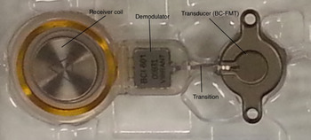
The Bonebridge system has two components: an external audio processor (Figure 1) and a bone conduction implant (Figure 2). The latter element consists of a receiver coil, a demodulator and a transducer. The signal from the audio processor is sent transcutaneously to the bone conduction implant, so that the transducer (more fully termed a bone conduction floating mass transducer) vibrates in a manner that is tailored to the patient's hearing requirements.
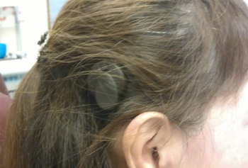
Photograph showing the external audio processor of the Bonebridge system.

Photograph showing the components of the bone conduction implant. BC-FMT = bone conduction floating mass transducer

The external audio processor is attached to the skin by magnetism. It contains microphones, a digital signal processor and a battery. Its audio output signal is transmitted transcutaneously via electromagnetic signals (instead of via a mechanical conduction system), obviating the need for an open pin tract site at the skin, as required in the BAHA system. Patients therefore do not need to perform daily peri-abutment wound care. In this way, the Bonebridge system aims to resolve the issue of post-operative peri-abutment pin tract wound infection.
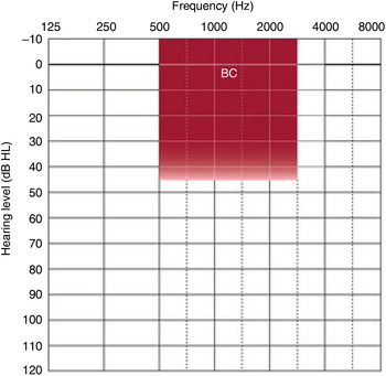
The therapeutic range of the Bonebridge system is similar to that of the Cochlear Baha Compact and Baha BP100 implants (Cochlear, Sydney, Australia). It is suitable for patients with conductive or mixed hearing loss, as indicated by bone conduction thresholds of 45 dB HL or better at 0.5, 1, 2 and 3 kHz, on audiometric testing (see Figure 3). The Bonebridge system can also provide sound re-routing for unilateral sensorineural deafness, provided the remaining, functioning ear is capable of detecting air-conducted sound at or below 20 dB HL at 0.5, 1, 2 and 3 kHz, on audiometric testing. The manufacturers recommend the following criteria for implant candidacy: patients should be 18 years of age or older, with anatomy that allows for appropriate placement of the Bonebridge system as determined by computed tomography (CT) scanning, and without retro-cochlear or central auditory disorders.3 Thus, the Bonebridge system appears to be suitable for patients with many types of hearing loss, provided their residual hearing is within the appropriate therapeutic range.
Audiological selection criteria for the Bonebridge system (area marked in red), for patients with conductive or mixed hearing loss. BC = bone conduction

In this report, we present the first Bonebridge implantation case performed in Asia. The patient was a 58-year-old woman with bilateral substantial conductive hearing loss. She was a singer and dancer and performed regularly in nightclubs. She had undergone a left mastoidectomy for cholesteatoma more than 10 years previously, while her right ear had been fitted with a traditional air conduction hearing aid following an unsuccessful tympanoplasty. She opted for a Bonebridge system implantation on her right side, as her current hearing aid produced much discomfort and gave poor quality sound amplification, especially in noisy environments. In addition, the occlusion effect of the ear mould produced considerable discomfort and recurrent episodes of otitis externa. In addition, stigmatisation by her peers, as the wearer of a visible hearing aid, caused her to wear the device less than recommended: her daily usage was never more than 6 hours.
Technique
Ethical approval
Ethical approval was obtained from the Joint Chinese University of Hong Kong New Territories East Cluster Clinical Research Ethics Committee, Hong Kong, China.
Pre-operative patient preparation
Computed tomography
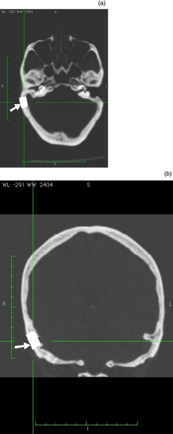
The patient was sent for temporal bone CT scanning; CT navigation planning was undertaken during the same session. The best placement site for the bone conduction implant was marked on the CT images (Figure 4).
(a) Axial and (b) coronal computed tomography images showing the position of implant placement (arrow). A = anterior; R = right; L = left; P = posterior; S = superior; I = inferior

Audiology
Before surgery, the patient underwent a series of audiological assessments, including the following: (1) pure tone audiometry (both air and bone conduction); (2) functional gain measurement in the sound field, using her usual right-sided hearing aid; (3) speech recognition testing in both quiet and noisy environments, using the Cantonese Hearing in Noise Test with and without the existing hearing aid.
Surgical preparation
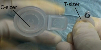
Sizers (dummies) provided by the manufacturer assisted with correct placement of the bone conduction implant. These comprised a designated coil sizer (C-sizer) and a transducer sizer (T-sizer) (Figure 5).
Photograph showing connection of the C-sizer to the T-sizer.

The polypropylene C-sizer helped determine the optimal bone conduction implant site on the head, prior to skin incision. The T-sizer was a template comprising a polypropylene body and two titanium drill guides. The functions of the T-sizer were: (1) to outline the exact size of the bone conduction implant seat before drilling; (2) to verify the size of the seat before implant placement; and (3) to act as a drilling guide to ensure the correct orientation and distance between the two anchor holes, and the correct depth of the anchoring holes.
The C- and the T-sizer could be connected together to represent the complete bone conduction implant and to assist identification of the best placement site.
Surgery
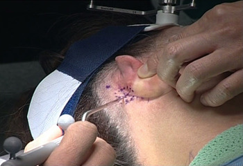
The surgery was performed at the otorhinolaryngology department, New Territories East Cluster hospital, Hong Kong, under general anaesthesia. A Brainlab navigation system (Brainlab, Feldkirchen, Germany) was employed intra-operatively to enable real-time anatomical monitoring and to facilitate location of a suitable implant site. Aided by the navigation system, and using the C- and T-sizers, an appropriate placement site was identified on the patient's head and marked with a surgical pen (Figure 6).
Surgical photograph showing marking of the implant placement site, with assistance from the surgical navigation system.

Surgery was commenced by making an incision along the post-auricular skin crease. Haemostasis was achieved with diathermy. Soft tissue dissection was carried out to create a surgical field with complete exposure of the whole mastoid, posterior bony canal wall and temporal line.
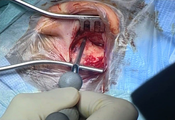
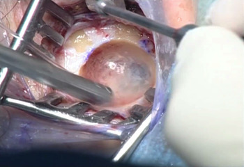
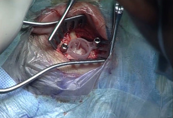
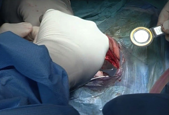
Before bone drilling, the C- and T-sizers were used to confirm the best placement site. The surgical navigation system was used to re-confirm the best drilling site (Figure 7). A bone recess was created near the sinodural angle of the mastoid (Figure 8). Special care was taken when drilling in proximity to vital structures such as the sigmoid sinus, the tegmen and the dura; a diamond burr was used when approaching these structures. As the bone conduction floating mass transducer was 8.7 mm thick and 15.8 mm in diameter, a bone recess of slightly larger dimensions was created, in which the T-sizer was positioned with its ‘arms’ resting on the skull (Figure 9). Oversize drilling would have affected the placement of the anchoring screws, hence the anchoring of the bone conduction floating mass transducer. The drilling position was checked regularly using the surgical navigation system, in order to minimise the risk of damage to nearby vital structures.
Surgical photograph showing confirmation of the implant placement site, using the surgical navigation system, following the initial postaural incision.

Surgical photograph showing the creation of a bone recess to house the bone conduction implant floating mass transducer. Note the position of the sigmoid sinus, appearing as a blue shadow.

Surgical photograph showing placement of the T-sizer within the bone recess, in order to establish the site of the fixing holes.

Using the T-sizer as a drilling guide, fixing points were created with a specially designed drill with a depth stop that ensured a hole depth of no more than 3.9 mm. We then prepared a sub-periosteal pocket to accommodate the coil and demodulator of the bone conduction implant. No drilling of the bone bed was needed for implantation of the demodulator.
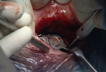
The bone conduction implant was oriented with the magnet protruding towards the skull, and was bent to fit its required final position (the angle of the bone conduction implant could be altered ±90° in the horizontal plane and −30° in the vertical plane). The implant coil and demodulator were placed under the periosteum, so that they would lie beneath the desired external location of the audio processor (Figure 10). The bone conduction floating mass transducer was then placed into the prepared bone recess and secured with cortical screws in each anchor hole (Figures 11 and 12). A torque wrench was used in the final stage of screw fixing to help prevent over-tightening.
Surgical photograph showing insertion of the bone conduction implant into the subperiosteal pocket.

Surgical photograph showing fixation of the bone conduction implant using cortical screws.

Surgical photograph showing the final position of the bone conduction implant.

Before closure, we ensured that the total skin flap thickness was no more than 7 mm, using the skin flap gauge provided. No trimming of the skin flap was required, but would have been performed if necessary. The skin wound was closed in double layers and a head pressure dressing was applied for 24 hours.
No intra- or post-operative surgical complications were encountered. The patient complained of mild wound pain and dizziness in the early post-operative period. All these symptoms were resolved with medical treatment. She was discharged home with antibiotics and analgesics, and reviewed one week after the surgery, by which time the post-auricular wound had completely healed.
The device was activated two weeks after surgery. The Amade audio processor (Med-El) was programmed using Connexx 6.4.5 software (Siemens Audiological Engineering, Munich, Germany) equipped with a Symfit database (Rev. 6.1; Med-El). The default desired sensation level amplification strategy was applied during the first fit.
The patient returned for review and fine tuning one month later, to ensure that her hearing was at the most comfortable (self-evaluated) level.
Results
Pre- and post-operative hearing thresholds
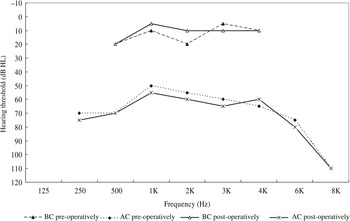
Any change in residual hearing was evaluated by comparing the pre-operative, unaided air and bone conduction thresholds with the post-operative, unaided air and bone conduction thresholds, using audiometric testing at frequencies from 250 Hz to 8 kHz. The results revealed no obvious change at any frequency. Figure 13 shows the pre- and post-operative air and bone conduction thresholds.
The patient's pre- and post-operative air conduction (AC) and bone conduction (BC) thresholds. K = 1000.

Functional gain improvement
Aided hearing benefit was evaluated by comparing the unaided and aided thresholds using warble tones in the sound field at frequencies from 500 Hz to 4 kHz, one month after surgery. The non-implanted ear was plugged and muffled during testing. An obvious improvement was noted at all the testing frequencies from 500 Hz to 4 kHz. Functional gain (i.e. the difference between the unaided and aided thresholds) varied from a minimum of 10 dB to a maximum of 40 dB (Figure 14). The aided improvements were also compared with the improvement obtained from the patient's previous hearing aid; results showed that the Bonebridge system out-performed the pre-operative hearing aid by at least approximately 5 dB at the majority of the test frequencies (Figure 14).
Graph showing sound field thresholds from 500 Hz to 4 kHz, for the right ear, under the following conditions: unaided (pre-operative); aided with a convention hearing aid (HA) (pre-operative); and aided with the Bonebridge system (BB) (one-month post-operative).

Speech recognition threshold improvement
Improvement in aided speech comprehension was evaluated using the Cantonese Hearing in Noise Test in both quiet and noisy situations.
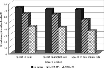
Figure 15 shows the patient's speech recognition threshold results in a quiet environment with speech presented from the front, from the implant side (right ear) and from the non-implant side (left ear), with and without the pre-operative hearing aid and the Bonebridge system aid (evaluated one month after activation). The non-implanted ear was plugged and muffled during testing. As shown, while the pre-operative hearing aid provided an average speech recognition threshold improvement of 12 dB across the various speech locations, the Bonebridge system produced an average speech recognition threshold improvement of 32 dB.
Graph showing mean speech recognition thresholds in quiet conditions when speech was presented from the front, from the implant side (right ear) and from the non-implant side (left ear), under the following conditions: unaided (pre-operative); aided with a conventional hearing aid (HA) (pre-operative); and aided with the Bonebridge system (BB) (one month post-operative).

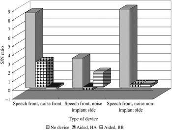
Figure 16 shows results (presented as signal to noise ratios) for speech testing in noisy conditions, for speech presented from the front together with noise presented either from the front, from the implant side or from the non-implant side. Use of the Bonebridge system improved binaural ‘squelch’ in all listening conditions, that is, the speech recognition threshold improved compared with the unaided condition. When noise was presented from the implant side, the use of the pre-operative hearing aid on the non-implant side assisted hearing, enabling a 2 dB improvement in speech recognition threshold. Overall, substantial improvement in speech recognition was observed.
Graph showing mean signal to noise (S/N) ratios for speech in noisy conditions, when speech was presented from the front together with noise presented from the front, from the implant side (right ear) or from the non-implant side (left ear), under the following conditions: unaided (pre-operative); aided with a conventional hearing aid (HA) (pre-operative); and aided with the Bonebridge system (BB) (one month post-operative).

At the time of writing, the patient was using the Bonebridge system for over 8 hours a day. She reported that the sound quality of the Bonebridge system was natural and clear, and that the system improved speech recognition in most listening situations. She was especially pleased with the Bonebridge to help her during singing and dancing performances, as well as its cosmetic acceptability.
Discussion
This paper presents the first case of implantation of a monaural Bonebridge system in an Asian, Cantonese-speaking patient with substantial bilateral conductive hearing loss. Preliminary results from this patient suggest that the Bonebridge system is a safe and effective treatment option for adults with conductive hearing loss. This patient had well preserved hearing thresholds after surgery and suffered no intra-operative or post-operative complications.
In our experience, surgical implantation of the Bonebridge system was simple and easy to master, in comparison with middle-ear implants such as the Vibrant Soundbridge and Carina systems, which require delicate and technically demanding surgical work with the ossicles or round window.Reference Tsang, Yu, Wong and Tong4, Reference Martin, Deveze, Richard, Lefebvre, Decat and Ibañez5
Careful CT planning was undertaken before the implantation procedure; this is highly recommended. We suggest that surgeons work closely with their radiology colleagues to identify the best implant placement site on the CT scan. During the operation, the surgeon must be vigilant in order to avoid damage to vital structures such as the dura and sigmoid sinus. Complication rates may be further minimised with the assistance of a CT surgical navigation system. In our patient, the only post-operative sequelae were wound pain and dizziness, which resolved without active intervention.
Our patient found the Bonebridge external audio processor easy to use and more appealing cosmetically. It was small enough to be well hidden under her hair (Figure 17). Having this type of concealable implant may allow patients to avoid the stigma associated with hearing aid usage, raising their self-esteem and improving the quality of their social lives.
Photograph showing the external audio sound processor, well hidden under the patient's hair.

In addition, unlike traditional percutaneous bone anchored hearing aid the use of the Bonebridge system eliminates post-operative pin tract infections, which greatly enhances the patient's quality of life as it obviates the need for daily implant wound care.
From an audiological point of view, our patient's Bonebridge system enabled substantial hearing improvement (10 dB–40 dB), relative to the unaided condition. Compared with the patient's pre-operative hearing aid, the Bonebridge system allowed a additional 5 dB functional gain, while speech recognition thresholds in quiet conditions were on average 2.7 times better. The speech recognition threshold improvement in noise was however slightly better for the original hearing aid, compared with the Bonebridge system. However, overall the patient benefited from additional speech understanding, especially in quiet conditions, after being fitted with the Bonebridge system.
Conclusion
This 58-year-old, Asian woman with bilateral substantial conductive hearing loss underwent successful implantation with a Bonebridge semi-implantable bone conduction hearing device. There were no peri- or post-operative complications. The patient received substantial hearing benefits from her new device.
These results support the recommended adult selection criteria for the Bonebridge system.
Acknowledgement
This study was partly funded by Med-El Hong Kong Asia Pacific Headquarters.