




No CrossRef data available.
Article contents
Heat shock and stress response of Taenia solium and T. crassiceps (Cestoda)
Published online by Cambridge University Press: 07 August 2001
Abstract
Heat shock and stress responses are documented for the first time in larval stages of the cestodes Taenia solium and Taenia crassiceps. Radioactive metabolic labelling after in vitro incubation of cysts at 43 °C, revealed the induction of heat shock proteins. In T. crassiceps, the major heat shock proteins were 80, 70 and 60 kDa. After prolonged incubation, a set of low molecular weight heat shock proteins (27, 31, 33 and 38 kDa), were also induced. In vitro incubation of cysts at 4 °C, induced the synthesis of stress proteins ranging from 31 to 80 kDa, indicating the parasite is also able to respond to cold shock. T. solium cysts exposure to temperature stress also resulted in an increased synthesis of 2 major heat shock proteins of 80 and 70 kDa. Western blots using the excretory–secretory products of T. solium showed that 2 heat shock proteins were recognized by antibodies in the sera of cysticercotic patients: one of 66 kDa and another migrating close to the run front. The T. solium 66 kDa protein was also recognized by specific antibodies directed to a 60 kDa bacterial heat shock protein, suggesting that it belongs to this family of proteins.
Keywords
Information
- Type
- Research Article
- Information
- Copyright
- 2001 Cambridge University Press
INTRODUCTION
Many parasites undergo sudden changes in temperature at some stage during their life-cycle (Newport, Culpepper & Agabian, 1988; Polla, 1991; Maresca & Carratù, 1992). For example, taeniid eggs in the environment suffer an increase in temperature when ingested by its warm-blooded intermediate host. The larvae in the intermediate host also undergo strong variations of temperature before reaching its final host. Moreover, larvae and adult worms may be exposed to sudden temperature changes due to host response to infection. Adaptation to these temperature variations is crucial for the maintenance of parasite transmission.
The heat shock response is a general homeostatic mechanism that protects cells and organisms from deleterious effects of environmental stress (Lindquist, 1986; Morimoto, 1990). It is characterized by a rapid transcriptional activation of a specific set of genes coding for proteins generically known as heat shock proteins (HSPs). These HSPs allow the adaptation of the organism to the new temperature (Morimoto et al. 1997; Feder & Hofmann, 1999). Similar adaptive responses are produced for a variety of other environmental stresses (Welch et al. 1991; De Maio, 1999). To our knowledge, no previous report is available on the heat shock and stress responses in taeniids, although some HSPs have been identified (Muhlschlegel et al. 1995; Benitez et al. 1998; Martinez et al. 1999). In Mesocestoides corti, Estes & Teale (1991) have reported the occurrence of HSPs in excretory–secretory (E–S) products. Here we describe a time-regulated induction of HSPs when Taenia solium and T. crassiceps larvae are maintained in culture at temperatures above 37 °C. Several proteins are excreted/secreted by T. solium larvae during the stress response, including 2 HSP, that were recognized by sera from patients with neurocysticercosis. The ES protein of 66 kDa was recognized by specific antibodies against bacterial HSP60.
MATERIALS AND METHODS
Larvae
T. solium cysticerci were dissected from the skeletal muscle of naturally infected pigs obtained from rural villages. The cysts were obtained at a maximum of 24 h after slaughter of pigs. The ORF strain of T. crassiceps is maintained in our laboratory by serial intraperitoneal passage of cysticerci in 6-week-old female BALB/c AnN mice (Sciutto et al. 1990). The larvae used in the experiments were obtained from mice with 2-month infections. Before use, all larvae were washed 5 times in a phosphate-buffered saline solution (PBS), pH 7.2, pre-warmed at 37 °C.
Radio-isotope labelling
Groups of 3 T. solium or 5 T. crassiceps cysts were placed into sterile polypropylene tubes (Falcon) containing 500 μl of a sterile methionine-free RPMI 1640 medium (GIBCO), pre-warmed at 37 °C (except for the group that was placed at 4 °C), and supplemented with 1% essential amino acids, pyruvate and antibiotic–antimycotic mixture (GIBCO). The groups were then incubated at 4, 37, 43 or 46 °C for 30 min and then metabolically labelled by addition of 100 μCi/ml of L-[35S]methionine (specific activity>1000 Ci/mmol, Amersham) for 4 h or overnight at the same temperatures as indicated above. To determine the kinetics of synthesis of HSPs in T. crassiceps, the cysts were maintained at 43 °C (optimal temperature for heat shock response, see results) for 15, 30 min, 1, 2, 4, 6 and 8 h, before adding the radio-isotope labelled methionine, and then maintained for 2 h at 37 °C.
After labelling, the groups of cysts were washed twice with 10 ml of PBS to remove free label and then placed in 500 μl of SDS–PAGE sample buffer for homogenization in a Soniprep 150 sonicator, for 40 s (20 s intervals), at 60 Watts. The culture medium containing E–S products was also collected, centrifuged at 1000 g for 10 min to remove any cellular debris and the supernatant was stored at −20 °C until use. Protein concentration in the crude extracts and culture medium was determined using a protein assay kit (BioRad).
Autoradiography
The crude extracts and the culture medium were analysed by sodium dodecyl sulphate–polyacrylamide gel electrophoresis (SDS–PAGE) (Laemmli, 1970). Samples of equal amounts of protein (5–20 μg/well) were run under reducing conditions and the gel was stained with Coomassie Brilliant Blue R-250 (BioRad) or Silver Stain (BioRad). The gel was prepared for fluorography by treatment with a 20% solution of PPO in dimethylsulfoxide (Laskey & Mills, 1975), dried on a BioRad slab gel drier and exposed to an X-ray film (Kodak X-OMAT) at −70 °C. Relative increases over controls for each HSP were determined by densitometric scanning of the protein bands by means of a microcomputer program (RFLP scan 3.0, Scanalytics Inc., Fairfax, VA, USA).
Western blotting
Western immunoblots were carried out according to the method of Towbin, Staehelin & Gordon (1979), using a rabbit polyclonal anti-HSP60, a mouse mAb anti-HSP70 (SPA-804 and SPA-822, respectively, from StressGen Biotechnologies Corp., Victoria B.C., Canada), and a mouse mAb IgGl anti-HSP60 (LK2, kindly provided by P. van Kooten (Boog et al. 1992), at a 1:100 dilution. Sera from patients with neurocysticercosis or other neurological disorders (kindly donated by D. Correa) were used at 1:30 dilution. Peroxidase-conjugated rabbit anti-mouse, goat anti-rabbit or goat anti-human antibodies (Zymed Laboratories) diluted 1:1000 were used followed by 0.1% diaminobenzidine with 0.015% hydrogen peroxide to develop the blots.
RESULTS
Effect of temperature on protein synthesis in T. solium and T. crassiceps
Autoradiograms of the L-[35S]methionine labelled crude extracts from cysts of both species, incubated at 37 °C, showed more than 30 protein bands ranging from 21 to 200 kDa (Fig. 1). When T. solium larvae were incubated at 43 °C for 4 h, the intensity of the protein bands of 80 (HSP 80) and 70 (HSP 70) kDa was highly increased, whereas the intensity of some labelled proteins observed at 37 °C decreased or disappeared (Fig. 1A). Densitometric analysis on the 2 major HSPs showed an increase over controls of 2.9 and 4 times respectively. At 4 °C and 46 °C no labelled proteins were observed in T. solium, suggesting that these temperatures are lethal for the parasite (not shown). Changes in the pattern of protein expression in response to temperature stress were also observed in T. crassiceps (Fig. 1B), including the 2 major HSPs described for T. solium (80 and 70 kDa). The relative increase over controls for HSP80 and HSP70 was 2.1 and 2.0 times, respectively. As in the case of T. solium, the synthesis of some proteins was diminished and in some cases stopped during incubation at 43 °C. Incubation of T. crassiceps cysts at 4 °C, induced the preferential synthesis of the 80 kDa protein, although some low molecular weight proteins ranging from 31 to 45 kDa were also observed (Fig. 1B), suggesting a cold shock response (Thieringer, Jones & Inouye, 1998). No label incorporation was detected when the larvae were incubated at 46 °C (not shown), indicating that this temperature also results in cyst killing. Microscopical observations of T. solium and T. crassiceps, confirmed that the cysts were non-motile after incubation at 46 °C.

Fig. 1. Autoradiogram showing SDS–PAGE profile of protein synthesis of Taenia solium (A) and T. crassiceps (B) cysticerci. Parasites were metabolically labelled in vitro with [35S]methionine for 4 h at the indicated temperatures, with the exception of the 4 °C lane, which was labelled for 24 h. Arrows denote major heat shock proteins.
Kinetics of synthesis of HSPs in T. crassiceps cysts
T. crassiceps cysts that were incubated at 43 °C for different periods reacted with a typical heat-shock response. In the initial 15–60 min of incubation, 2 proteins of 80 and 70 kDa were the most prominent (Fig. 2). After 2 h, a larger number of proteins were visible, a major protein of 60 kDa and several others ranging from 14 to 200 kDa, including some proteins that were present before the heat shock. After 8 h, the most prominent bands corresponded to 80, 70 and 60 kDa, followed by other proteins of 50, 38 kDa and a small protein (LMWP) migrating close to the run front (Fig. 2).

Fig. 2. Kinetics of protein synthesis in Taenia crassiceps cysticerci in response to heat shock. After treatment at 43 °C during the indicated time, the cysts were transferred to 37 °C and metabolically labelled in vitro with [35S]methionine for 2 h. Control parasites were maintained at 37 °C which is the normal growth temperature. Arrows denote major heat shock proteins.
Excreted–secreted heat shock products
The culture media where T. solium cysts were maintained overnight at different temperatures were collected, analysed by SDS–PAGE and silver-stained for E–S stress products. At 37 °C, the most prominent E–S proteins were 66, 58, 54, 48, 45, 38, 33 and 31 kDa (Fig. 3A). This pattern of E–S products is similar to that described for T. solium by (Ko & Ng 1998). Several high molecular weight proteins are observed in the E–S products from cultures incubated at 43 and 46 °C (Fig. 3A). Autoradiograms of the slab gel revealed a 5-fold increase of a major band of 66 kDa at 43 °C that was also present at 37 °C (Fig. 3B). Protein bands of 97, 74, 54, 52 and 47 kDa are also excreted/secreted at 43 °C. T. crassiceps also showed some E–S products in the supernatant (not shown). Some of these products have been described to cross-react with well-known antigens of T. solium (Tsang, Brand & Boyer, 1989; Ko & Ng, 1998).

Fig. 3. Excretory–secretory products of Taenia solium cysticerci exposed to temperature stress. The cysts were incubated at the indicated temperatures and metabolically labelled with [35S]methionine, overnight. (A) SDS–PAGE profile of the culture medium containing the excretory–secretory products using silver stain. (B) Autoradiogram of the same slab gel.
Western blotting using specific anti-heat shock protein antibodies
The cyst extracts and culture media of T. solium cysts maintained overnight at 37 or 43 °C, were analysed by Western blot using specific antibodies to HSP60 and HSP70. In the crude extracts, both antibodies revealed protein bands of 60 and 70 kDa, respectively (Fig. 4A, lanes 1–4). However, they failed to reveal differences in amount, between cysts incubated at 37 and 43 °C, suggesting that at least this 70 kDa protein is different from the HSP70 described above, that is clearly induced during heat shock. This is not surprising, as constitutive expression of a HSP homologue has been reported in Schistosoma mansoni (Tielens, Van Den Heuvel & Van Eden, 1993). When the culture media were blotted and reacted with the anti-HSP60 antibody, 2 bands of 66 and 60 kDa were revealed (Fig. 4A, lanes 5 and 7). In contrast, no band was observed when the culture media were probed with the anti-HSP70 monoclonal antibody (Fig. 4A, lanes 6 and 8).

Fig. 4. Antibody recognition of HSP60 and HSP70 in crude extracts and excreted–secreted (ES) products of Taenia solium (A) or T. crassiceps (B) cysticerci. Lanes 1, 3, 5 and 7 were probed with rabbit polyclonal anti-HSP60. Lanes 2, 4, 6 and 8 were probed with anti-HSP70 mAb.
In the T. crassiceps crude extracts, similar but thinner protein bands of 60 and 70 kDa were revealed by anti-HSP60 and anti-HSP70 antibodies, respectively (Fig. 4B, lanes 1–4). Also in this case, no quantitative variation was detected between cysticerci incubated at 37 and 43 °C. None of the antibodies reacted with the culture media of T. crassiceps cysts (Fig. 4B, lanes 5–8).
Western blotting using sera from patients with neurocysticercosis
Western blots were also performed on the crude extract and culture medium of T. solium cysticerci incubated at 43 °C, using sera from patients with neurocysticercosis. When the extract was probed, the sera recognized several antigens including a protein of 80 kDa that may correspond to the HSP 80 (Fig. 5A). When T. solium culture medium was reacted with the patient sera, all sera recognized a 66 kDa protein and the LMWP (Fig. 5B). The 66 kDa protein, was confirmed to be a HSP by autoradiography, showing that this protein was synthesized during the stress response. Antibody LK2, directed against human HSP60, recognized in the crude extract, a protein band of 60 kDa, as expected, as well as another band of 75 kDa. In contrast, no protein was recognized by the LK2 antibody in the culture medium, indicating that no protein leaked from the cysts and that the integrity of the bladder wall was preserved during incubation at 43 °C.

Fig. 5. Western blots of crude extracts and excreted–secreted products of Taenia solium cysticerci probed with sera from neurocysticercotic patients. Cysticerci were metabolically labelled with [35S]methionine, at 43 °C, overnight. Crude extracts (A) and excreted–secreted products (B), were reacted with neurocysticercotic patient sera (lanes 1–15), or with sera from patients with other neurological disorders (lanes 16–20). Lane 22 was reacted with a monoclonal antibody directed to HSP60 (LK2) and lane 21 with the second antibody alone. Lane X shows the autoradiogram of the blots.
Similar results were obtained when crude extracts of T. crassiceps were reacted with the patient sera (not shown), confirming the extensive antigenic cross-reactivity between these two taeniid cysticerci (Larralde et al. 1990; Sciutto et al. 1990; Ko & Ng, 1998). However, in the culture medium of T. crassiceps cysts, the patient sera failed to detect the 66 kDa band (not shown).
DISCUSSION
The present study documents for the first time the response to temperature stress in cysticerci of T. solium and T. crassiceps. Both parasites share 2 major proteins in response to heat shock (HSP80 and HSP70), while only T. crassiceps synthesized another major 60 kDa protein. The family of HSP80 is known to be immunogenic in other parasites (Newport et al.1988; Polla, 1991). In Schistosoma mansoni, an open reading frame of a 86 kDa antigen was shown to be highly homologous to HSP90 of Saccharomyces cerevisiae and HSP83 of Drosophila melanogaster (Johnson et al. 1989). The family of HSP70 is associated with molecular chaperones that facilitate protein transport across membranes, remove denatured proteins, and allow cellular recovery from glucose starvation (Burdon, 1986; Newport et al. 1988; Hartl, Martin & Neupert, 1992). HSP70 is also a major immunogen in several parasite infections, including Brugia spp., Onchocerca volvulus and S. mansoni (Hedstrom et al. 1988; Rothstein et al. 1989; Selkirk et al. 1989). HSP60 has also been shown to function as molecular chaperone (Hemmingsen et al. 1988; Zeilstra-Ryalls, Fayet & Georgopoulos, 1991) and has been identified in several parasites (Maresca & Carratù, 1992).
Differences in the profile of response to temperature stress observed between the two species of cysticerci, can be due to interspecific variations, or to differences in the way both parasites are obtained before being used in the experiments. While T. crassiceps were freshly obtained from infected mice maintained in the laboratory, T. solium cysts were obtained at least 5–6 h after slaughter, from naturally infected pigs detected in abattoirs. It is conceivable that the lack of response to low temperature in T. solium, might be due to the longer time the cysts remained in the pig carcass before being collected for the experiments.
The heat shock response observed in T. crassiceps, resulting in the recovery of several proteins expressed by cysts at 37 °C, suggests that early HSPs play an efficient role in the protection of other proteins, in agreement with some examples of response to stress. Thus, temperature stress response may play a role in the parasite adaptation to sudden changes in environmental temperature.
The presence of HSP66 and LMWP in the E–S products of taeniids has not been reported before. The labelled proteins detected in E–S products of T. solium are regarded as HSPs because both became metabolically labelled during heat-shock treatment. To our knowledge, only 2 other reports have described the occurrence of HSPs among E–S products of helminth parasites. Two E–S proteins of Mesocestoides corti, that are homologous to the HSP70 and HSP60, stimulate proliferative responses of splenic lymphocytes of infected mice and antibody responses restricted to IgM and IgGl isotypes, suggesting that they play a role in the immune response to M. corti (Estes & Teale, 1991). Additionally, 2 well-known E–S antigens from the nematode Trichinella spiralis and T. pseudospiralis, that are commonly employed for serodiagnosis, have been suggested to correspond to stress proteins because of their similar molecular masses and isolelectric points (Ko & Fan, 1996).
Our results indicate that human neurocysticercotic patients recognize at least two excreted/secreted HSPs products. The 66 kDa product that is recognized by a specific antibody against HSP60, appears to correspond to a well-known 66 kDa antigen (Ko & Ng, 1998). It has been reported that HSP60 has to bind a low molecular weight protein of about 10 kDa in order to perform its chaperone function (Gragerov et al. 1992). The LMWP that is also recognized by our patient sera, might correspond to the HSP66 partner, explaining why these 2 proteins appear together among the E–S products of cysticerci.
This work was supported in part by grants from CONACYT, LOO42-M9607 and from DGAPA-UNAM, IN-207195 (J.P.L.). L.V.P. was supported by scholarships from CONACYT and DGAPA-UNAM. C.F.S. is supported by scholarships from CONACYT and DGEP-UNAM. We would like to thank T. Govezensky for the densitometric analysis.
References
BENITEZ, L., HARRISON, L. J., PARKHOUSE, R. M. & GARATE, T. (1998). Sequence and preliminary characterisation of a Taenia saginata oncosphere gene homologue of the small heat-shock protein family. Parasitology Research 84, 423–425.CrossRefGoogle Scholar
BOOG, C. J. P., DE GRAEFF-MEEDER, E. R., LUCASSEN, M. A., VAN DER ZEE, R., VOORHOST-OGINK, M. M., VAN KOOTEN, P. J. S., GEUZE, H. J. & VAN EDEN, W. (1992). Two monoclonal antibodies generated against human hsp60 show reactivity with synovial membranes of patients with juvenile chronic arthritis. Journal of Experimental Medicine 175, 1805–1810.CrossRefGoogle Scholar
BURDON, R. H. (1986). Heat shock and the heat shock proteins. Biochemistry Journal 240, 313–324.CrossRefGoogle Scholar
DE MAIO, A. (1999). Heat shock proteins: facts, thoughts, and dreams. Shock 11, 1–12.CrossRefGoogle Scholar
ESTES, D. M. & TEALE, J. M. (1991). Biochemical and functional analysis of extracellular stress proteins of Mesocestoides corti. Journal of Immunology 147, 3926–3934.Google Scholar
FEDER, M. E. & HOFMANN, G. E. (1999). Heat-shock proteins, molecular chaperones, and the stress response: evolutionary and ecological physiology. Annual Reviews in Physiology 61, 243–282.CrossRefGoogle Scholar
GRAGEROV, A., NUDLER, E., KOMMISAROVA, N., GAITANARIS, G. A., GOTTESMAN, M. E. & NIKIFOROV, V. (1992). Cooperation of GroEL/GroES and DnaK/DnaJ heatshock proteins in preventing protein misfolding in Escherichia coli. Proceedings of the National Academy of Sciences, USA 89, 10341–10344.CrossRefGoogle Scholar
HARTL, F. U., MARTIN, J. & NEUPERT, W. (1992). Protein folding in the cell: the role of molecular chaperones Hsp70 and Hsp60. Annual Review of Biophysics and Biomolecular Structure 21, 293–322.CrossRefGoogle Scholar
HEDSTROM, R., CULPEPPER, J., SCHINSKI, V., AGABIAN, N. & NEWPORT, G. (1988). Schistosome heat-shock proteins are immunologically distinct host-like antigens. Molecular and Biochemical Parasitology 29, 275–282.CrossRefGoogle Scholar
HEMMINGSEN, S. M., WOOLFORD, C., VAN, D. V. S. M., TILLY, K., DENNIS, D. T., GEORGOPOULOS, C. P., HENDRIX, R. W. & ELLIS, R. J. (1988). Homologous plant and bacterial proteins chaperone oligomeric protein assembly. Nature, London 333, 330–334.CrossRefGoogle Scholar
JOHNSON, K. K., WELLS, K., BOCK, J. V., NENE, V., TAYLOR, D. W. & CORDINGLEY, J. S. (1989). The 86-kilodalton antigen from Schistosoma mansoni is a heat-shock protein homologous to yeast HSP-90. Molecular and Biochemical Parasitology 33, 19–28.CrossRefGoogle Scholar
KO, R. C. & FAN, L. (1996). Heat shock response of Trichinella spiralis and T. pseudospiralis. Parasitology 112, 89–95.CrossRefGoogle Scholar
KO, R. C. & NG, T. F. (1998). Evaluation of excretory/secretory products of larval Taenia solium as diagnostic antigens for porcine and human cysticercosis. Journal of Helminthology 72, 147–152.CrossRefGoogle Scholar
LAEMMLI, U. K. (1970). Cleavage of structural proteins during the assembly of the head of bacteriophage T4. Nature, London 227, 680–685.CrossRefGoogle Scholar
LARRALDE, C., SOTELO, J., MONTOYA, R. M., PALENCIA, G., PADILLA, A., GOVEZENSKY, T., DIAZ, M. L. & SCIUTTO, E. (1990). Immunodiagnosis of human cysticercosis in cerebrospinal fluid. Antigens from murine Taenia crassiceps cysticerci effectively substitute those from porcine Taenia solium. Archives of Pathological Laboratory Medicine 114, 926–928.Google Scholar
LASKEY, R. A. & MILLS, A. D. (1975). Quantitative film detection of 3H and 14C in polyacrylamide gels by fluorography. European Journal of Biochemistry 56, 335–341.CrossRefGoogle Scholar
LINDQUIST, S. (1986). The heat-shock response. Annual Reviews of Biochemistry 55, 1151–1191.CrossRefGoogle Scholar
MARESCA, B. & CARRATÙ, L. (1992). The biology of the heat shock response in parasites. Parasitology Today 8, 260–266.CrossRefGoogle Scholar
MARTINEZ, J., PEREZ-SERRANO, J., BODEGA, G., CASADO, N. & RODRIGUEZ-CAABEIRO, F. (1999). Heat shock proteins HSP70 and HSP60 in Echinococcus granulosus protoscolices. Folia Parasitologia (Praha) 46, 76–78.Google Scholar
MORIMOTO, R. I. (1990). The stress response, function of the proteins, and perspectives. In Stress Proteins in Biology and Medicine (ed. MORIMOTO, R. I. TISSIÈRES, A. & GEORGOPOULOS, C.), pp. 1–36. Cold Spring Harbor Laboratory Press, Cold Spring Harbor, NY.
MORIMOTO, R. I., KLINE, M. P., BIMSTON, D. N. & COTTO, J. J. (1997). The heat shock response: regulation and function of heat-shock proteins and molecular chaperones. Essays in Biochemistry 32, 17–29.Google Scholar
MUHLSCHLEGEL, F., FRISCG, P., CASTRO, A., APFEL, H., MULLER, A. & FROSCH, M. (1995). Molecular cloning and characterization of an Echinococcus multilocularis and Echinococcus granulosus stress protein homologous to the mammalian 78 kD glucose regulated protein. Molecular and Biochemical Parasitology 74, 245–250.CrossRefGoogle Scholar
NEWPORT, G., CULPEPPER, J. & AGABIAN, N. (1988). Parasite heat-shock proteins. Parasitology Today 4, 306–312.CrossRefGoogle Scholar
POLLA, B. S. (1991). Heat shock proteins in host–parasite interactions. Immunology Today 12, A38–41.CrossRefGoogle Scholar
ROTHSTEIN, N. M., HIGASHI, G., YATES, J. & RAJAN, T. V. (1989). Onchocerca volvulus heat shock protein 70 is a major immunogen in amicrofilaremic individuals from a filariasis-endemic area. Molecular and Biochemical Parasitology 33, 229–236.CrossRefGoogle Scholar
SCIUTTO, E., FRAGOSO, G., TRUEBA, L., LEMUS, D., MONTOYA, R. M., DIAZ, M. L., GOVEZENSKY, T., LOMELI, C., TAPIA, G. & LARRALDE, C. (1990). Cysticercosis vaccine: cross protecting immunity with Taenia solium antigens against experimental murine Taenia crassiceps cysticercosis. Parasite Immunology 12, 687–689.CrossRefGoogle Scholar
SELKIRK, M. E., DENHAM, D. A., PARTONO, F. & MAIZELS, R. (1989). Heat shock cognate 70 is a prominent immunogen in brugian filariasis. Journal of Immunology 143, 299–308.Google Scholar
THIERINGER, H. A., JONES, P. G. & INOUYE, M. (1998). Cold shock and adaptation. Bioessays 20, 49–57.3.0.CO;2-N>CrossRefGoogle Scholar
TIELENS, A. G. M., VAN DEN HEUVEL, J. M. & VAN EDEN, W. (1993). Schistosoma mansoni: An hsp60 homologue is constitutively expressed in cercariae, adults, and sporocysts. Experimental Parasitology 77, 495–497.CrossRefGoogle Scholar
TOWBIN, H., STAEHELIN, T. & GORDON, J. (1979). Electrophoretic transfer of proteins from polyacrylamide gels to nitrocellulose sheets. Procedure and some applications. Proceedings of the National academy of Sciences, USA 76, 4350–4354.CrossRefGoogle Scholar
TSANG, V., BRAND, J. & BOYER, A. (1989). An enzyme-linked immunoelectrotransfer blot and glycoprotein antigens for diagnosing human cysticercosis (Taenia solium). Journal of Infectious Diseases 159, 50–59.CrossRefGoogle Scholar
WELCH, W. J., KANG, H. S., BECKMAN, R. P. & MIZZEN, L. A. (1991). Response of mammalian cells to metabolic stress; changes in cell physiology and structure/function of stress proteins. In Heat Shock Proteins and Immune Response (ed. KAUFMANN, S. H. E.), pp. 31–55. Springer-Verlag, Berlin–Heidelberg.CrossRef
ZEILSTRA-RYALLS, J., FAYET, O. & GEORGOPOULOS, C. (1991). The universally conserved GroE (Hsp60) chaperonins. Annual Review of Microbiology 45, 301–325.CrossRefGoogle Scholar

Fig. 1. Autoradiogram showing SDS–PAGE profile of protein synthesis of Taenia solium (A) and T. crassiceps (B) cysticerci. Parasites were metabolically labelled in vitro with [35S]methionine for 4 h at the indicated temperatures, with the exception of the 4 °C lane, which was labelled for 24 h. Arrows denote major heat shock proteins.

Fig. 2. Kinetics of protein synthesis in Taenia crassiceps cysticerci in response to heat shock. After treatment at 43 °C during the indicated time, the cysts were transferred to 37 °C and metabolically labelled in vitro with [35S]methionine for 2 h. Control parasites were maintained at 37 °C which is the normal growth temperature. Arrows denote major heat shock proteins.

Fig. 3. Excretory–secretory products of Taenia solium cysticerci exposed to temperature stress. The cysts were incubated at the indicated temperatures and metabolically labelled with [35S]methionine, overnight. (A) SDS–PAGE profile of the culture medium containing the excretory–secretory products using silver stain. (B) Autoradiogram of the same slab gel.

Fig. 4. Antibody recognition of HSP60 and HSP70 in crude extracts and excreted–secreted (ES) products of Taenia solium (A) or T. crassiceps (B) cysticerci. Lanes 1, 3, 5 and 7 were probed with rabbit polyclonal anti-HSP60. Lanes 2, 4, 6 and 8 were probed with anti-HSP70 mAb.
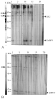
Fig. 5. Western blots of crude extracts and excreted–secreted products of Taenia solium cysticerci probed with sera from neurocysticercotic patients. Cysticerci were metabolically labelled with [35S]methionine, at 43 °C, overnight. Crude extracts (A) and excreted–secreted products (B), were reacted with neurocysticercotic patient sera (lanes 1–15), or with sera from patients with other neurological disorders (lanes 16–20). Lane 22 was reacted with a monoclonal antibody directed to HSP60 (LK2) and lane 21 with the second antibody alone. Lane X shows the autoradiogram of the blots.
 You have
Access
- 28
- Cited by