Introduction
Logopenic progressive aphasia (LPA) is a progressive neurodegenerative syndrome which affects the language network and is part of the primary progressive aphasias (Gorno-Tempini et al., Reference Gorno-Tempini, Hillis, Weintraub, Kertesz, Mendez, Cappa and Grossman2011). This syndrome is characterized by hesitant speech with major difficulties in word finding and sentence repetition, with relative preservation of motor speech, grammar, and single word comprehension (Gorno-Tempini et al., Reference Gorno-Tempini, Dronkers, Rankin, Ogar, Phengrasamy, Rosen and Miller2004, Reference Gorno-Tempini, Hillis, Weintraub, Kertesz, Mendez, Cappa and Grossman2011). Recent evidence indicates that LPA is predominantly, but not exclusively, associated with Alzheimer pathology (Mesulam et al., Reference Mesulam, Wicklund, Johnson, Rogalski, Leger, Rademaker and Bigio2008; Rabinovici et al., Reference Rabinovici, Jagust, Furst, Ogar, Racine, Mormino and Gorno-Tempini2008), leading some to claim that LPA may represent an atypical presentation of Alzheimer's disease (AD) (Leyton et al., Reference Leyton, Villemagne, Savage, Pike, Ballard, Piguet and Hodges2011; Rohrer, Rossor, & Warren, Reference Rohrer, Rossor and Warren2012).
Despite this shared pathology, the clinical presentation of LPA and AD are distinct early in the disease process. Briefly, LPA patients typically exhibit language difficulties, in contrast to AD patients whose main complaint relates to a decline in recent episodic memory, although language and episodic memory can be affected in both conditions (Dubois et al., Reference Dubois, Feldman, Jacova, Dekosky, Barberger-Gateau, Cummings and Scheltens2007; Gorno-Tempini et al., Reference Gorno-Tempini, Hillis, Weintraub, Kertesz, Mendez, Cappa and Grossman2011; Hodges, Salmon, & Butters, Reference Hodges, Salmon and Butters1991, Reference Hodges, Salmon and Butters1992; Weintraub, Wicklund, & Salmon, Reference Weintraub, Wicklund and Salmon2012). These clinical phenotypes are also associated with distinct patterns of brain atrophy on imaging. Patients with LPA show early atrophy in the left posterior perisylvian and temporoparietal regions, including the superior and middle temporal gyri (Gorno-Tempini et al., Reference Gorno-Tempini, Brambati, Ginex, Ogar, Dronkers, Marcone and Miller2008; Migliaccio et al., Reference Migliaccio, Agosta, Rascovsky, Karydas, Bonasera, Rabinovici and Gorno-Tempini2009; Rohrer, Ridgway, et al., Reference Rohrer, Ridgway, Crutch, Hailstone, Goll, Clarkson and Warren2010). By contrast, atrophy in early AD is typically found in and surrounding the medial temporal lobe region, including the hippocampus, entorhinal cortex, and amygdala bilaterally (Braak & Braak, Reference Braak and Braak1995; Desikan et al., Reference Desikan, Cabral, Hess, Dillon, Glastonbury and Weiner2009; Dubois et al., Reference Dubois, Feldman, Jacova, Dekosky, Barberger-Gateau, Cummings and Scheltens2007).
A cardinal feature in LPA is reduced verbal short-term memory, a finding consistently demonstrated across tasks, including digit and word span, and sentence repetition (Gorno-Tempini et al., Reference Gorno-Tempini, Brambati, Ginex, Ogar, Dronkers, Marcone and Miller2004, Reference Gorno-Tempini, Dronkers, Rankin, Ogar, Phengrasamy, Rosen and Miller2008; Rohrer, Ridgway, et al., Reference Rohrer, Ridgway, Crutch, Hailstone, Goll, Clarkson and Warren2010). This deficit appears to be due to a breakdown of the storage and rehearsal processes of the phonological system, which is supported by the left temporoparietal region (Baddeley, Reference Baddeley2003; Gorno-Tempini et al., Reference Gorno-Tempini, Brambati, Ginex, Ogar, Dronkers, Marcone and Miller2008; Rohrer, Ridgway, et al., Reference Rohrer, Ridgway, Crutch, Hailstone, Goll, Clarkson and Warren2010). In contrast, visuospatial short-term memory and ability, commonly associated with right parietal and frontal regions (Baddeley, Reference Baddeley2003; D'Esposito et al., Reference D'Esposito, Aguirre, Zarahn, Ballard, Shin and Lease1998; Smith & Jonides, Reference Smith and Jonides1997; Smith, Jonides, & Koeppe, Reference Smith, Jonides and Koeppe1996), is comparatively spared in the early stages of LPA (Gorno-Tempini et al., Reference Gorno-Tempini, Dronkers, Rankin, Ogar, Phengrasamy, Rosen and Miller2004; Migliaccio et al., Reference Migliaccio, Agosta, Rascovsky, Karydas, Bonasera, Rabinovici and Gorno-Tempini2009; Rohrer, Rossor, et al., Reference Rohrer, Rossor and Warren2012). Conversely, recent evidence suggests AD patients show more difficulty on visuospatial compared to verbal short-term memory tasks, particularly on more simple tasks, such as forward span, than on tasks with an executive or working memory component, such as backward span (Carlesimo et al., Reference Carlesimo, Mauri, Graceffa, Fadda, Loasses, Lorusso and Caltagirone1998; Huntley & Howard, Reference Huntley and Howard2010; Kertesz, Davidson, McCabe, Takagi, & Munoz, Reference Kertesz, Davidson, McCabe, Takagi and Munoz2003; Toepper, Beblo, Thomas, & Driessen, Reference Toepper, Beblo, Thomas and Driessen2008).
To our knowledge, no study has directly compared verbal and visuospatial short-term memory systems in LPA and AD patients with equivalent dementia severity. This study addresses this issue by administering two tests commonly used in clinical practice that measure short-term memory in the auditory and visuospatial modality: the WMS3 Digit and Spatial Span Forward and Backward tasks. We hypothesized that Digit Span Forward performance would be impaired in LPA compared to AD, but that the converse profile would be evident on the Spatial Span Forward task, with LPA outperforming AD. We further predicted that both patient groups would be equally impaired on the backward conditions of the span tasks, attributable to the executive components of the tasks.
Methods
Participants
Twenty-four dementia patients (LPA = 12; AD = 12) and 12 age- and education-matched healthy control volunteers were recruited from FRONTIER, a clinical dementia research group in Sydney, Australia. All patients were seen by a senior neurologist (J.R.H.) and underwent a comprehensive neuropsychological evaluation and structural MRI scan, and met current clinical diagnostic criteria for LPA and AD (Gorno-Tempini et al., Reference Gorno-Tempini, Hillis, Weintraub, Kertesz, Mendez, Cappa and Grossman2011; McKhann et al., Reference McKhann, Knopman, Chertkow, Hyman, Jack, Kawas and Phelps2011). Briefly, LPA patients presented with impaired single word retrieval, phonological errors in spontaneous speech and naming, and impaired repetition of sentences; in the context of preserved single word comprehension and object knowledge, and the absence of motor speech or frank agrammatism. MRI scans were reviewed by the senior neurologist for predominant left posterior perisylvian or parietal cortical atrophy. Eight of the 12 LPA patients (66%) underwent a Pittsburgh compound B (PiB) positron emission tomography scan, which binds to the amyloid protein (Klunk et al., Reference Klunk, Engler, Nordberg, Wang, Blomqvist, Holt and Langstrom2004). All eight patients (100%) showed a positive uptake to the PiB tracer, confirming the presence of Alzheimer pathology (Leyton et al., Reference Leyton, Piguet, Savage, Burrell and Hodges2012). Patients with AD showed episodic memory loss in the context of relatively preserved language function, and predominant bilateral atrophy of the medial temporal and parietal lobes on MRI. All clinical and neuropsychological assessments were completed within 3 months of the MRI scan. Diagnosis was established by consensus between the neurologist, neuropsychologist and the occupational therapist after reviewing the clinical, cognitive and imaging data. Control participants were selected from a healthy volunteer panel or were spouses/carers of patients. None of the participants had a history of substance abuse, major depression, schizophrenia, traumatic brain injury or other neurological conditions.
This study was approved by the South Eastern Sydney and Illawarra Area Health Service and the University of New South Wales Ethics committees. Consent to take part in the study was obtained from all participants.
General Cognition and Dementia Severity Assessment
All participants completed the Addenbrooke's Cognitive Examination (ACE-R) (Mioshi, Dawson, Mitchell, Arnold, & Hodges, Reference Mioshi, Dawson, Mitchell, Arnold and Hodges2006) as a general cognitive measure to examine the integrity of Attention, Memory (immediate and delayed episodic, and semantic), Fluency, Language, and Visuospatial cognitive domains. Participants also completed the following tests: (i) a Picture Naming task measuring verbal output and comprehension based on the Repeat and Point task (Hodges, Martinos, Woollams, Patterson, & Adlam, Reference Hodges, Martinos, Woollams, Patterson and Adlam2008); (ii) a single-Word Repetition task which uses the same items as the Picture Naming task; (iii) the Sentence Repetition task from the Multilingual Aphasia Examination (Benton, Hamsher, & Sivian, Reference Benton, Hamsher and Sivian1994); (iv) the Rey Complex Figure Test Copy and Recall (RCFT) (Meyers & Meyers, Reference Meyers and Meyers1995); (v) Doors Test A from the Doors and People memory battery (Baddeley, Emslie, & Nimmo-Smith, Reference Baddeley, Emslie and Nimmo-Smith1994); and (vi) the Trail Making Test (Trail B—Trail A in seconds) (Reitan, Reference Reitan1955). These tests were indices of language comprehension and expression, verbal short-term memory and syntax comprehension, delayed visuospatial memory, delayed visual recognition, and executive (inhibitory) processes, respectively.
Dementia severity was established with the Disability Assessment for Dementia (DAD) (Gelinas, Gauthier, McIntyre, & Gauthier, Reference Gelinas, Gauthier, McIntyre and Gauthier1999). The DAD is a carer-based questionnaire measuring functional independence for 40 activities of daily living, which are either basic (e.g., dressing, eating, hygiene) or instrumental activities of daily living (ADL) (e.g., meal preparation, managing finances, medication). Lower scores on the DAD denote greater impairment.
Assessment of Short-Term Memory
Integrity of verbal short-term memory was assessed using the Digit Span subtest of the Wechsler Memory Scale-III (WMS3) (Weschler, Reference Weschler1997). The Digit Span task was selected over comparable verbal span tasks as it circumvents language difficulties (Martin & Ayala, Reference Martin and Ayala2004), such as those observed in LPA. Visuospatial short-term memory was examined with the Spatial Span subtest of the WMS3 (Weschler, Reference Weschler1997).
Statistical Analyses
Before analyses, all variables were checked for normality of distribution using Kolmogorov-Smirnov tests. Overall group differences on the neuropsychological and experimental tests were investigated using analyses of variance (ANOVA) with Tukey post hoc tests. Within-group differences were measured using paired-sample t-tests corrected for multiple comparisons. Effect sizes partial eta-squared (ηp2) were calculated for all significant group comparisons on the experimental variables (Rosenthal, Reference Rosenthal1991). Finally, correlations were obtained to investigate the relationship between the experimental variables and neuropsychological test performance.
Results
Demographics
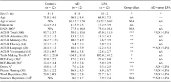
Participants were matched for age (p = .309) and years of education (p = .367) but not for sex, χ 2 (2) = 6.545, p = .038, with a higher female:male ratio observed in the AD compared to the other groups (Table 1). Overall group differences were present for general cognitive functioning on the ACE-R, F(2,33) = 53.505, p < .0001, with LPA and AD patients scoring significantly lower than Controls (all p values < .0001). In addition, the LPA group scored lower than the AD group on this measure (p = .048), reflecting the large language component of this task. Disease severity as measured by the DAD did not differ significantly between AD and LPA (t(19) = .27; p = .268).
Demographic and neuropsychological test scores in healthy controls, AD and LPA patients (means ± SD)

aNumber of missing values: Disability Assessment for Dementia (DAD): AD = 3; Trail Making Test: Controls = 1, AD = 3, LPA = 5; Rey Complex Figure: Controls = 7, AD = 4, LPA = 2; Doors A: Controls = 1, AD = 1, LPA = 2.
bNormative scores for Sentence Repetition taken from Schum and Sivan (Reference Schum and Sivan1997).
*p < .05; **p < .001; ***p < .0001.
AD = Alzheimer's disease; LPA = logopenic progressive aphasia; ACE = Addenbrooke's Cognitive Examination; RCF = Rey Complex Figure Test Copy and Recall (RCFT); N/A = not applicable.
Analysis of the ACE-R subscales revealed significant overall group differences across all cognitive domains (all p values < .001). Different profiles of impairment were evident contingent on patient group. LPA patients displayed significant impairments with respect to Controls in Attention, Memory, Fluency, and Language (all p values < .001), with no impairments evident on the Visuospatial subscale (p = .066). In contrast, AD patients showed significant impairments across all subscales of the ACE-R (Attention: p = .008; Memory: p < .001; Fluency: p < .0001; Language: p = .002; Visuospatial: p = .001). LPA patients performed significantly worse than the AD group on the language subscale of the ACE-R (p = .001), with no other differences evident between the patient groups (all p values > .05) (Table 1).
Profiles of Cognitive Impairment
Significant overall group differences were observed on all cognitive tasks (Table 1): Trail Making Test, F(2,24) =7.784, p = .002; Rey Complex Figure-Copy, F(2,20) = 4.172, p = .031; Rey Complex Figure-Recall, F(2,20) = 14.481, p < .0001; Doors Test, F(2,29) = 7.973, p = .002; Picture Naming, F(2,33) = 33.409, p < .0001; and Word Repetition F(2,33) = 11.159, p < .0001. In addition, while the Sentence Repetition task was not administered to Controls, both AD and LPA groups were impaired on this task, scoring 2 standard deviations below age-matched normative scores (mean score = 12.1 ± 1.2); (Schum & Sivan, Reference Schum and Sivan1997).
Post hoc tests revealed cognitive profiles characteristic of each dementia subtype. Compared to controls, the AD group was impaired on Picture Naming (p < .0001), Copy (p = .043) and Recall (p < .0001) of the Rey Figure, and Doors Test (p = .001), but not on single word Repetition (p = .854) or on the Trail Making Test (p = .206).
In contrast, the LPA group was impaired relative to controls on the Trail Making Test (p = .002), Picture Naming (p < .0001), single word Repetition task (p < .0001), and recall of the Rey Complex Figure (p = .002), but not the copy of the Rey Figure or the Doors Test (both p values > .20). Finally, the LPA group was significantly more impaired than the AD group on Picture Naming (p = .001) and Sentence Repetition (p < .0001) tasks while the AD group was more impaired than the LPA group on the Doors Tests (p = .041).
Short-Term Memory Measures—Overall Group Differences
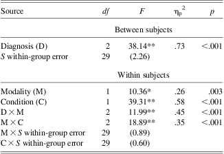
A mixed design repeated measures analysis of variance revealed significant main effects of diagnosis, task modality, and testing condition. In addition, significant interactions of task modality with both diagnosis and testing condition were also present (Table 2).
Multivariate analysis of variance for the Digit and Visuospatial spans in healthy controls, AD, and LPA patients
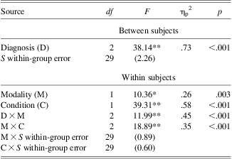
Note. Values enclosed in parentheses represent mean square errors. S = subjects.
AD = Alzheimer's disease; LPA = logopenic progressive aphasia.
*p < .005. **p < .001.
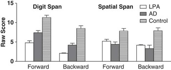
Paired-sample t tests were conducted to investigate these interactions further. Digit Span Forward was significantly higher than Spatial Span Forward in both Controls, t(10) = 4.944, p = .001, r = .36, and in AD, t(11) = 5.196, p < .001, r = .51. No differences between modality were found on the backward span tasks in these groups (both p values > .30). The LPA group, however, showed no difference on the forward condition between the Digit and Spatial Span tasks (t < 1, ns) but a significantly higher backward span on the Spatial Span task compared to the Digit Span task, t(11) = 3.317, p = .007, (Figure 1).
Performance on the Wechsler Memory Scale-III (WMS3) Digit Span and Spatial Span Forward and Backward tasks. Error bars represent standard errors of the mean.

In contrast, while all groups demonstrated higher Digit Forward than Digit Backward spans (Controls: t(10) = 5.871, p < .001, r = .54; AD: t(10) = 5.487, p < .001, r = .51; LPA: t(11) = 3.386, p = .006, r = .31), only AD also showed a significantly higher Spatial Forward than Spatial Backward Span, t(9) = 2.449, p = .037, r = .85.
Relationship Between Span Performance and Neuropsychological Tests
In the AD group, Sentence Repetition performance correlated with Digit Span Forward, rs = .84, p = .002, and Backward, rs = .87, p = .002. The visuospatial subscale of the ACE-R correlated with Spatial Span Forward, rs = .86, p < .0001, and Backward, rs = .90, p < .001. In addition, Spatial Span Backward correlated with the language subscale of the ACE, rs = .83, p = .003.
In the LPA group, Digit Span Forward correlated with Sentence Repetition, rs = .79, p = .006, and single word repetition, rs = .88, p < .001. No other significant correlations were evident in the LPA group (all p values > .05).
Discussion
This study investigated the verbal and visuospatial short-term memory systems in LPA and AD using the Digit and Spatial Span tasks. While LPA and AD patients showed significant impairments on both span tasks compared to controls, their profiles of performance differed across tasks. In AD patients, Digit Span Forward was significantly better than Spatial Span Forward, but the performance on backward span tasks did not differ. In contrast, LPA patients performed significantly better on Spatial Span Backward than Digit Span Backward, with no difference between the Spatial and Digit Span Forward tasks. To our knowledge, this investigation represents one of the first studies to directly compare the verbal and visuospatial short-term memory profiles of LPA and AD. These findings demonstrate that, although LPA and AD patients commonly share the same underlying pathological process, their profiles on short-term memory tasks vary considerably.
Digit Span
Both LPA and AD groups were impaired on Digit Span Forward with LPA being more affected than AD. The LPA group's impaired Digit Span Forward performance was consistent with our prediction and supports the position of a compromised phonological system in this group. Unfortunately, as Digit Span was the only test used to measure verbal span in LPA, we were not in a position to establish which of the components within the phonological system (store, rehearsal, and/or buffer processes) were compromised; an issue which will be investigated in a future study. Surprisingly, the AD group also showed reduced performance on the Digit Span tasks. While the AD group's performance was less severe than the LPA group, the findings suggest verbal span is compromised in typical AD. Reduced forward verbal span in AD has been previously reported but the extent of this deficit remains equivocal (Collette, Van der Linden, Bechet, & Salmon, Reference Collette, Van der Linden, Bechet and Salmon1999; Huntley & Howard, Reference Huntley and Howard2010; Peters et al., Reference Peters, Majerus, Olivier, van der Linden, Salmon and Collette2007). Digit Span Backward is a more difficult task than Digit Span Forward (Carlesimo et al., Reference Carlesimo, Mauri, Graceffa, Fadda, Loasses, Lorusso and Caltagirone1998; Wilde & Strauss, Reference Wilde and Strauss2002). In addition to storage and rehearsal processes, Digit Span Backward requires the manipulation of verbal information, which is modulated in part by executive processes supported by the prefrontal cortex (Hartley & Speer, Reference Hartley and Speer2000; Owen, Reference Owen2000). Not surprisingly, all groups showed a significantly lower performance on the Backward than the Forward component of this task. Furthermore, patient groups were significantly impaired on Digit Span Backward compared to controls, with LPA patients being more impaired compared to the AD patients. Whether reduced test performance on Digit Span Backward was due to (i) a primary disintegration of the temporary storage system, mediated by the temporoparietal region; (ii) a disturbance of frontal executive processes supporting manipulation of verbal information; or (iii) a combination of both, remains unresolved. These accounts warrant further investigation in LPA and AD as the neurocognitive mechanisms underlying impaired Digit Span Backward performance may differ between, or even within, patient groups.
Spatial Span
Based on existing literature, we predicted that visuospatial short-term memory would be relatively spared in LPA compared to AD. Although LPA patients scored higher than AD patients on both Spatial Span tasks, these differences were not statistically significant. A similar pattern was observed on other tasks containing a visuospatial component (Rey Complex figure, ACE-R Visuospatial). It is possible that visuospatial ability is compromised later in the disease process of LPA. Although few studies have investigated the cognitive profile of later stage LPA (Goll et al., Reference Goll, Kim, Hailstone, Lehmann, Buckley, Crutch and Warren2011; Rohrer, Rossor, et al., Reference Rohrer, Rossor and Warren2012), the report that severe (mean MMSE: 9.4) LPA patients show similar deficits on the Spatial and Digit Span Forward tasks would suggest visuospatial ability is compromised with disease progression (Goll et al., Reference Goll, Kim, Hailstone, Lehmann, Buckley, Crutch and Warren2011). Thus, the dissociation between (impaired) verbal and (preserved) visuospatial short-term memory may be prominent only in the early stages of the disease.
Because of the integration of information from multiple modalities (visuospatial, temporal, motor), Spatial Span tasks are considered more difficult than Digit Span tasks (Parmentier, Andres, Elford, & Jones, Reference Parmentier, Andres, Elford and Jones2006), with the suggestion that both Forward and Backward conditions require not only visuospatial short-term memory but also executive processes (Bor, Duncan, Lee, Parr, & Owen, Reference Bor, Duncan, Lee, Parr and Owen2006; Carlesimo, Fadda, Lorusso, & Caltagirone, Reference Carlesimo, Fadda, Lorusso and Caltagirone1994). Of interest, LPA patients performed significantly better on Spatial Span Backward compared to Digit Span Backward yet displayed similar performances on the Forward tasks. This performance contrasted with the AD and Control groups, who both showed similar performances across the Backward components of the two tasks and a higher performance on the Digit Span Forward than on the Spatial Span Forward tasks. This finding supports the position for a phonological, rather than a central, short-term memory deficit in LPA (Gorno-Tempini et al., Reference Gorno-Tempini, Hillis, Weintraub, Kertesz, Mendez, Cappa and Grossman2011). The reported interactions with task modality further strengthens this position: both Controls and LPA patients showed similar performances on the Forward and Backward conditions of the Spatial Span task. In contrast, AD patients performed better on the Forward than the Backward components of the Span tasks, irrespective of modality. Taken together, the findings suggest the AD patients are vulnerable to the “executive” requirement of the task, regardless of modality, whereas performance in LPA patients, as well as Controls, is contingent upon task modality.
The independence of verbal and visual short-term memory systems is supported by neuroimaging and lesion studies. In healthy adults, verbal short-term memory tasks, such as Digit Span, tend to recruit the left temporoparietal junction and prefrontal regions (Henson, Burgess, & Frith, Reference Henson, Burgess and Frith2000; Ravizza, Hazeltine, Ruiz, & Zhu, Reference Ravizza, Hazeltine, Ruiz and Zhu2011; Smith, Jonides, Marshuetz, & Koeppe, Reference Smith, Jonides, Marshuetz and Koeppe1998), with similar regions in the right hemisphere showing increased activation during visuospatial short-term memory tasks, such as spatial span tasks (Owen et al., Reference Owen, Herrod, Menon, Clark, Downey, Carpenter and Pickard1999; Smith & Jonides, Reference Smith and Jonides1997; Smith et al., Reference Smith, Jonides and Koeppe1996). Furthermore, impaired verbal or visuospatial short-term memory follows left or right stroke in these regions respectively (Laures-Gore, Marshall, & Verner, Reference Laures-Gore, Marshall and Verner2011; van Asselen et al., Reference van Asselen, Kessels, Neggers, Kappelle, Frijns and Postma2006). Imaging studies have shown that these brain regions are affected in LPA and in AD (Leyton et al., Reference Leyton, Piguet, Savage, Burrell and Hodges2012; Migliaccio et al., Reference Migliaccio, Agosta, Rascovsky, Karydas, Bonasera, Rabinovici and Gorno-Tempini2009; Rohrer, Ridgway, et al., Reference Rohrer, Ridgway, Crutch, Hailstone, Goll, Clarkson and Warren2010; Salmon et al., Reference Salmon, Sadzot, Maquet, Degueldre, Lemaire, Rigo and Franck1994). This evidence suggests that the short-term memory verbal/visuospatial dissociation found in LPA and AD reflect lateralized patterns of brain atrophy, although this will need to receive neuroimaging confirmation.
In conclusion, the current study demonstrates the specificity of the verbal short-term memory disturbance with a comparatively preserved visuospatial short-term memory in LPA. This deficit, which arises from a breakdown in phonological processing, may be related to specific atrophy of the left superior temporal region typically shown in LPA (Gorno-Tempini et al., Reference Gorno-Tempini, Brambati, Ginex, Ogar, Dronkers, Marcone and Miller2004, Reference Gorno-Tempini, Dronkers, Rankin, Ogar, Phengrasamy, Rosen and Miller2008; Leyton et al., Reference Leyton, Piguet, Savage, Burrell and Hodges2012). From a clinical perspective, our results demonstrate that short-term memory tasks have an important role to play in the cognitive assessment of dementia syndromes and that the combination of verbal and visuospatial span tasks prove a valuable combination when examining short-term memory integrity in patients with suspected logopenic progressive aphasia.
Acknowledgments
We thank all the participants for their time and contribution to this study. We also thank Sharon Savage, Felicity Leslie, and Naomi Daveson for their help in test administration and scoring. This research was supported in part by the Australian Research Council (ARC) Centre of Excellence in Cognition and its Disorders (CE110001021); M.I. is supported by an ARC Discovery Project grant (DP10933279); JRH is supported by an ARC Federation Fellowship (FF0776229); O.P. is supported by a National Health and Medical Research Council of Australia Career Development Fellowship (APP1022684). The authors certify that no actual or potential conflict of interest in relation to this study exists.