Primary cardiac tumours of the heart are rare, with their incidence cited at from 0.0017% to 0.28% in autopsy series. In a multicentric review of such tumours encountered prenatally, the overall prevalence in pregnancies referred for fetal echocardiography was 0.14%, with almost all being rhabdomyomas, and the remainder being fibromas or haemangiomas.Reference Holley, Martin, Huhta and Kleinman1 Less than one-twentieth of all primary cardiac tumours are haemangiomas, most being diagnosed in the neonatal period, with very few having been reported in children. We have now successfully removed right atrial haemangioma in three neonates, who were diagnosed prenatally with varied presentations.
Case 1
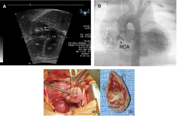
Routine prenatal ultrasound at 32nd gestation week, in a 19-year-old woman during her first pregnancy showed a large mass involving the right atrial free wall. The mass was irregular and lobulated, with evidence of calcification, and occupied more than half of the right atrial cavity (Fig. 1A). Spontaneous vaginal delivery took place at 39 gestation weeks. The baby boy weighed 2870 grams, and Apgar scores were 8 at 1 minute, and 9 at 5 minutes. Cardiac catheterization was performed on the third day of life, revealing the mass to communicate with the right coronary artery (Fig. 1B). Surgical removal through a median sternotomy took place on the 5th day of life. The wall of the right atrial appendage was somewhat discolored (Fig. 1Ca). Palpation revealed a gelatinous mass that could be elevated almost completely into the atrial appendage. We noted 2 branches of the right coronary artery feeding the tumour. Placement of a vascular clamp from outside the heart permitted encirclement of the mass without cardiopulmonary bypass. The mass was excised along with a portion of the right atrial wall (Fig. 1Cb), and the atriotomy was oversewn. The histopathologic examination confirmed the presence of a capillary haemangioma with calcification. The patient was discharged on the 6th postoperative day without any complication. Follow-up echocardiography at 2 years of age showed no residual tumour.
A mass with mixed echogenicity is seen (A) arising from the right atrial free wall and engulfing more than half of right atrium. Preoperative angiography (B) showed that the tumour was supplied by a branch of the right coronary artery. White arrow head: tumour stain. In panel C, we show the haemangioma as seen in the right atrium (a), and subsequent to its resection (b).

Case 2
A right atrial tumour was diagnosed at 33 weeks of gestation, the mother being 28 years old. There was severe hydrops and atrial flutter, which was treated with digoxin and flecainide, providing partial control of the ventricular rate in range from 140 to 180 beats per minute. Elective caesarean section was then performed at 34 weeks because of worsening fetal cardiac failure. The baby, a boy, weighed 2 kilograms, and had an initial Apgar score of 2. He was intubated for respiratory acidosis. After stabilization with high frequency ventilation, the patient underwent surgery on the 3rd day of life. Through a median sternotomy, inspection showed the right atrium to be almost completely replaced internally with tumour, which was visible through the right atrial wall as a shiny pearly white mass. Cardiopulmonary bypass was established using ascending aortic and right atrial cannulation, sufficient room being obtained posteriorly within the right atrium to achieve bypass. Opening the right atrium showed the tumour to be adherent to the antero-superior leaflet of the tricuspid valve and the right atrial wall. We were able to remove the greater part of the tumour, albeit leaving a portion on the tricuspid valvar annulus, and also a small portion beneath the right coronary artery. Histopathologic examination confirmed the diagnosis of a capillary haemangioma. The postoperative course was complicated by respiratory failure and a brief episode of atrial flutter, but the patient was extubated on the 6th postoperative day, and discharged 3 weeks after the operation. Follow-up echocardiography 1 year after the surgery revealed no residual tumour in the right atrium, the patient doing well in sinus rhythm.
Case 3
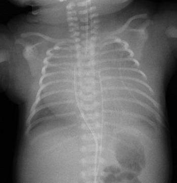
Ultrasound during the 20th week of gestation in a 30-year-old woman during her second pregnancy revealed a right atrial mass and significant pericardial effusion. Fetal pericardiocentesis was performed, and the aspirated fluid was serosanguineous. After pericardiocentesis, the residual pericardial effusion was small, and there was good expansion of the lungs. At 35 weeks of gestation, the fetus was noted to have exaggerated reversal of flow with atrial systole in the inferior caval vein, with an increase in the size of the tumour. The mother was transferred to our hospital, and delivered by caesarean section. The baby girl weighed 2840 grams, with Apgar scores of 4 at 1 minute, and 9 at 5 minutes. The patient was initially apnoeic and bradycardic, requiring intubation. The chest X-ray showed massive cardiomegaly, with the tumour obstructing the right lung (Fig. 2). She underwent surgery on the 5th day of life. An approach through a midline sternotomy showed the tumour to be completely obliterating the anterior part of the right atrium, and extending into the right chest. The right coronary artery was contiguous with the tumour as it coursed in the atrioventricular groove. Cardiopulmonary bypass was established using aortic cannulation, the single venous cannula being inserted at the junction between the inferior caval vein and the right atrium. The tumour was subtotally extirpated, leaving a small rim of tumour adjacent to the right coronary artery. A patent oval foramen was closed with a single mattressed suture. A piece of bovine pericardium was used to reconstruct the atrial free wall. Histopathological examination confirmed the diagnosis of capillary haemangioma. The postoperative course was complicated by respiratory failure due to ateletctasis of the right lung. The patient is now doing well, with follow-up echocardiography after 18 months showing no residual tumour.
The chest X-ray shows a round mass occupying the right half of the thorax.

Discussion
Cardiac tumours, though rare, are now being diagnosed with increasing frequency and great accuracy by prenatal ultrasound.Reference Zhou, Fan, Peng, Zhang, Fu and Wang2 Of the cardiac tumours found in the fetus, three-fifths are rhabdomyomas, with one-fifth being teratomas, and one-eighth fibromas.Reference Laga, Gewilling, Van Schoubroeck and Daenen3 Cardiac haemangiomas are amongst the rarest types, and are usually diagnosed in the neonatal period.Reference Tongsong, Sirichotiyakul, Sittiwangkul and Wanapirak4 Cardiac tumours are often benign, and can regress spontaneously.Reference Holley, Martin, Huhta and Kleinman1 Malignancy, associated malformations, and aneuploidy are infrequently observed. The combination of sonographic features and their location allows reliable prediction of histological type in the majority of tumours diagnosed prenatally.Reference Zhou, Fan, Peng, Zhang, Fu and Wang2 Differential diagnosis is important, as this affects prognosis and subsequent management.
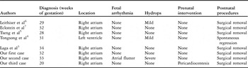
The most common site of origin of cardiac haemangiomas is the base of the heart adjacent to the right atrium.Reference Tseng, Chou, Lee and Ho5 Rhabdomyomas are usually multiple, while fibromas are always single, but both are usually located in ventricular myocardium, and have uniform echogenicity. Teratomas are usually extracardiac, being attached at the base of the arterial trunks, with heterogenous echogenicity. The diagnostic sonographic features of haemangiomas include presence of a solitary sessile noncapsulated mass with mixed echogenicity, at times associated with pericardial effusion, arrhythmia, calcification and hydrops. Cardiac haemangiomas are secretory in nature, in contrast to rhabdomyomas and fibromas, which explains their frequent association with pericardial effusions. Their vascular nature in most cases is not demonstrable with colour flow mapping, as the abnormal vessels are typically microscopic, albeit that sometimes, the main feeding artery is readily demonstrable.Reference Tongsong, Sirichotiyakul, Sittiwangkul and Wanapirak4 They can be either capillary or cavernous in nature. Most grow as gestation advances, as in 2 of our cases. Hence, they require serial assessment after initial diagnosis. Doppler interrogation is important to assess their effects on central venous pressure. In Table 1, we have summarized those cases previously diagnosed prenatally, along with postnatal follow up.Reference Laga, Gewilling, Van Schoubroeck and Daenen3–Reference Eckstein, Heinemann, Mielke, Greschniok, Bader and Ziemer7
Summary of cardiac haemangiomas reported in the literature with fetal diagnosis and postnatal follow-up.

The diagnosis is often made only after serious complications have become evident, including congestive cardiac failure, pericardial effusions, or cardiac arrhythmias, so early fetal echocardiographic diagnosis is advantageous.Reference Laga, Gewilling, Van Schoubroeck and Daenen3, Reference Eckstein, Heinemann, Mielke, Greschniok, Bader and Ziemer7 When hydrops is present, antenatal management of the fetus with a cardiac tumour becomes challenging. Such fetuses have a high rate of mortality, with overall survival reported for no more than one-fifth.Reference Hou, Chao, Wang, Chao and Hsueh8, Reference Isaacs9 Our second patient had hydrops secondary to atrial flutter, but this was successfully controlled with anti-arrhythmic medications, followed by elective delivery.
Surgical intervention is only required for children with relevant clinical symptoms.Reference Stiller, Hetzer and Meyer10 The relationship of the right atrial haemangiomas with the right coronary artery and the sinus node has to be considered prior to surgical removal. In 2 of our cases, the close proximity of the tumour to the right coronary artery precluded its complete resection, albeit with a significant haemodynamic improvement. Follow-up echocardiography revealed regression of the tumour in all the cases. So, prognosis is usually good even after partial removal. In tumours proving to be unresectable, the options of radiotherapy and use of steroids have been reported.
Acknowledgement
We thank Drs Jan Quaegebeur, Charles Kleinman, and Jonathan Chen for their valuable contributions and guidance.