Anomalous origin of one branch pulmonary artery from the aorta is a rare anomaly that accounts for <0.1% of all CHD.Reference Kutsche and Van Mierop 1 Physiologically, one lung is exposed to unrestricted systemic blood flow from the anomalous aorto-pulmonary connection, whereas the contralateral lung receives the entire right ventricular output. Thus, many patients develop pulmonary vascular disease and subsequent pulmonary arterial hypertension. We describe a surprising case of an infant with this CHD who presented with exceptionally early and severe pulmonary arterial hypertension caused instead by a more unusual consequence of this anatomy.
Case report
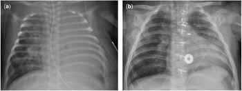
The patient was a previously healthy full-term infant who presented with symptoms of feeding intolerance, tachycardia, and respiratory distress at 1 month of age. Her physical examination revealed significant respiratory distress with diminished left lung sounds and hepatomegaly with the liver edge 4 cm below the costal margin. Her cardiac examination was notable for an increased praecordial impulse, a normal S1 and loud S2, and a 2/6 continuous murmur along the right sternal border. Her echocardiogram demonstrated moderate tricuspid regurgitation, a dilated right ventricle with an estimated systolic pressure of 60 mmHg, and an additional finding of an anomalous origin of the right pulmonary artery from the aorta (Fig 1). The left pulmonary artery, arising from the right ventricle, was severely dilated. By chest radiograph, the patient’s left lung field appeared completely opacified (Fig 2a), and a subsequent chest CT scan revealed extensive left-sided atelectasis. Bronchoscopy, performed for further evaluation of the left lung, demonstrated severe left-sided bronchomalacia with pulsatile compression anteriorly from the left pulmonary artery.
Three-dimensional reconstruction of chest CT angiogram, with a posterior view demonstrating anomalous origin of the right pulmonary artery from the ascending aorta and dilated left pulmonary artery arising from the main pulmonary artery. LPA=left pulmonary artery; MPA=main pulmonary artery; RPA=right pulmonary artery.

Serial chest radiographs of the patient performed at ( a ) the time of admission to our hospital demonstrating severe atelectasis of the left lung and ( b ) 2 months after surgical repair demonstrating complete resolution of left lung atelectasis.

Clinically, the patient’s respiratory status quickly deteriorated, requiring full ventilatory support with 20 ppm of inhaled nitric oxide. She ultimately underwent surgical re-implantation of the right pulmonary artery to the main pulmonary artery. Her left pulmonary artery, which was found to be ~2½ times the size of the ascending aorta, was translocated anterior to the aorta – LeCompte Manoeuver – to relieve compression on the left bronchus. The surgery was performed on cardiopulmonary bypass with the heart beating. She recovered uneventfully and her atelectasis resolved without additional therapies (Fig 2b). A follow-up cardiac catheterisation performed 3 months later demonstrated significant improvement of her pulmonary arterial hypertension, with a near-normal right ventricular systolic pressure of 33 mmHg.
Discussion
Anomalous origin of one branch pulmonary artery from the aorta, occasionally referred to as hemitruncus arteriosus, is a rare congenital defect first described by Oscar Fraentzel in 1868.Reference Fraentzel 2 Owing to the physiological consequences of continuous augmented blood flow to the lungs, the natural history of this defect includes development of pulmonary vascular disease leading to pulmonary arterial hypertension. Clinically, these patients initially present with progressive respiratory distress and congestive heart failure as the pulmonary vascular resistance drops during the neonatal period. Pulmonary vascular disease does not typically develop until after 6 months of age in patients with this defect, and is progressive as patients grow older.Reference Keane, Maltz, Bernhard, Corwin and Nadas 3 – Reference Nathan, Rimmer and Piercey 5
Our patient’s presentation of severe pulmonary arterial hypertension and right heart failure at such a young age was atypical for this cardiac defect. Another unusual finding was her persistent and severe left-sided atelectasis, which led to further investigation before surgery. Bronchoscopic evaluation ultimately determined that she had left-sided bronchomalacia related to chronic compression by her left pulmonary artery, which had become progressively dilated from receiving all of the right ventricular cardiac output.
Tracheo-bronchial compression is a well-described complication of infants with certain forms of CHD. It is most often encountered in the setting of vascular rings and slings, as well as defects with large left-to-right shunts that may result in dilated pulmonary arteries or cardiomegaly.Reference Robotin, Bruniaux and Serraf 6 , Reference Kussman, Geva and McGowan 7 Prolonged extrinsic compression may result in tracheomalacia and/or bronchomalacia, and such lower airway obstruction may in turn lead to pulmonary arterial hypertension.Reference Kussman, Geva and McGowan 7 , Reference Park, Park and Kim 8 Symptoms typically improve after relief of external compression, but may not resolve completely during the immediate perioperative period. Careful anaesthetic considerations and postoperative management of respiratory status are essential in these patients.
In the past few decades, there have only been rare reports of anomalous origin of one pulmonary artery from the aorta associated with tracheobronchial compression.Reference Ben-Shachar, Beder, Liebman and Van Heeckeren 9 , Reference Rosa and Wade 10 The large obligate left-to-right – aorta-to-pulmonary artery – shunt that occurs in this cardiac lesion as well as a frequently dilated contralateral pulmonary artery together create a perfect setting for tracheobronchial compression to occur. The apparent rarity of tracheobronchial compression reported with anomalous origin of one pulmonary artery from the aorta calls into question whether this particular association may be under-recognised. Perhaps, because pulmonary arterial hypertension is an expected consequence of this CHD, other potential contributors of pulmonary arterial hypertension, such as tracheobronchial compression, are infrequently considered.
In summary, pulmonary arterial hypertension may develop in patients with an anomalous origin of one pulmonary artery from the aorta due to the progression of pulmonary vascular disease; however, in patients who demonstrate unusually early- and severe-onset pulmonary arterial hypertension, evaluation of other potential causes is essential. Associated co-morbidities such as tracheobronchial compression are important to identify as these may critically impact surgical approach, prognostication, and postoperative management.
Acknowledgement
None.
Financial Support
This research received no specific grant from any funding agency, commercial, or not-for-profit sectors.
Conflicts of Interest
None.