Introduction
Surgical management is the mainstay of treatment for tumours in the parapharyngeal space. In a cumulative series, 95 per cent of patients underwent surgery.Reference Riffat, Dwivedi, Palme, Fish and Jani1 Access to the parapharyngeal space, because of its deep location and adjacent vital structures, remains a challenging issue in the head and neck region. Various surgical routes have been used to resect parapharyngeal space tumours, including the traditional surgical approaches (transcervical, transparotid, mandibulotomy and infratemporal fossa approaches) and the endoscope-assisted transoral, transnasal or transvestibular corridors.Reference Paderno, Piazza and Nicolai2–Reference Sun, Yan, Truong, Borghei-Razavi, Snyderman and Fernandez-Miranda8 Each approach has its own advantages and disadvantages. The ideal surgical approach can not only remove tumours completely without injury to vital structures but is also acceptable cosmetically and functionally.
The classical transoral approach is an ideal surgical approach in highly selected cases, even in some cases with large parapharyngeal space tumours.Reference Hussain, Ah-See and Shakeel9 However, most surgeons do not advocate routine use of this approach. In 2014, Mong-Loon et al. conducted a systematic review of 1293 cases reported over 25 years and found that the transoral approach was used in only 3 per cent of the surgical cases.Reference Kuet, Kasbekar, Masterson and Jani10 It has traditionally been limited to only small benign neoplasms (less than 3 cm) that occupy the anterior parapharyngeal space.Reference Myatt and Remedios11 The principal weakness of classical transoral approach is poor illumination, which is possibly associated with difficult manoeuvrability, capsule disruption, incomplete removal, uncontrollable blood loss and vital nerve injuries.
Endoscopic surgery, which has been adopted in other areas recently,Reference Castelnuovo, Dallan, Battaglia and Bignami12 has made wider neoplasm resections possible with more conservative approaches. Compared with the classical transoral approach to the parapharyngeal space, the endoscopic transoral approach, with its adequate illumination and magnified view, overcomes the limit of ‘blind surgery’. In this study, we evaluated the indications, the limits and the technical nuances of the endoscopic transoral approach based on our clinical experience in the management of parapharyngeal space tumours.
Materials and methods
A retrospective review of 13 patients with parapharyngeal space neoplasms from May 2017 to November 2020 was carried out. All the patients underwent surgery via the endoscopic transoral approach at the Eye and ENT Hospital, Fudan University, Shanghai. Demographic and clinical characteristics including gender, age, symptoms, radiographical findings, surgical approaches, post-operative pathological findings, complications and follow-up results were collected.
The features of the lesion and its relationship with the adjacent vital structures were evaluated by computed tomography (CT), magnetic resonance imaging (MRI), or both CT and MRI pre-operatively. The decision regarding surgical approach was made on the basis of the characteristics of the tumour (including biological behaviour, position, size, vascularity and the neighbouring anatomical structures) and the patient's preferences. All the patients were fully informed about the surgical procedures and risks. In addition, they were informed and counselled about the possible need of a conversion to external approaches. The study was approved by the institutional review board of Fudan University.
Surgical procedures
All the surgical procedures were performed with the patient in the supine position under general anaesthesia. The patient was intubated by the orotracheal route. At the beginning of the operation, a Davis mouth gag was inserted to retract the tongue and expose the lateral wall of oropharynx. A tonsillectomy was not routinely required and was not performed in this series.
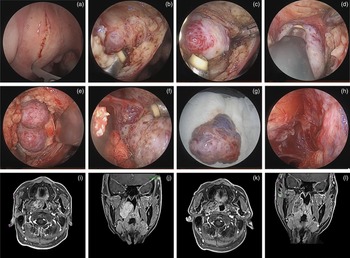
According to the location of the tumour, a vertical mucosal and submucosal incision was routinely made over the bulge (the tumour) on the soft palate with a knife blade (Figure 1a). The fascia and superior constrictor muscle were cut and separated (Figure 1b), and the parapharyngeal tumour was exposed (Figure 1c). In order to reduce bleeding and detach the tumour easily, dissection is best performed along the capsule. The anterior aspect of the mass was firstly dissected free from the surrounding tissues (Figure 1c). The superior and inferior margins of the tumour were gradually separated under endoscopy (Figure 1d). The tumour's posterior margin was usually difficult to expose. In order to deal with this, gauze was packed carefully around the tumour to detach and isolate the tumour from the nearby tissues and to prevent possible haemorrhage (Figure 1e). Then the tumour was pushed into the oral cavity, and the surrounding tissues of the tumour were pushed to the opposite side. Lastly, the posterior margin of the tumour was gently exposed and separated from the surrounding vital structures with the help of the endoscope (Figure 1f). Eventually, the tumour could be mobilised and removed transorally (Figure 1g–k).
(a–f) Images of the surgical procedure using the endoscopic transoral approach to the parapharyngeal space (example of case 1: en-bloc excision of a haemangioma); (g) endoscopic image showing completely resected haemangioma; (h) endoscopic image of the operative cavity after tumour removal; pre-operative enhanced magnetic resonance imaging (MRI) scans in the (i) axial and (j) coronal planes; and post-operative enhanced MRI scans in the (k) axial and (l) coronal planes.

After the excision of the mass, a haemostasis was achieved carefully by coagulation or packing oxidised cellulose sheets. The surgical field was thoroughly irrigated with warm saline to identify that there was no bleeding site and to wash out any possible residual tumour cells. At the end of surgery, a rubber drainage strip or a thin negative-pressure drainage tube was placed in the surgical cavity to drain effusion if necessary. The incision was sutured layer by layer with 3-0 absorbable stitches. Lastly, a nasogastric feeding tube was inserted according to the expected post-operative swallowing difficulties. Antibiotics were used routinely for about one week to prevent infection. Steroids were administrated as needed for 2–3 days to alleviate local oedema. Except for the patient who underwent biopsy, all patients underwent post-surgical surveillance with an electrocardiogram (ECG) monitor in the intensive care unit for 24–48 hours. Urgent tracheostomy equipment was prepared and left beside the bed for patients at a high risk of airway failure.
Enhanced MRI scans were performed on patients with radical resection on the first post-operative day to evaluate the extent of resection. The drainage strip was usually removed on the second or third day, and the drainage tube was removed when the daily drainage was less than 10 ml. The nasogastric tube could be removed in the out-patient clinic according to the swallowing rehabilitation (usually on days 5–14 post-operatively).
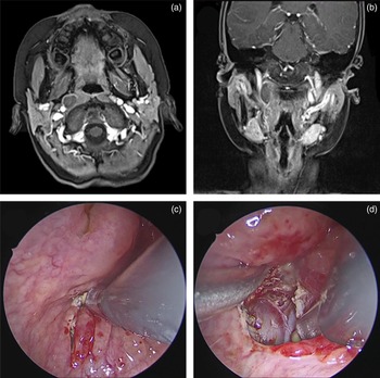
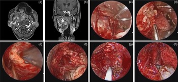
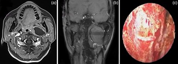
If there was no obvious mass in the oropharyngeal cavity, the incision would not be made on the soft palate. It could be made on the lateral pharyngeal wall to expose the tumour easily and to make a short corridor according to the radiological findings (Figure 2). In order to avoid injury to surrounding vital structures and to avoid the risk of an uncontrolled spillage, an internal debulking was performed in large or fragile lesions (Figure 3a–d). The further dissection along the capsule would then be easier and still under visual control (Figure 3e and f). Sometimes, the adjacent vital structures were recognised and checked again. In the images shown in Figure 3 (g and h), the internal carotid artery and the styloid process were recognised. In the dissection procedure, small vessels were cauterised by Coblator™ or bipolar coagulation forceps, whereas vital neurovascular structures were preserved cautiously (Figure 4). If the superior boundary of the mass was difficult to expose by the endoscopic transoral approach, the endoscopic transnasal transmaxillary approach to the upper parapharyngeal space could be used as well to facilitate exposure.Reference Liu, Wang, Zhao, Zhao, Song, Sun and Yu13
Pre-operative enhanced magnetic resonance imaging scans in (a) axial and (b) coronal planes showing that the tumour was near the lateral pharyngeal wall; (c and d) endoscopic images showing the incision was made on the lateral pharyngeal wall to expose the tumour easily and to make a short corridor.

Pre-operative enhanced magnetic resonance imaging scans in (a) axial and (b) coronal planes showing a huge pleomorphic adenoma (case 5). (c–f) Endoscopic images showing an internal debulking that was performed to avoid injury to surrounding vital structures and to avoid the risk of an unrecognised spillage, with (c) showing exposure of the tumour, (d) showing internal debulking after excision of the tumour capsule, (e) showing complete removal of the tumour and (f) showing haemostasis after tumour removal. (g) Endoscopic image showing the internal carotid artery, which was recognised by the ultrasonic Doppler system after tumour removal. (h) Endoscopic image showing the styloid process that was recognised in the operative cavity.

Pre-operative enhanced magnetic resonance imaging scans in (a) axial and (b) coronal planes showing a huge schwannoma (case 13), which displaced the internal carotid artery medially and (c) endoscopic image showing preservation of the internal carotid artery after it was cautiously dissected from the tumour.

Results
Thirteen consecutive patients (7 males and 6 females) were enrolled in this study with a median age of 50 years (range, 10 to 69 years). The demographic data and the patients’ clinical profiles are shown in Table 1. The complaints reported by the patients were: discomfort in the throat (3 of 13), dysphagia (2 of 13), headache (2 of 13), snoring (2 of 13), sensation of ear fullness (2 of 13), nasal obstruction (1 of 13), hypoglossal nerve palsy (1 of 13) and hoarseness (1 of 13). Asymptomatic neoplasm found by physical examination was found in two patients. The most common clinical findings were oropharyngeal mass (11 of 13) and nasopharyngeal mass (4 of 13), which pushed the pharyngeal structures medially (11 of 13). In two cases, there was no obvious mass in the oropharyngeal or nasopharyngeal cavity.
Clinical characteristics of 13 cases of parapharyngeal space tumours

M = male; A = alive; NED = no evidence of disease; F = female; LR = local recurrence
All the patients underwent surgery for complete oncological resection except one patient who received treatment for diagnostic purposes. The median maximum diameter of the excised tumours was 5.0 cm (range, 2.1–6.8 cm). The mass was located on the right side in six cases and on the left side in seven cases. Nine patients underwent surgery via a transoral approach, and four patients underwent a combined endoscopic transoral and endoscopic transnasal transmaxillary approach. Three tumours were removed en bloc in this cohort. An intra-operative internal debulking of the tumour was performed in nine cases. Nine of the tumours were benign and four were malignant. In this series, salivary gland tumours were the most common primary lesion (6 of 13), followed by neurogenic lesions (3 of 13).
No major complications were reported intra-operatively, with excellent control of the vital structures of the parapharyngeal space. The wound surface was directly sutured in 13 cases and drained in 9 cases. A nasogastric feeding tube was placed in seven patients. All patients were extubated after surgery, and no patients needed temporary tracheostomy. The most common post-operative complication (5 of 13) in this series was dehiscence of the suture, which spontaneously healed in all patients. Three patients reported mild oral tenderness that resolved after conservative treatment. One patient had a haematoma in the surgical cavity requiring evacuation under general anaesthesia.
No conversion to an external approach was required. Enhanced MRI on post-operative day 1 confirmed total tumour resection in all cases with complete oncological resection. Nine patients with benign tumours were followed for 17 to 59 months, with a mean follow-up time of 23 months. Physical examination, CT, and MRI showed no recurrent lesions at the last follow up. In the malignant tumour group, one patient with poorly differentiated squamous cell carcinoma underwent chemoradiotherapy in the oncology department. Two patients were alive and well. One patient with chondrosarcoma was alive with local recurrence at the follow-up visit after 24 months.
Discussion
In clinical practice, accurate diagnosis of parapharyngeal space tumours is difficult before surgery because of their deep location. The complaints and clinical characteristics, as reported by Prasad et al., are usually diverse and non-specific.Reference Prasad, Piccirillo, Chovanec, La Melia, De Donato and Sanna3 In our study, each patient's main complaints differed from each other. As some patients may be asymptomatic for a long time, tumours in the parapharyngeal space can be occasionally diagnosed during routine physical check-up or after imaging examination.Reference Bradley, Bradley and Olsen14 In our series, 2 tumours (case 5 for 40 months, case 8 for 3 months) grew without showing symptoms and were found during routine examination. Besides the clinical symptoms and signs, the pre-operative diagnosis relies mainly on radiological findings. Computed tomography and MRI scans not only allow demarcation of the position and border of the tumour but also provide important information related to the type and origin of parapharyngeal space tumours and the adjacent neurovascular structures. Computed tomography can clearly depict the bone destruction and the relationship between the tumour and bone structures (the mandible, skull base, pterygoid process). However, MRI is more sensitive and specific for differentiating benign and malignant disease than CT.Reference Iseri, Ozturk, Kara, Ucar, Aydin and Keskin15 In addition, magnetic resonance angiography is a useful tool, which can provide sufficient imaging of tumour vascularisation and carotid profiles.Reference Carrau, Myers and Johnson16 Radiological findings are crucial for pursuing the appropriate surgical approach.
The traditional transoral approach to the parapharyngeal space is the most controversial approach because of many disadvantages. Its use has always been limited. However, the surgical scenario of the transoral approach has substantially changed with the assistance of endoscopy and surgical robotics.Reference Dallan, Fiacchini, Turri-Zanoni, Seccia, Battaglia and Casani17–Reference Chen, Chen, Yu, Zhou, Bao and Shang19 High medical costs, being time-consuming, a lack of tactile sensation and the extensive training for hand-eye co-ordination preclude the widespread use of surgical robotics.Reference Iavazzo, Papadopoulou and Gkegkes20 Compared with the limited availability of robotic surgery, the endoscopic transoral approach, with its widespread familiarity, has become a more promising route for the general population.
With technical innovations and an in-depth understanding of the structures in parapharyngeal space, we have tried to use the endoscopic transoral approach to resect more parapharyngeal space tumours in recent years. We have found that the indications were much wider than the traditional transoral approach. The traditional transoral approach has routinely been limited to small benign neoplasms (less than 3 cm).Reference Myatt and Remedios11 However, our patients (11 tumours of more than 3 cm) all received complete tumour extirpation with the assistance of endoscopy and without overt complications. The technique yielded an excellent outcome, as reported by other studies.Reference Chen, Chen, Yu, Zhou, Bao and Shang19,Reference Wang, Gong, Lu, Pan, Luo and Sun21–Reference Wang, Li, Yang, Jin and Niu23 Large tumours usually cause a smooth bulge in the lateral pharyngeal wall because the space is limited superiorly and laterally by the rigid bones of skull base and mandible, respectively. The bulging tumours, with their mass effect, create a natural access for surgery (cases 1, 3, 4, 5, 6, 10–13). Therefore, the anteromedial margin of large tumours can be easily exposed through the endoscopic transoral approach. Instead, the exposure of a small tumour is relatively difficult (cases 7 and 9). It is hard to expose the lateral extent of the tumour via this approach. Therefore, we consider that the indication of the endoscopic transoral approach is associated with the difficulty in visualising and dissecting the lateral extent of the tumour.
In our series, not only common benign neoplasms but also a selected vascular tumour and three malignant tumours were resected completely. One of the important eligibility criteria for complete excision in this study was parapharyngeal space tumours with clear margins and an intact capsule without adhesion to vital structures. Therefore, the tumour can be dissected along the capsule without injury to surrounding vital structures. Highly vascularised tumours and malignant tumours were contraindications to classical transoral access.Reference Ijichi and Murakami24 In this situation, external surgical approaches with or without a mandibulotomy were advised in the past.Reference Rzepakowska, Osuch-Wójcikiewicz, Krupa, Durmaj and Niemczyk25 Splitting the mandible can ensure wide access but can relate to great surgical trauma.Reference Riffat, Dwivedi, Palme, Fish and Jani1 Since the endoscopically assisted transoral approach was used to excise parapharyngeal space tumours in 2014, at least 104 parapharyngeal space tumours excised through this approach have been described.Reference Dallan, Fiacchini, Turri-Zanoni, Seccia, Battaglia and Casani17,Reference Chen, Chen, Yu, Zhou, Bao and Shang19,Reference Wang, Li, Yang, Jin and Niu23,Reference Meng, Zhong, Fang, Ma, Wang and Wei26 Of these tumours, five were haemangiomas, three were primary malignant tumours and six were secondary malignant tumours. No evidence of disease recurrence was observed in any of the eight primary tumours at the last follow up. During this research, major complications were not found either intra-operatively or post-operatively. The results in this study were similar to those reports except for local recurrence in one patient with chondrosarcoma. Even so, owing to the complexity of malignant tumour recurrence, we still agree with Meng et al.Reference Meng, Zhong, Fang, Ma, Wang and Wei26 that we can try to use this approach to resect some malignancies with an intact capsule. Some malignant tumours in the parapharyngeal space are surrounded by an intact fibrous capsule, which are different from those in other parts of the body.
With endoscope-assisted visualisation and magnification on the monitor, the surgical indication of the endoscopic transoral approach is much wider than the traditional transoral approach. According to our previous cadaveric study,Reference Sun, Yan, Truong, Borghei-Razavi, Snyderman and Fernandez-Miranda8 all the important structures in the parapharyngeal space can be identified through the endoscope. The inclusion criteria for using the endoscopic transoral approach to the parapharyngeal space based on our own clinical practice were: main body (exceeding 50 per cent) of parapharyngeal space tumour below the level of the hard palate; the lateral boundary not exceeding the mandibular ramus; an intact capsule without adhesion to vital structures; not a paraganglioma; and no contraindications to surgery. This approach can be used alone in selected cases or be combined with the transnasal approach (a multiportal endoscopic approach). Hence, the clinical indications of the endoscopic transoral approach have been extended.
Successful operations depend on the selection of appropriate patients as well as surgical techniques. Here, we provide our surgical experience to demonstrate the technical nuances. First, absolute haemostasis is very crucial for clear vision of the operative field and accurate identification of vital structures. When bleeding, coagulation by haemostatic techniques or compression by gauze or haemostatic agents can be applied. In addition, a suction is always placed in the surgical field by the assistant to maintain a clear working field. Second, the tumour can be separated with a combination of blunt and sharp dissection after identification of the tumour and the critical structures. In the surgery, we usually use the Coblator Surgery SystemTM for sharp dissection, and we also pack gauze for blunt separation. The Coblator Surgery System is a powerful technique with many functions, such as ablation, resection, suction, coagulation of soft tissue and haemostasis of blood vessels. The system also dissolves tissues with minimal thermal effect on surrounding healthy tissues. Third, we agree with other surgeonsReference Hussain, Ah-See and Shakeel9,Reference Dallan, Fiacchini, Turri-Zanoni, Seccia, Battaglia and Casani17,Reference Chen, Chen, Yu, Zhou, Bao and Shang19,Reference De Virgilio, Park, Kim, Byeon, Lee and Kim27 that large tumours, except vascular tumours, should be debulked from their inner part to avoid the risk of an unrecognised spillage; they can then be dissected along the capsule under visual control. In our series, we did not intentionally seek the integrity of the capsule, but sought the complete excision along the capsule from an endoscopic point of view. Fourth, preventive tracheostomy was not a regular procedure, but patients at high risk of airway failure should undergo surveillance with an ECG-monitor in the intensive care unit, and urgent tracheostomy equipment should be prepared beside the bed. Fifth, nasogastric tube and drainage tube (strip) were recommended in large operative cavities. Finally, oral health education for the patients should be strengthened to prevent infection.
A key challenge of this approach is managing the internal carotid artery (ICA) in the parapharyngeal space. To avoid ICA injury, surgeons should comprehensively master the anatomical knowledge of the parapharyngeal space and know the anatomical variation of parapharyngeal ICA. In addition, it is crucial to understand the relationship between the tumour and ICA on the basis of images before operation. During the surgery, image guidance systems and an ultrasonic Doppler system can also be implemented to localise the major vessels accurately. Most surgeons consider that the transoral approach is not suitable for tumours that are lateral or posterolateral to the carotid sheath.Reference Wang, Gong, Lu, Pan, Luo and Sun21,Reference Liu, Yu and Zhen28,Reference Boyce, Curry, Luginbuhl and Cognetti29 However, Ducic et al.Reference Ducic, Oxford and Pontius30 reported adequate exposure and control of the ICA, and there are also other similar isolated case reports described in the literature.Reference Van Rompaey, Suruliraj, Carrau, Panizza and Solares6,Reference Iseri, Ozturk, Kara, Ucar, Aydin and Keskin15,Reference Ducic, Oxford and Pontius30 We agree that tumours that are lateral or posterolateral to the carotid sheath are a relative contraindication for an endoscopic approach.Reference Van Rompaey, Suruliraj, Carrau, Panizza and Solares6 Manipulation of the ICA should only be attempted by experienced surgeons. We have extensive experience in this field.Reference Liu, Sun, Liu, Wang, Wang and Ma31–Reference Liu, Yu, Sun, Wang, Gu and Liu33 Hence, a schwannoma (case 13) in the post-styloid space displaced the ICA medially in our study and was removed by a combined endoscopic transoral and endoscopic transnasal transmaxillary approach.
• Surgical management is the mainstay of treatment for tumours in the parapharyngeal space
• The clinical indications of the endoscopic transoral approach to the parapharyngeal space have been extended
• Endoscopic transoral approach to the parapharyngeal space is a promising alternative approach for selected parapharyngeal space tumours with satisfactory outcomes
Although the early results from our study are reassuring, it is a retrospective study with a relatively small number of cases with a short follow-up period. Longer and greater experience is needed to validate the real indications and long-term efficacy of this procedure. However, we feel that the endoscopic transoral approach to the parapharyngeal space is an acceptable alternative approach that is less invasive, and the associated morbidity is not significant compared with the external approaches.
Conclusion
In conclusion, the endoscopic transoral approach to the parapharyngeal space is a promising alternative approach for selected parapharyngeal space tumours with satisfactory outcomes.
Acknowledgements
This study was supported by the youth fund by Shanghai Municipal Health Commission (number: 20204Y0057), Shanghai Municipal Health Commission (number: 201940143), New Technologies of Endoscopic Surgery in Skull Base Tumours, Chinese Academy of Medical Sciences Innovation Fund for Medical Science (2019-I2M-5-003), and the Shanghai Science and Technology Committee Foundation (number: 19411950600).
Competing interests
None declared