INTRODUCTION
Syllidae Grube, 1850 is an exceptionally diversified family of polychaetes, usually abundant in almost all types of marine environments, with representatives found from intertidal to abyssal depths (San Martín, Reference San Martín, Ramos, Alba Tercedor, Bellés i Ros, Gosálbez i Noguera, Guerra Sierra, Macpherson Mayol, Martin Piera, Serrano Marino and Templado González2003). The family includes some rare taxa with bizarre appearance, if compared with the typical syllids (Aguado & San Martín, Reference Aguado and San Martín2008). In some cases, the only known specimens of these strange taxa are those of the type series, with no additional specimens ever found; this is the case of the genus Nuchalosyllis Rullier & Amoureux, Reference Rullier and Amoureux1979.
Nuchalosyllis was described based on a single specimen, collected off Salvador (State of Bahia, Brazil). It is an atypical genus of syllids by having some interesting combination of characters and, especially, by the unique shape of the nuchal epaulettes (see below). Recently, a redescription of N. lamellicornis Rullier & Amoureux, Reference Rullier and Amoureux1979 was provided by Aguado & San Martín (Reference Aguado and San Martín2008).
We herein describe a second species of Nuchalosyllis, N. maiteae sp. nov., which, as happens with the type species, is based on a single specimen collected from off the Brazilian coast. For the task, the use of confocal laser scanning microscopy, a technique not commonly used in taxonomic studies (see Valdecasas, Reference Valdecasas2008), proved to be helpful especially to elucidate the morphology of the anterior body appendages, since we could not examine the material under the scanning electron microscope, as we have a single specimen.
MATERIALS AND METHODS
The specimen was collected in a large survey off the State of Rio de Janeiro, Habitats Project–Campos Basin Environmental Heterogeneity by CENPES/PETROBRAS, funded by the Brazilian energy company Petrobras and carried out through partnerships with many Brazilian institutions. Material was received sorted to family and preserved in 70% ethanol. Identification was based on morphological characters; illustrations were done with the aid of a drawing tube and photographs were taken with an Olympus C-7070® camera, both attached to an Olympus BX-51® microscope. Type material is deposited at the Museu de Zoologia, Universidade de São Paulo, São Paulo, Brazil (MZUSP).
SYSTEMATICS
Family SYLLIDAE Grube, 1850
Subfamily SYLLINAE Grube, 1850
Genus Nuchalosyllis Rullier & Amoureux, Reference Rullier and Amoureux1979
TYPE SPECIES
Nuchalosyllis lamellicornis Rullier & Amoureux, Reference Rullier and Amoureux1979, by monotypy.
DIAGNOSIS
Relatively large syllids, with long, slightly dorso-ventrally flattened body. Palps ventrally bent, triangular to kidney-shaped, free from each other or only fused basally. Prostomium with 2 pairs of eyes, 3 antennae and 1 pair of nuchal epaulettes, with transverse lamellae. Peristomium with 2 pairs of peristomial cirri. Articulated antennae, peristomial and dorsal cirri throughout, with conspicuous cirrophores; ventral cirri conical, as long as parapodial lobes or slightly exceeding them. Parapodia distally bilobed, dorsal lobe longer than ventral lobe. Compound chaetae as heterogomph falcigers; simple chaetae not known. Aciculae straight, distally tapering. Pharynx with anterior tooth and crown of papillae on anterior border. Reproduction by schizogamy.
REMARKS
The diagnosis above is based on the original description and on a recent redescription of N. lamellicornis Rullier & Amoureux, Reference Rullier and Amoureux1979 (Aguado & San Martín, Reference Aguado and San Martín2008), since some features, such as indications of the reproduction mode, could not be assessed on the material analysed for the present study.
Nuchalosyllis presents some characters which are typical of the subfamily Autolytinae Langerhans, 1879, such as the ventrally bent palps and nuchal epaulettes; on the other hand, different to the pattern in autolytines, Nuchalosyllis has conspicuous ventral cirri, articulated dorsal cirri and straight pharynx, without a trepan. In fact, as already pointed out by Aguado & San Martín (Reference Aguado and San Martín2008), Nuchalosyllis resembles the sylline genus Trypanosyllis Claparède, 1864 in the dorso-ventrally flattened body and especially in the shape of falcigers and aciculae, leading to the placement of the genus inside the subfamily Syllinae Grube, 1850 in a recent proposal of phylogenetic reconstruction of the family (Aguado & San Martín, Reference Aguado and San Martín2009; see also Aguado et al., Reference Aguado, San Martín and Siddall2011).
Nuchalosyllis maiteae sp. nov.
(Figures 1–3)
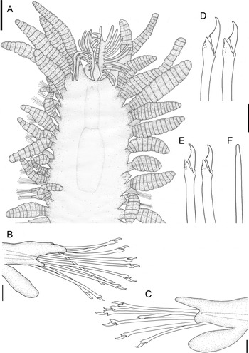
Nuchalosyllis maiteae sp. nov. (holotype, MZUSP 01016). (A) Anterior body, dorsal view; (B) parapodium, anterior or mid-body chaetiger; (C) parapodium, posterior chaetiger; (D) falcigers, anterior body chaetiger; (E) falcigers, posterior body chaetiger; (F) acicula, mid-body to posterior body chaetiger. Scale bars: A, 500 µm; B, 50 µm; C–F, 25 µm.

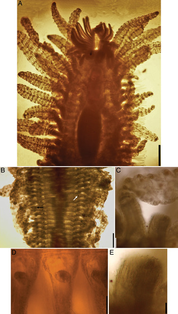
Nuchalosyllis maiteae sp. nov. (holotype, MZUSP 01016). (A) Anterior body, dorsal view; (B) posterior body, ventral view—white arrow points to parapodial gland, dark arrow points to ventral cirrus; (C) cirrophores and base of one dorsal cirrus, mid-body to posterior body, dorsal view; (D) bases of posterior body parapodia, ventral view; (E) detail of one cirrophore from Figure 2C. Scale bars: A, 400 µm; B, 300 µm; C and D, 50 µm; E, 25 µm.

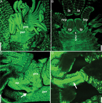
Nuchalosyllis maiteae sp. nov. (holotype, MZUSP 01016), confocal microscopy. (A) Anterior end, dorsal view; (B) anterior end, ventral view—numbers refer to chaetigers; (C) prostomium, peristomium and left epaulette—arrow points to origin of epaulette; (D) detail of cirrophore, midbody chaetiger—arrow points to internal canal: ep, epaulette; la, lateral antennae; lp, left palp; lvp, left ventral peristomial cirrus; per, peristomium; pro, prostomium; rp, right palp; rvp, right ventral peristomial cirrus. Scale bars: A, 200 µm; B, 100 µm; C & D, 50 µm.

TYPE MATERIAL
Holotype (MZUSP 01016): Brazil, State of Rio de Janeiro, offshore (Campos Basin), 22°03′45.395″S 40°09′59.684″W, 75 m depth, van Veen grab, coll. 25 February 2009.
DESCRIPTION
Relatively large syllid, holotype broken in two pieces, anterior fragment 3.45 mm long, 1.90 mm wide, with 24 chaetigers, posterior fragment 13.5 mm long, 1.90 mm wide, with 120 chaetigers, posteriorly incomplete. Body dorso-ventrally flattened, yellowish, pigmentation restricted to dark-red inclusions in articles of antennae and cirri (Figures 1A & 2A–C). Palps long, kidney-shaped, free from each other, ventrally bent below prostomium, reaching half-length of chaetiger 1 (Figure 3B). Prostomium oval, shorter than palps (Figures 1A, 2A & 3A–C); 2 pairs of eyes in open trapezoidal arrangement (Figures 1A & 2A); median antenna absent, scar of insertion between anterior pair of eyes, lateral antennae inserted close to anterior border of prostomium, with 9–12 wide articles each (Figure 3A, B); 1 pair of nuchal epaulettes inserted on posterior border of prostomium, anteriorly directed, each with ~12 separate transverse lamellae (Figures 1A, 2A & 3A, C). Peristomium dorsally short with 2 pairs of peristomial cirri, dorsal peristomial cirri with ~25 articles each, ventral peristomial cirri ~1/3 length of dorsal peristomial cirri, ~11 articles each (Figures 1A, 2A & 3B). Antennae, peristomial and dorsal cirri throughout with distinct cirrophores (Figures 1A, 2C, E & 3D), cirrophores of mid-body and posterior body dorsal cirri covered by numerous triangular, spinose papillae (Figures 1A & 2C, E), with internal canal (Figure 3D); cirrostyles with dorso-ventrally flattened articles, basal articles more closely compacted than following articles (Figures 1A & 2A); peristomial and dorsal cirri throughout with distinct internal longitudinal canal (Figures 1A & 2A); dorsal cirri loosely alternating longer cirri, with ~12–15 articles, and shorter cirri, with ~9–10 more compacted articles (Figures 1A & 2A); ventral cirri flattened, oval, reaching tip of dorsal parapodial lobe or slightly beyond (Figures 1B, C, 2B & 3B), progressively slightly longer towards posterior body. Parapodia conical, distally bilobed, dorsal lobe larger than ventral lobe, differences between lobes progressively increasing towards posterior body (Figure 1B, C); small, rounded parapodial glands with granular material ventrally located, close to base of parapodia (Figure 2B, D). Anterior body parapodia with 12–15 falcigers each, 10–12 on mid-body, ~6 falcigers on each posterior body parapodium; falcigers heterogomph, shafts slightly spinulated subdistally (Figure 1D, E); blades smooth to slightly spinulated, usually unidentate (Figure 1D), some blades of posterior body dorsalmost falcigers sub-bidentate, with enlarged subdistal spine (Figure 1E); blades 33–28 µm long on anterior body, 32–27 µm long on mid-body, 27–22 µm long on posterior body. Simple chaetae not observed. Aciculae straight, distally tapering throughout, 2–3 aciculae per parapodium on anterior body chaetigers, number of aciculae per parapodium progressively diminishing towards posterior end, posterior body parapodia with single acicula each (Figure 1F). Pygidium unknown. Pharynx extending through ~7 segments, with single, conical tooth close to anterior border; proventricle through 7–8 segments, with ~33 rows of muscle-cells (Figure 1A).
REMARKS
Nuchalosyllis maiteae sp. nov. resembles N. lamellicornis by the dorso-ventrally flattened body, by the presence of the lamellate, comb-shaped nuchal epaulettes, articulated antennae, peristomial and dorsal cirri throughout, with pigment inclusions inside articles, and by the similar morphology of falcigers and aciculae. However, N. lamellicornis differs from N. maiteae sp. nov. by presenting transverse dorsal grooves and crests on mid-body and posterior body chaetigers, instead of smooth dorsum as in N. maiteae sp. nov.; and by having smooth cirrophores, while those of N. maiteae sp. nov. present triangular, spinose papillae (see Rullier & Amoureux, Reference Rullier and Amoureux1979; Aguado & San Martín, Reference Aguado and San Martín2008).
Furthermore, differing not only from N. lamellicornis, but also from all other known species of Syllidae which present nuchal epaulettes (such as species of the genus Amblyosyllis Grube, 1857, and most of the members of the subfamily Autolytinae), N. maiteae sp. nov. has the nuchal epaulettes directed towards anterior end, instead of posteriorly directed. Although we have examined a single specimen, it is in good state of preservation, and the shape and position of the epaulettes are clear.
ETYMOLOGY
The epithet ‘maiteae’ is attributed to this species in honour of Dr María Teresa (‘Maite’) Aguado (Universidad Autónoma de Madrid, Spain), a devoted researcher who has contributed to great advances in the state of knowledge about Syllidae in recent years.
ACKNOWLEDGEMENTS
We are grateful to Petrobras, namely Ana Paula Falcão (CENPES/PETROBRAS) and Professor Dr Helena Lavrado (Universidade Federal do Rio de Janeiro), on behalf of all the collection and triage teams of the Habitats Project, for the opportunity to work with this material. We are also grateful to Professor Dr Gláucia Machado Santelli and the technician Roberto Cabado M. Junior (Laboratório de Microscopia Confocal, Instituto de Ciências Biomédicas/USP) for the use of the confocal microscope. M.V.F. receives a postdoctoral fellowship from FAPESP—Fundação de Amparo à Pesquisa do Estado de São Paulo (proc. 10/19424-7). J.M.M.N. receives a productivity fellowship from CNPq—Conselho Nacional de Desenvolvimento Científico e Tecnológico.