Introduction
Obstructive sleep apnoea (OSA) occurs as a consequence of partial or complete upper airway obstruction. Airway obstruction is mostly caused by the collapse of musculature, and less frequently by the hypertrophy of upper airway tissue. A narrow and flaccid upper airway usually poses no problem in an awake individual, but provides a pathophysiological and anatomical basis for OSA. In some cases, the associated loss of muscle tone makes the airway even more narrowed and flaccid, in particular during rapid eye movement sleep.
The disorder can be acquired or congenital. The upper airway obstruction or collapse in OSA may manifest at one level or more frequently at multiple levels, with upper airway collapse or obstruction from the palate level to the epiglottis level considered the main cause of the disorder.Reference Borek, Thaler, Kim, Jackson, Mandel and Schwab1 Drug-induced sleep endoscopy is the ‘gold standard’ in identifying the obstruction site, and in planning conservative and surgical treatment, contributing significantly to operative success.Reference De Vito, Carrasco, Vanni, Bosi, Braghiroli and Campanini2
Case reports
Case one
A 42-year-old female patient underwent clinical investigation for intensive snoring and recurrent episodes of sleep apnoea. At the age of two years, the patient had undergone tracheostomy for extraction of a bronchial aspirated foreign body. The patient reported sleep problems persisting for the past 10 years, characterised by recurrent episodes of prolonged apnoea, frequent waking and intensive snoring. She also noticed increased weariness and inattentiveness, with minimal diurnal drowsiness.
Three years before, she had undergone uvulopalatopharyngoplasty for intensive snoring at another hospital. Neither polysomnography nor drug-induced sleep endoscopy was performed pre-operatively. At 12 months post-operatively, snoring was less intensive, but the characteristics of the sleep apnoea episodes remained unchanged.
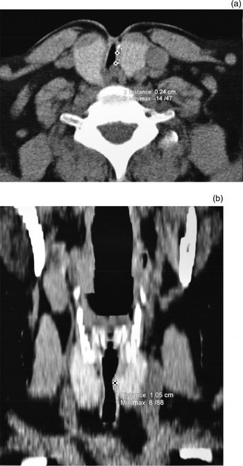
The patient's body mass index was 27 kg/m2. Her Epworth Sleepiness Scale score was 14. Otorhinolaryngological examination revealed a shortened soft palate with complete resection of the uvula, a post-tonsillectomy cicatricle tonsillar area, and a Friedman grade III tongue position. Fibre-optic nasopharyngoscopy (performed when the patient was awake) revealed lingual tonsil hypertrophy and tongue base mucosa overgrowing both vallecular (grade IV), and semicircular lumen stenosis of up to 20 per cent at the level of tracheal rings 2–3. Computed tomography showed cervical tracheal obstruction, 21 mm high, at the level of tracheal rings 2–4 (Figure 1).
Reformatted (a) axial and (b) coronal multi-slice computed tomography images, showing tracheal stenosis following tracheostomy.

Polysomnography, recorded on a SomnoLab 2 device (Weinmann, Hamburg, Germany), yielded an apnoea hypopnea index of 111.6 episodes per hour: central apnoea = 6.4 episodes per hour, obstructive apnoea = 34.6 episodes per hour, mixed apnoea = 5.1 episodes per hour and hypopnea = 65.4 episodes per hour. The average peripheral oxygen saturation was 96 per cent and the oxygen desaturation index was 64.9 episodes per hour. A total of 971 snoring episodes, or 160.1 episodes per hour, were recorded during sleep.
For drug-induced sleep endoscopy, sleep was induced by slow intravenous titration of propofol 10 mg/ml. Depth of sedation was monitored by a bispectral index monitor (A-2000 BIS Monitor; Aspect Medical Systems, Newton, Massachusetts, USA).
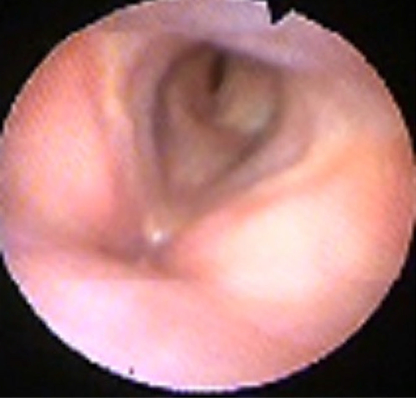
Endoscopy showed normal findings at the level of the soft palate and oropharynx, with an enlarged tongue base overcast by a hypertrophic lingual tonsil, which completely covered the epiglottis and obstructed the airway. The airway showed normal patency at the level of the vocal folds and subglottis. Partial collapse of the laterolateral type was present at the level of the cervical trachea obstruction, with 80–90 per cent obstruction of the tracheal circumference (Figure 2; see also the short video (Appendix 1), available on The Journal of Laryngology & Otology website).
Still image showing partial cervical trachea collapse, taken from supplementary video 1 (Appendix 1).

At the end of drug-induced sleep endoscopy, a modified jaw thrust (or Esmarch manoeuvre) was performed to predict the treatment outcome of an oral appliance. The mandible was gently advanced, up to 4–5 mm, and there was no significant reopening of the upper airway at the level of the tongue base.
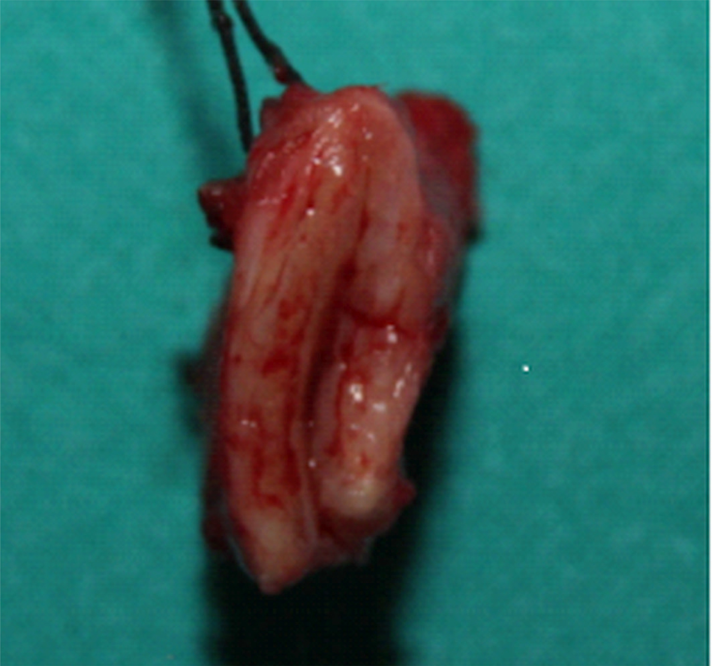
Operative treatment included resection of the hypertrophic tissue of the tongue base via lateral pharyngectomy, and resection of the collapsed segment of the trachea with primary reconstruction of the trachea by end-to-end anastomosis (Figure 3).
Resected segment of the collapsed cervical trachea.

On follow-up polysomnography, performed one-month post-operatively, the apnoea hypopnea index was 17.2: central apnoea = 3.4 episodes per hour, obstructive apnoea = 1.6 episodes per hour, mixed apnoea = 1 episode per hour and hypopnea = 11.1 episodes per hour. The parameters of peripheral oxygenation were normal. The snoring index was 1.3 episodes per hour.
Case two
A 73-year-old male patient with laryngeal squamous cell carcinoma (tumour–node–metastasis (TNM) staging of T3,N0,M0) underwent partial vertical laryngectomy and selective neck dissection (regions II, III and IV), with tracheostomy and the creation of a temporary tracheostomy. The patient had long-standing hypertension, diabetes mellitus type 2 and chronic obstructive pulmonary disease, and had suffered a stroke four years previously.
The post-operative course was uneventful and the cannula was removed two weeks post-operatively. Eight months after the operation, the patient experienced apnoeic episodes in sleep, accompanied by breathlessness and choking, frequent waking, pronounced diurnal fatigue, and drowsiness (Epworth Sleepiness Scale score of 18).
The patient's body mass index was 31 kg/m2. Endoscopic examination (performed awake) revealed thickened soft palate mucosa and an enlarged tongue base that partially overcast the epiglottis. The mucosa and other anatomical structures of the endolarynx were free from tumour or other pathological lesions. A small cicatricle lesion of the mucosa from a previous tracheostomy was observed on the anterior wall of the cervical trachea, at the level of tracheal rings 2 and 3. Mucosa of the lower part of the trachea was normal.
Polysomnography indicated severe OSA, with an apnoea hypopnea index of 67.4 episodes per hour: central apnoea = 12.4 episodes per hour, obstructive apnoea = 28.7 episodes per hour and hypopnea = 16.9 episodes per hour. The oxygen desaturation index was 74.2 episodes per hour, with an average peripheral oxygen saturation of 88 per cent.
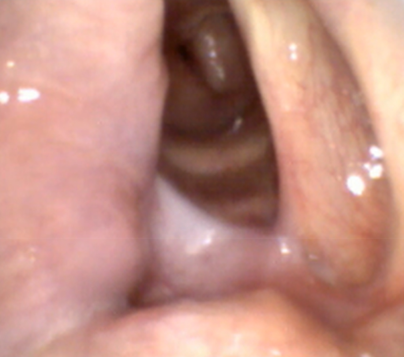
Drug-induced sleep endoscopy revealed complete circular obstruction of the soft palate and partial airway obstruction at the tongue base level. The rest of the larynx was free from collapsing anatomical structures. On inspiration, collapse with complete obstruction was observed in the area of tracheal rings 2 and 3 (Figure 4; see also the short video (Appendix 2), available on The Journal of Laryngology & Otology website).
Still image showing the post partial laryngectomy state, with complete collapse of the cervical trachea in sleep following tracheostomy, taken from supplementary video 2 (Appendix 2).

The patient was recommended continuous positive airway pressure therapy. However, the patient died from myocardial infarction nine months after the diagnosis of severe OSA.
Discussion
Good knowledge of the aetiology of airway obstruction, and its localisation and quantification, are of utmost importance for choosing the appropriate therapeutic procedure and for disease prognosis. Pathological lesions of the lower airway as the cause of OSA, in the cervical trachea in particular, are rarely considered.
• This paper presents two cases of severe obstructive sleep apnoea caused by cervical trachea collapse in sleep following previous tracheostomy
• Drug-induced sleep endoscopy should be used for visualising anatomical structures below the vocal folds in patients with a history of cervical trachea surgery
• Drug-induced sleep endoscopy is important in planning conservative and surgical treatment, contributing significantly to operative success
Stenosis and tracheomalacia are late complications of tracheostomy that lead to narrowing of the tracheal lumen.Reference Epstein3 Tracheomalacia in the broadest sense refers to the abnormal collapse of the tracheal wall as an extreme grade of airway dynamic narrowing on expiration. Primary idiopathic tracheomalacia is rarely seen. Secondary tracheomalacia generally occurs consequent to long-lasting intubation, or, less frequently, as a tracheostomy related complication. It can occur as an isolated lesion or in combination with other pathological changes of the trachea.
In patients with classic tracheomalacia, the collapse of tracheal structures manifests as contact between the anterior and posterior walls, both when awake and in sleep. Collapse always occurs on expiration, with varying grades of respiratory insufficiency. The accompanying symptoms predominantly include expiratory stridor, dyspnoea, tachypnoea and recurrent cough.
Obstructive sleep apnoea as a sequelae of tracheomalacia is usually recorded in the paediatric population.4–6 Qubty et al. reported tracheomalacia in one-third of neonates with verified OSA.Reference Qubty, Mrelashvili, Kotagal and Lloyd7 Cervical trachea collapse in sleep as the cause of apnoea is rare in adults. It is challenging to diagnose because patients have subclinical symptoms or the symptoms are masked by other predominant discomforts.
In our two patients, OSA was not caused exclusively by upper airway collapse, but in part by cervical trachea collapse as a late complication of tracheostomy. When awake, the patients had almost unchanged tracheal lumen, without signs of collapse or other symptoms of breathing difficulty when awake. However, their anatomical and clinical picture changed significantly during sleep. In both patients, the pathophysiological process of cervical trachea collapse was induced by: airway obstruction at the tongue base level, with the formation of negative pressure caudally; destabilisation of the semicircular cartilaginous skeleton associated with resection of the frontal third; and probably change in the cervical trachea musculature tone accompanying higher collapsibility of the upper airway musculature, which was objectively demonstrated by endoscopy during sleep. Studies by Rubenstein et al.Reference Rubenstein, Bradeley, Zamel and Hoffstein8 and Katz et al.Reference Katz, Zamel, Slutsky, Rebuck and Hoffstein9 emphasised the association of impaired upper and lower airway musculature tone in patients with OSA.
Inadequate diagnostic investigation is the main reason for the inappropriate selection of OSA patients for surgical treatment, and thus for the poor success of surgical treatment in the management of OSA patients. Careful patient selection, according to disease severity, co-morbidities and choice of surgical method, is of great importance. The proper choice of surgical method requires endoscopic analysis of the level, form and size of collapse, when the patient is awake and during sleep.
In 2014, a European position paper issued recommendations on the indications and levels of endoscopic visualisation of upper airway obstruction, suggesting that local status be inspected at four levels: the palate, the oropharynx and tonsil, the tongue base, and the epiglottis.Reference De Vito, Carrasco, Vanni, Bosi, Braghiroli and Campanini2 The European experts agreed that the nose, nasopharynx and glottis are not relevant for endoscopic analysis during sleep, considering that obstruction at these levels does not change during sleep versus when awake, and therefore does not contribute significantly to OSA development.Reference De Vito, Carrasco, Vanni, Bosi, Braghiroli and Campanini2 Vicini et al. proposed the inspection of a fifth level: the supraglottis and glottis area.Reference Vicini, De Vito, Benazzo, Frassineti, Campanini and Frasconi10 They demonstrated that the cause of OSA could also be located at lower airway levels, in the cervical trachea.
Our experience, based on the two patients described, confirmed all the advantages of dynamic visualisation of pathological airway lesions by endoscopy during sleep. This method should be used for anatomical structures below the vocal folds, at the sixth level, in patients who have undergone previous surgical procedures on the cervical trachea. It was demonstrated once again that the anatomical picture of the airway obtained when the patient is awake may not be similar or identical to the findings obtained during sleep.
Conclusion
In our patients, OSA developed as a consequence of upper and lower airway obstruction and collapse. Collapse of anatomical structures of the cervical trachea in sleep, with impaired breathing, is a rare clinical entity. It is frequently asymptomatic and often diagnosed incidentally. Airway endoscopy performed during sleep has a critical diagnostic role in all cases of suspected cervical trachea collapse. The cases presented highlight the importance of extensive diagnostic investigation in OSA patients, as diagnosis can be highly challenging for clinicians.
Competing interests
None declared
Appendix 1 Supplementary video material of patient one
A short video demonstrating partial collapse (of the laterolateral type) in patient one, at the level of cervical trachea obstruction, with 80–90 per cent obstruction of the tracheal circumference. This video is available online at The Journal of Laryngology & Otology website, at https://doi.org/10.1017/S0022215119001166.
Appendix 2 Supplementary video material of patient two
A short video demonstrating collapse with complete obstruction in patient two on inspiration, in the area of tracheal rings 2 and 3. This video is available online at The Journal of Laryngology & Otology website, at https://doi.org/10.1017/S0022215119001166.