In 1925, an intriguing electrocardiogram trace was published in a book by Welsh cardiologist Sir Thomas Lewis. Reference Riera, Barros, de Soussa and Baranchuk1 This rhythm was then known as accelerated idioventricular rhythm, a rare rhythm with much to learn about its epidemiology and progression. Reference Freire and Dubrow2 This case illustrates the diagnostic and management challenges of accelerated idioventricular rhythm in a well neonate.
Case presentation
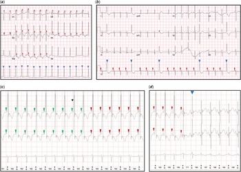
An 11-day-old female neonate was referred to the paediatric cardiology service for advice because of a regular broad (QRS) complex rhythm as shown in Figure 1a. This was captured on a 12-lead electrocardiogram during neonatal follow-up for an irregular heart rhythm at birth. Antenatal anomaly scan was normal with no mention of arrhythmia in the fetus, and the family history was negative for inherited arrhythmia syndromes and congenital cardiovascular abnormalities. She was delivered at 40+2 weeks’ gestation via an emergency caesarean section due to failure to progress of the labour, in the presence of a fetal tachycardia at a rate of 170 beats per minute. The heart rate at birth was 160 beats per minute. A 12-lead electrocardiogram showed intermittent broad QRS complexes with preceding sinus beats (Fig 1b). Apart from transiently elevated serum sodium of 147 mmol/L (which normalised later spontaneously), other biochemistry tests and clinical examination were all normal. These intermittent wide QRS complexes were initially interpreted as ventricular ectopic beats. She was discharged home with a follow-up planned in 1 week’s time.
(a) Electrocardiogram on day 11 of life during initial neonatal outpatient showing a regular broad QRS complex rhythm with isorhythmic atrioventricular dissociation and T wave inversion across precordial leads. (b) Electrocardiogram at birth with baseline heart rate of 125 beats per minute and intermittent broad QRS complexes, absence of convincing preceding P waves and inverted T waves with a compensatory pause. (c and d) Holter monitoring on day 11 documenting a regular broad QRS complex rhythm with intermittently preceding P waves and short PR intervals with sudden resolution.

On a subsequent assessment by the cardiology team, she appeared slightly jaundiced but the serum bilirubin level was below phototherapy treatment. An isolated serum magnesium level of 0.71 mmol/L was noted. An echocardiogram showed normal heart structures and good myocardial function. A diagnosis of Wolff-Parkinson-White syndrome was suspected initially and owing to frequent and persistent arrhythmia, oral flecainide at 1 mg/kg/dose twice daily and magnesium supplements were commenced. This was in view of a regular broad QRS complex rhythm with possible slurred upstrokes suggestive of pre-excitation and Holter recordings of a 12-minute episode of tachycardia at a rate of 180 beats per minute (Fig 1c). Preceding P waves and short PR intervals were seen in parts of the Holter recording (Fig 1c and d). This broad QRS complex rhythm terminated suddenly as evidenced by changes in PR and QRS morphologies (Fig 1d). This led to the initial diagnosis of intermittent Wolff-Parkinson-White syndrome.
A review of the recordings by a paediatric electrophysiologist revealed the occasional absence of the P waves preceding the broad QRS complexes (Fig 1a) and as in Figure 1b, the baseline heart rate was noted to be in sinus rhythm at 125 beats per minute. This supported a rhythm that was not of the sinoatrial node origin. There was isorhythmic atrioventricular dissociation with a P wave occurring immediately after each QRS complex. The P waves occurring after the QRS complexes raised the suspicion of possible retrograde conduction. However, regular broad QRS complexes favoured a ventricular source and prompted the possibility of ventricular tachycardia. In this case, the ventricular rate of 150 beats per minute was within normal limits for a well neonate which rendered ventricular tachycardia less likely. A complete heart block could also be considered, but atrioventricular dissociation in a complete heart block is not isorhythmic with atrial rate being typically faster than the ventricular rate.
A short history of arrhythmia and inverted T waves across precordial leads (Fig 1a) in keeping with abnormal myocardial depolarisation and repolarisation also favoured a ventricular source. It became clearer that the presumed delta waves initially identified were a fusion of P waves and QRS complexes. These findings of a monomorphic broad QRS complex rhythm at 150 beats per minute (approximately 15% above preceding sinus rhythm) with isorhythmic dissociation in an asymptomatic neonate supported a diagnosis of an accelerated idioventricular rhythm.
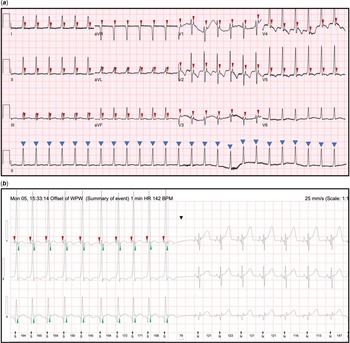
Owing to persistent documentation of the accelerated idioventricular rhythm despite being on flecainide treatment for 24 hours (Fig 2a), propranolol 1 mg/kg/dose twice daily was added. This decision was taken due to the incessant arrhythmia and to achieve satisfactory rate control. A scheduled echocardiogram on day 18 of life demonstrated a new finding of mild mitral regurgitation. There was also ongoing intermittent broad QRS complex rhythm and isorhythmic dissociation captured on a Holter monitoring (Fig 2b). Therefore, the flecainide was increased to 2 mg/kg/dose twice daily. Magnesium supplements were stopped as the serum levels had normalised. She remained on propranolol 1 mg/kg/dose twice daily. She was asymptomatic throughout from birth and tolerated the antiarrhythmic treatment well. Successful rate and rhythm control were achieved as demonstrated by sinus rhythm on an electrocardiogram obtained on day 23 of life. An echocardiogram at 26 days of life showed improved myocardial function evidenced by resolving mitral regurgitation. Care of the neonate was subsequently transferred to another unit following relocation from our institution.
(a and b) Electrocardiogram and Holter monitoring repeated 24 hours after commencing oral flecainide showing a monomorphic broad QRS complex rhythm with P waves occurring after each QR complex and T wave inversion across precordial leads.

Discussion
This case illustrates the importance of being vigilant in well neonates exhibiting slurred or wide QRS complexes on their electrocardiograms. The presence of broad QRS complexes should prompt urgent assessment and exclusion of not only sinister rhythms such as supraventricular tachycardia with aberrancy and ventricular tachycardia but also accelerated idioventricular rhythm.
Accelerated idioventricular rhythm is relatively rare with or without congenital cardiac disease. Reference Reynolds and Pickoffrid3 When encountered in the neonatal period as in this case, it often manifests shortly after birth. Reference Freire and Dubrow2 Electrocardiogram findings include tachycardia at 10–15% of the normal sinus rate Reference Fouron4–Reference Bisset, Janos and Gaum7 with a pattern of three or more consecutive monomorphic beats. Reference Riera, Barros, de Soussa and Baranchuk1 As the ectopic focus fires at a rate similar to the intrinsic sinoatrial rate, isorhythmic dissociation along with fusion and capture beats are seen. Reference Riera, Barros, de Soussa and Baranchuk1 Ventricular tachycardia where fusion and capture beats are also seen is an equally as important to alternative diagnosis exclude. Reference Kothari8,Reference Ban9 Although typically presenting with faster ventricular rates (age dependent in children), Reference Kothari8,Reference Ban9 slow ventricular tachycardia should also be considered, and diagnosis should be made in context of the clinical picture. Adenosine test or transoesophageal diagnostic electrophysiology could be utilised in cases where pre-excitation cannot be confidently excluded.
Reynolds and Beach et al have reported the accelerated idioventricular rhythm to be relatively benign and the patients being typically asymptomatic, not requiring treatment but the risk of degenerating into sinister rhythms was unclear. Reference Reynolds and Pickoffrid3,Reference Beach, Marcuccio, Beerman and Arora10 A consensus statement in 2014 suggested that in the absence of symptoms, haemodynamic compromise, or underlying cardiac abnormalities, treatment is not indicated. Reference Crosson, Callans and Bradley12 On the contrary, a recent case report by Ergul et al described degeneration of accelerated idioventricular rhythm into torsades de pointes during sleep, requiring cardiac resuscitation in a previously asymptomatic 11-year-old boy. Reference Ergul, Kafali and Uysal11 Isolated case intervention may be warranted in the presence of syncope or sustained arrhythmia due to risk of sudden death, and emerging abnormal echocardiography findings like in our case. Reference Ergul, Kafali and Uysal11,Reference Hohnloser, Zabel, Olschewski, Kasper and Just13 Notably, this rhythm may be resistant to pharmacological treatment. Reference Freire and Dubrow2
Freire and Dubrow described five neonatal cases of accelerated idioventricular rhythm, all being asymptomatic with varied presentations and eventual resolution of arrhythmia. Reference Freire and Dubrow2 Spontaneous resolution was seen in two cases, and monotherapy with propranolol or amiodarone was necessary in the other three cases due to frequent arrhythmia. Amiodarone was preferred in one case with a significant ventricular septal defect to avoid negative inotropy. Reference Freire and Dubrow2 This previous report and our case both supported a good prognosis of accelerated idioventricular rhythm even though an unpredictable response to treatment can be rarely seen. Reference Freire and Dubrow2
Conclusion
Accelerated idioventricular rhythm in neonates is rare and typically encountered on the first few days of life. Albeit mostly benign and resolves spontaneously, it requires close monitoring and may necessitate even antiarrhythmic treatment based on individual risk assessment. It is pertinent to consider accelerated idioventricular rhythm in the differential diagnosis of broad complex rhythms as the clinical management may differ in other aetiologies.
Acknowledgements
We would like to thank the parents of this child for their enthusiasism and continuous support in sharing of this case to improve our knowledge in presentation and management of this arrhythmia. We also thank all healthcare team members involved in the management of this case for their hardwork and perseverance. Finally, we would like to extend our gratitude to the paediatric cardiology secretaries for consolidating all documentation required for preparation of this case report.
Financial support
This research received no specific grant from any funding agency, commercial, or not-for-profit sectors.
Conflicts of interest
None.
Ethical standards
The authors assert that all procedures contributing to this work comply with the ethical standards of the relevant national guidelines and Helsinki Declaration of 1975, as revised in 2008.
Patient consent
Obtained.