Introduction
Intracytoplasmic sperm injection (ICSI) (Palermo et al., Reference Palermo, Joris, Devroey and Van Steirteghem1992) has revolutionized the treatment of male factor infertility. Most studies have reported that ICSI with ejaculated spermatozoa results in a mean fertilization rate (FR) of 70–75% (Flaherty et al., Reference Flaherty, Payne and Matthews1998; Pujol et al., Reference Pujol, García, Obradors, Rodríguez and Vassena2018). However, total fertilization failure (FF) after ICSI, i.e. the absence of normally fertilized zygotes (containing two polar bodies and two pronuclei) at 16–19 h post-insemination, is a persistent problem in fertility clinics. In many cases, FF is associated with a low oocyte yield: FF occurs in ∼17% of cycles with fewer than three oocytes, and in ∼31% of cycles when only one mature oocyte is available (Flaherty et al., Reference Flaherty, Payne and Matthews1998; Hojnik and Kovačič, Reference Hojnik, Kovačič, Bin and Feng2019). However, its incidence remains at 1–3% in all cycles even when inseminating a high number of oocytes (Flaherty et al., Reference Flaherty, Payne and Matthews1998; Palermo et al., Reference Palermo, O’Neill, Chow, Cheung, Parrella, Pereira and Rosenwaks2017). FF can be repetitive, and it can happen even when morphologically normal sperm and oocytes are used for ICSI.
The most common aetiology of FF is by far oocyte activation failure (OAF; the failure to elicit the release from the meiotic block in response to sperm entry in the oocyte), accounting for ∼40–80% of non-fertilized oocytes and characterized by the absence of pronuclei (Sousa and Tesarik, Reference Sousa and Tesarik1994; Flaherty et al., Reference Flaherty, Payne and Matthews1998; Rawe et al., Reference Rawe, Olmedo, Nodar, Doncel, Acosta and Vitullo2000). Abnormal fertilization is also a common problem, characterized by the presence of an abnormal number of pronuclei (1PN, 3PN, or ≥3PN), representing ∼10% of all inseminated oocytes (Balakier, Reference Balakier1993). In some cases, oocyte activation occurs normally and 2PN zygotes are formed, but the embryo fails to undergo the first mitotic division and arrests its development at the pronuclear stage, something commonly referred to as zygote arrest (Zamora et al., Reference Zamora, Sánchez, Pérez, Díaz, Quintana and Bethencourt2011).
Independently of their origin, all these situations result in a lack of viable embryos before 24 h post-insemination. Often, FF and zygotic arrest are unexplained and unexpected, and cannot be easily resolved using routine in vitro fertilization (IVF) methods. They invariably imply a high economic and psychological effect for the patients and are associated with difficult clinical management and counselling. Currently, treatment options for these patients are limited to the use of donor gametes, assisted oocyte activation (AOA) in some cases, or trying a new cycle using the same gametes. Nevertheless, this last option is usually accompanied by low expectations of success.
Some of the cellular alterations underlying FF are oocyte cytoplasmic immaturity (Balakier et al., Reference Balakier, Sojecki, Motamedi and Librach2004), lack of sperm nucleus decondensation and/or aster formation (Rawe et al., Reference Rawe, Díaz, Abdelmassih, Wójcik, Morales, Sutovsky and Chemes2008), defects in oocyte spindle and cytoskeletal function (Rawe et al., Reference Rawe, Olmedo, Nodar, Doncel, Acosta and Vitullo2000), the sperm’s inability to trigger calcium oscillations (Yoon et al., Reference Yoon, Jellerette, Salicioni, Lee, Yoo, Coward, Parrington, Grow, Cibelli, Visconti, Mager and Fissore2008), or the inability of the oocyte machinery to respond to its stimuli (Yeste et al., Reference Yeste, Jones, Amdani, Patel and Coward2016; Ferrer-Buitrago et al., Reference Ferrer-Buitrago, Bonte, Dhaenens, Vermorgen, Lu, De Sutter and Heindryckx2019). Similarly, zygote arrest seems to be associated with specific morphological features in the zygote, such as uneven pronuclear size or reduced organization of nucleolar precursor bodies (Sadowy et al., Reference Sadowy, Tomkin, Munné, Ferrara-Congedo and Cohen1998; Zamora et al., Reference Zamora, Sánchez, Pérez, Díaz, Quintana and Bethencourt2011), and chromosomal abnormalities (Benkhalifa et al., Reference Benkhalifa, Kahraman, Caserta, Domez and Qumsiyeh2003). Several proteins participate in oocyte activation and early fertilization events, including sperm PLCζ, as well as oocyte IP3 receptors, kinases such as CAMKII, PKC, WEE2, MLCK, MOS and MAPK, calcium channels such as TRPM7 and CaV3.2, factors involved in sperm decondensation such as NPM2, HIRA, and the recently discovered SRPK1, and proteins of the subcortical maternal complex (SCMC), among many others (Moos et al., Reference Moos, Visconti, Moore, Schultz and Kopf1995; Ducibella and Fissore, Reference Ducibella and Fissore2008; Oh et al., Reference Oh, Susor and Conti2011; Yeste et al., Reference Yeste, Jones, Amdani, Patel and Coward2016; Bernhardt et al., Reference Bernhardt, Padilla-Banks, Stein, Zhang and Williams2017; Gou et al., Reference Gou, Lim, Ma, Aubol, Hao, Wang, Zhao, Liang, Shao, Zhang, Meng, Li, Zhang, Xu, Li, Rosenfeld, Mellon, Adams, Liu and Fu2020). However, a causal link between genetic alterations in these factors and FF has remained largely speculative.
Fortunately, during the last decade, multiple genetic variants have been associated with FF or zygote arrest after ICSI. The objective of this review was to compile all the genetic variants identified in infertile patients experiencing FF, abnormal fertilization, and zygotic arrest after ICSI, and comprehensively analyze them in terms of pathogenesis, inheritance, frequency, molecular effects on protein function and fertilization and treatment options. Our objective is to provide a tool for the diagnosis and counselling of infertile patients.
Materials and methods
This systematic review was conducted according to the Preferred Reporting Items for Systematic Reviews and Meta-Analyses (PRISMA) guidelines (Liberati et al., Reference Liberati, Altman, Tetzlaff, Mulrow, Gøtzsche, Ioannidis, Clarke, Devereaux, Kleijnen and Moher2009).
Search strategy
A systematic literature search was performed using the PubMed database to identify research articles reporting genetic variants affecting genes with a demonstrated or suspected role during fertilization through ICSI (from insemination until first mitotic division), identified in infertile patients. The results were restricted to studies published in the English language from 2006 to 2021. Some factors participate in a wide range of cellular processes (from oocyte maturation to first embryonic cellular divisions) and their alterations can result in diverse clinical consequences. For this reason two terms associated with phenotypes downstream and upstream from the zygotic stage were included in the systematic search: oocyte maturation arrest and embryonic arrest. In total, seven terms were considered to identify the infertility phenotypes compatible with the objective of the present review, and key words were combined in the following search equation:
((((fertilization failure [Title/Abstract] OR oocyte activation [Title/Abstract] OR zygotic arrest [Title/Abstract] OR cleavage failure [Title/Abstract] OR abnormal fertilization [Title/Abstract] OR oocyte maturation arrest [Title/Abstract] OR embryonic arrest [Title/Abstract]) AND (mutation [Title/Abstract] OR sequencing [Title/Abstract] OR variant [Title/Abstract] OR variation [Title/Abstract]) AND (ICSI [Title/Abstract] OR in vitro fertilization [Title/Abstract] OR infertility [Title/Abstract] OR patient [Title/Abstract]))) AND English [Language]).
The reports matching the search question were retrieved, and their reference lists were manually reviewed to identify additional studies. In addition, using the same terms, additional studies were manually identified from peer-reviewed abstracts presented in ESHRE Annual Meetings from 2014 to 2021.
Study selection
Studies were screened for eligibility by titles and abstracts. Importantly, we only considered genetic variants identified in patients presenting gametes without severe morphological alterations (i.e. globozoospermia, oocytes with giant polar bodies, etc.), therefore the cases in which the FF or zygotic arrest problem are usually unexpected. All studies including infertile men or women undergoing ICSI in which sequencing was used to identify genetic variants in genes associated with fertilization were included. The studies that (1) did not use gene sequencing to report genetic variants, (2) exclusively included patients with phenotypes not matching the research question, or (3) used other insemination techniques (such as conventional IVF), were discarded. Study selection and data extraction (i.e. building the lists of genetic variants and subsequent analysis) were performed and verified by two independent investigators.
Study outcomes, data extraction and analysis
Data concerning infertility phenotype/s, experimental or in silico validation of the variant effect on fertilization, affected protein domains, inheritance, zygosity, treatment options, and genetic counselling were manually extracted from tables and/or text, collected and analyzed. To identify clear cases of FF that are likely to have been caused by alterations in the gametes, the inclusion criteria for patients in the studies included in this review were: four or more mature oocytes [morphologically normal and recognizable as PB1/metaphase II (MII) oocytes] inseminated by ICSI, and total FF or low FR (≤25% 2PN zygotes) and/or ≤25% cleavage rate among fertilized oocytes (2PN).
To analyze the population frequency of the identified variants, the following public databases were considered: GnomAD_exome (https://gnomad.broadinstitute.org/) and TOPMED.
Results
Results of a systematic search of the literature
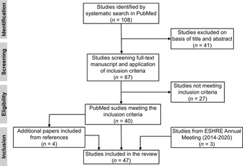
The initial systematic search of the PubMed database identified 108 studies. After screening of titles and abstracts, 67 studies were selected for detailed analysis of the full-text manuscript, 40 of which met the inclusion criteria. Analysis of the bibliographical references in each manuscript allowed for the inclusion of four additional studies, and three studies were identified from ESHRE Annual Meeting (2014–2020). In total, 47 studies were finally included in the present review (Figure 1).
PRISMA flow diagram of identification, screening, eligibility, and inclusion steps.

The 47 studies reported 121 exonic variants in 141 infertile patients. Specifically, 38 variants were identified in 61 men, while 83 variants were identified in 80 women. These variants affected 16 genes: PLCZ1, WBP2NL, ACTL9, ACTLA7 and DNAH17 in infertile men, and WEE2, TUBB8, PATL2, TLE6, PADI6, TRIP13, BGT4, NLRP5, NLRP7, CDC20 and ZAR1 in infertile women. Among the 121 variants reported, 58 (47.9%) were not present in the public databases analyzed and, except for some cases, the Minor Allele Frequency (MAF) reported for most of the genetic variants present in the dbSNP database was very low (Table S1).
Most variants included in this review (98 out of 121; 81.0%) directly affect essential domains for protein function; these include missense variants directly affecting a specific domain, or variants affecting the whole protein structure (such as frameshift or nonsense) located upstream of reported domains. The specific effect of each mutation on protein function and early fertilization events will be detailed for each factor in the following sections.
Genetic variants in PLCZ1: the sperm-borne oocyte-activating factor
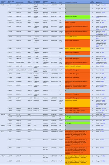
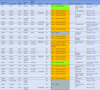
In total, 27 PLCZ1 exonic variants were identified in 14 studies, including 50 men who experienced FF or low FR after ICSI associated with oocyte activation deficiencies (Table 1). PLCZ1 is located on chromosome 12, consists of 15 exons, and codes for the sperm-specific enzyme phospholipase C zeta (PLCζ). Since its discovery in 2002 (Saunders et al., Reference Saunders, Larman, Parrington, Cox, Royse, Blayney, Swann and Lai2002), PLCζ has emerged as the main sperm-borne oocyte activation factor (SOAF), a soluble protein conserved through several taxonomic groups that is essential for eliciting the intracellular calcium oscillations observed shortly after the sperm enters the oocyte (Saunders et al., Reference Saunders, Larman, Parrington, Cox, Royse, Blayney, Swann and Lai2002; Coward et al., Reference Coward, Ponting, Chang, Hibbitt, Savolainen, Jones and Parrington2005). In mature sperm, PLCζ localizes in the perinuclear theca, a condensed cytosolic fraction situated between the acrosome and the nuclear envelope, which first contacts the oocyte cytoplasm (Escoffier et al., Reference Escoffier, Yassine, Lee, Martinez, Delaroche, Coutton, Karaouzène, Zouari, Metzler-Guillemain, Pernet-Gallay, Hennebicq, Ray, Fissore and Arnoult2015). Once inside the oocyte, PLCζ functions by hydrolyzing PIP2 into IP3 and DAG. Subsequently, PIP2 is distributed in vesicles throughout the oocyte cytoplasm (Sanders et al., Reference Sanders, Ashley, Moon, Woolley and Swann2018).
PLCZ1, WBP2NL, DNAH17 and ACTL9 genetic variants detected in non-globozoospermic men presenting fertilization failure or low fertilization rates after ICSI. The table shows the specific exons and protein domains affected, the type of mutation, the dbSNP ID (if any), the gene dosage (Homo: homozygosity, Het: heterozygosity, Het*: compound heterozygosity) and the total number of patients in which the specific variant has been identified. The information about experimental validation for each genetic variant is also indicated, and the colour code provides information on their pathogenicity: no experimental validation reported (grey), reported as benign (green), reported as pathogenic (orange), potentially pathogenic or unclear according to existing data (yellow)

The essential role of PLCζ in fertilization has been revealed by numerous findings. Depleting PLCζ from sperm extracts by specific antibodies abolishes the induction of Ca2+ oscillations in the oocyte (Saunders et al., Reference Saunders, Larman, Parrington, Cox, Royse, Blayney, Swann and Lai2002). Microinjection of human PLCZ1 cRNA or recombinant PLCζ protein into human oocytes causes parthenogenetic activation and development to the pseudo-blastocyst stage, inducing Ca2+ oscillations that are very similar to those observed after sperm fertilization (Rogers et al., Reference Rogers, Hobson, Pickering, Lai, Braude and Swann2004; Yoon et al., Reference Yoon, Eum, Lee, Lee, Kim, Han, Won, Park, Shim, Lee, Fissore, Lee and Yoon2012). Moreover, recent studies have demonstrated that sperm from Plcz1 knockout mice did not induce calcium oscillations after ICSI, causing fertilization failure (FF; Hachem et al., Reference Hachem, Godwin, Ruas, Lee, Ferrer Buitrago, Ardestani, Bassett, Fox, Navarrete, de Sutter, Heindryckx, Fissore and Parrington2017; Nozawa et al., Reference Nozawa, Satouh, Fujimoto, Oji and Ikawa2018).
Approximately a decade ago, the two first PLCZ1 pathogenic variants associated with FF after ICSI were reported; p.H233L and p.H398P were identified in a non-globozoospermic patient in compound heterozygosity, and were demonstrated to compromise PLCζ function (Heytens et al., Reference Heytens, Parrington, Coward, Young, Lambrecht, Yoon, Fissore, Hamer, Deane, Ruas, Grasa, Soleimani, Cuvelier, Gerris, Dhont, Deforce, Leybaert and De Sutter2009; Kashir et al., Reference Kashir, Konstantinidis, Jones, Lemmon, Lee, Hamer, Heindryckx, Deane, De Sutter, Fissore, Parrington, Wells and Coward2012). Since then, different studies have sequenced PLCZ1 in men experiencing FF after ICSI (and, in many cases, their close relatives), significantly broadening the list of PLCZ1 variants with clinical relevance. Of the 27 PLCZ1 variants compiled in this review, 19 have been reported in the last 3 years (Table 1). While the number of patients carrying PLCZ1 variants is very low, some small cohort studies may give an idea of the incidence of PLCZ1 variants among infertile men experiencing FF after ICSI. This incidence is around one-third of the reported cases (33.6%) and seems to be similar across other studies reported in the literature (range 25–35%; Table 2).
List of studies screening for PLCZ1 or WEE2 genetic variants in multiple male or female patients (≥3 unrelated individuals) who experienced fertilization failure or low fertilization rates after ICSI. The population of patients as defined in each study is indicated. The frequency of patients carrying at least one variant in PLCZ1 or WEE2 among all FF patients is indicated for each study and overall

Functional and protein structure assays, either experimentally or in silico, were performed for 21 out of the 27 (77.8%) PLCZ1 genetic variants identified so far. Altogether, 18 variants had a demonstrated deleterious effect on PLCζ structure and/or function, one did not cause a negative effect on PLCζ, while two resulted in unclear results (Table 1). In most cases, the frequency of these variants in the general population was below 0.1% (Table S1). However, a higher MAF was reported for some PLCZ1 variants that were reported to be benign or with unclear effects, such as p.I120M and p.S500L, in accordance with the null or little effect of these genetic variants on fertilization success through ICSI.
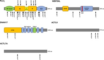
As indicated in Table 1 and Figure 2, all PLCZ1 variants directly affect at least one of the domains required for PLCζ catalytic and regulatory activity, causing a partial or complete failure for the enzyme to generate the calcium oscillations essential for oocyte activation. These domains are tandem EF hands, conferring calcium sensitivity to PLCζ; X and Y core domains, required for enzymatic catalytic activity; the C2 domain, essential for PLCζ to bind to two phosphoinositides (PI(3)P and PI(5)P) present in liposomes, therefore facilitating substrate – PI(4,5)P2 – accessibility; and the XY-linker region, involved in both enzymatic and regulatory functions (Saunders et al., Reference Saunders, Larman, Parrington, Cox, Royse, Blayney, Swann and Lai2002; Kouchi et al., Reference Kouchi, Shikano, Nakamura, Shirakawa, Fukami and Miyazaki2005; Nomikos et al., Reference Nomikos, Elgmati, Theodoridou, Calver, Nounesis, Swann and Lai2011, Reference Nomikos, Stamatiadis, Sanders, Beck, Calver, Buntwal, Lofty, Sideratou, Swann and Lai2017).
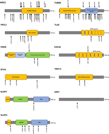
Location of genetic variants associated with male-related fertilization failure within the protein structure. The main domains and the total number of amino acids are indicated for each protein.

Most PLCZ1 variants affect the catalytic domains X and Y (15; 55.6%). As previously demonstrated, missense variants affecting the C2 domain (such as the p.I489F) can affect PLCζ ability to bind to liposomes containing its substrate (Nomikos et al., Reference Nomikos, Stamatiadis, Sanders, Beck, Calver, Buntwal, Lofty, Sideratou, Swann and Lai2017) and, perhaps due to the regulatory nature of this domain, the in vitro effect of these variants can be compensated by increasing the amount of mutated PLCζ. For instance, for p.I489F and p.R553P variants, microinjection of physiological levels of mutated cRNA failed to trigger calcium oscillations in mice oocytes, but a two-fold increase in the same cRNA led to successful oocyte activation (Nomikos et al., Reference Nomikos, Stamatiadis, Sanders, Beck, Calver, Buntwal, Lofty, Sideratou, Swann and Lai2017; Yuan et al., Reference Yuan, Yang, Ren, Yan, Nie, Yan and Qiao2020). In contrast, mutations such as p.V326Kfs*25 and p.R412Efs*15 resulted in truncated and completely non-functional forms of PLCζ that lacked the C2 domain and part of the catalytic domain (Torra-Massana et al., Reference Torra-Massana, Cornet-Bartolomé, Barragán, Durban, Ferrer-Vaquer, Zambelli, Rodriguez, Oliva and Vassena2019; Mu et al., Reference Mu, Zhang, Wu, Fu, Chen, Yan, Li, Zhou, Wang, Zhao, Dong, Kuang, Sun, He, Wang and Sang2020). Regarding the EF hands domains, no variants identified in this region have been proven as pathogenic by in vitro experimental procedures, and one of them (p.I120M) was characterized as benign; while this region helps PLCζ gain its enzymatic activity by binding calcium, its role is not critical in fertilization through ICSI.
An altered enzymatic function is not the only alteration caused by PLCZ1, as this problem is usually accompanied by abnormal levels and subcellular localization of PLCζ protein (Escoffier et al., Reference Escoffier, Lee, Yassine, Zouari, Martinez, Karaouzène, Coutton, Kherraf, Halouani, Triki, Nef, Thierry-Mieg, Savinov, Fissore, Ray and Arnoult2016; Wang et al., Reference Wang, Zhang, Kong, Li, Zhang, He, Wu, Tang, Zha, Tan, Duan, Cao and Zhu2020; Yan et al., Reference Yan, Fan, Wang, Yan, Li, Ouyang, Wu, Yin, Zhao, Kuang, Li and Lyu2020), often confirmed in somatic cells transfected with mutant PLCZ1 (Mu et al., Reference Mu, Zhang, Wu, Fu, Chen, Yan, Li, Zhou, Wang, Zhao, Dong, Kuang, Sun, He, Wang and Sang2020). Nevertheless, some patients carrying PLCZ1 variants with apparently normal levels and subcellular localization of PLCζ are reported to experience FF even in ICSI cycles using fertility-proven oocytes from donors (Torra-Massana et al., Reference Torra-Massana, Cornet-Bartolomé, Barragán, Durban, Ferrer-Vaquer, Zambelli, Rodriguez, Oliva and Vassena2019). It is tempting to speculate that the effect of PLCZ1 variants on protein expression and subcellular localization could be specific for each variant depending on additional unknown factors. The differences observed between studies could be attributed to the large variability in levels and localization of PLCζ among patients, even in normozoospermic men with good FR (Ferrer-Vaquer et al., Reference Ferrer-Vaquer, Barragan, Freour, Vernaeve and Vassena2016).
Additional genetic variants associated with male-related fertilization failure
PLCZ1 is not the only gene that has been associated with FF; for example, this is also the case for WBP2NL. WBP2NL gene is located on chromosome 22 in humans, consists of six exons and codes for the post-acrosomal WW domain-binding protein (PAWP), a sperm-specific protein expressed in elongated spermatids and located in the post-acrosomal sheath in the mature sperm (Wu et al., Reference Wu, Sutovsky, Manandhar, Xu, Katayama, Day, Park, Yi, Xi, Prather and Oko2007). PAWP was reported to promote meiotic resumption and pronuclear formation when injected in porcine, bovine, macaque, and Xenopus oocytes (Wu et al., Reference Wu, Sutovsky, Manandhar, Xu, Katayama, Day, Park, Yi, Xi, Prather and Oko2007). PAWP contains PPXY motifs that would interact with the WW domain present in PLCϒ, in turn hydrolyzing PIP2 and triggering oocyte activation following a non-canonical pathway (Aarabi et al., Reference Aarabi, Balakier, Bashar, Moskovtsev, Sutovsky, Librach and Oko2014). However, the PAWP role in human oocyte activation is not clear, as PLCϒ is not required to generate calcium oscillations in the human oocyte (Kline et al., Reference Kline, Mehlmann, Fox and Terasaki1999). While some reports propose that PAWP (either cRNA or recombinant protein) can trigger calcium oscillations and oocyte activation (Aarabi et al., Reference Aarabi, Balakier, Bashar, Moskovtsev, Sutovsky, Librach and Oko2014), other authors failed to confirm these findings when repeating the same experiment in mouse oocytes (Nomikos et al., Reference Nomikos, Sanders, Theodoridou, Kashir, Matthews, Nounesis, Lai and Swann2014). Additional data reported normal fertility in Wbp2nl −/− mice (Satouh et al., Reference Satouh, Nozawa and Ikawa2015).
Nonetheless, some attempts have been made to find the genetic alterations in WBP2NL associated with FF after ICSI. Two studies did not find WBP2NL variants in patients with FF after ICSI (Escoffier et al., Reference Escoffier, Lee, Yassine, Zouari, Martinez, Karaouzène, Coutton, Kherraf, Halouani, Triki, Nef, Thierry-Mieg, Savinov, Fissore, Ray and Arnoult2016; Wang et al., Reference Wang, Zhang, Kong, Li, Zhang, He, Wu, Tang, Zha, Tan, Duan, Cao and Zhu2020). Freour and colleagues found four different missense variants in WBP2NL in six patients with FF after ICSI (Freour et al., Reference Freour, Barragan, Torra-Massana, Ferrer-Vaquer and Vassena2018). Two of these variants (p.D121G and p.Q285H) were not likely to affect sperm fertilization ability, as they were also found in men with good FR, while two variants (p.Q5E and p.C170F) were identified in homozygosity exclusively in FF patients (Table 1). As no functional tests were performed, the effect of these variants on sperm fertilization ability remains uncertain. The reported MAF for all four WBP2NL gene variants indicates that its frequency is very high in the general population, reinforcing the idea that these common genetic variants do not affect the sperm’s ability to trigger oocyte activation (Table S1).
PAWP protein contains a C-terminal region rich in proline, with a PPXY consensus sequence that can interact with WW group I domain-containing proteins, as well as a repeated motif (YGXPPXG), and a GRAM domain in the N-terminus (Wu et al., Reference Wu, Sutovsky, Manandhar, Xu, Katayama, Day, Park, Yi, Xi, Prather and Oko2007). However, none of the two variants detected exclusively in FF patients (p.Q5E and p.C170F) affects these domains directly (Figure 2). So far, contrary to what happens with PLCζ, the role of PAWP in human fertilization has not been demonstrated and should not be considered a diagnostic target; and the same seems to be true for WBP2 (the ortholog of WBP2NL) (Hamilton et al., Reference Hamilton, Suzuki, Acteau, Shi, Xu, Meinsohn, Sutovsky and Oko2018).
Recently, three additional genes have broadened the spectrum of factors associated with male-related FF after ICSI, historically limited to PLCZ1. The first one is ACTL9, which encodes actin-like protein 9. Dai and colleagues reported three infertile patients carrying ACTL9 pathogenic variants in homozygosity, all of them experiencing total FF due to OAF after ICSI (Dai et al., Reference Dai, Zhang, Guo, Zhou, Gu, Zhang, Hu, Zong, Song, Zhang, Dai, Gong, Lu, Zheng and Lin2021; Table 1). These variants led to ultrastructural defects in the sperm as analyzed by TEM, in which the acrosome is detached from the nuclear envelope causing perinuclear theca abnormalities. Apparently, mutant sperm lose the interaction between ACTL9 and ACTL7A (its paralog, another actin-like protein required to maintain the ultrastructure of the acrosome), which led to lower levels and altered localization of PLCζ within the sperm head, therefore explaining the inability to trigger calcium oscillations (Dai et al., Reference Dai, Zhang, Guo, Zhou, Gu, Zhang, Hu, Zong, Song, Zhang, Dai, Gong, Lu, Zheng and Lin2021). For this reason, ACTL7A could be considered as an additional male-related FF target and, indeed, two variants in compound heterozygosity were recently identified in this gene in a patient with FF after ICSI (Wang et al., Reference Wang, Zhang, Sun, Lin, Cai, Cui, Liu, Liu and Yang2021). Interestingly, ACTL7A genetic variants can also cause acrosomal defects and altered PLCζ localization.
Finally, two genetic variants affecting the DNAH17 gene were identified in a compound heterozygous infertile man who experienced FF after ICSI and ICSI-AOA (Jia et al., Reference Jia, Shi and Xue2021). DNAH17 encodes dynein axonemal heavy chain 17, a protein specifically located in the sperm flagellum. Inner and outer dynein arm proteins are essential for sperm motility as they are structural components of the axoneme and allow beating of the sperm flagellum. Both variants were reported by Jia and colleagues, the first case report associating this kind of variant with FF, affecting the STEM domain (Table 1 and Figure 2). At least for the p.R350* variant, a truncated non-functional form of the protein is predicted; however, no functional tests were performed and further research is needed to characterize the effect of these variants on protein function and the FF phenotype.
Previous reports have associated the presence of DNAH1, DNAH2, DNAH7, DNAH8 and DNAH10 mutations (other dynein arm proteins) with asthenoteratozoospermia and multiple morphological abnormalities of the flagella (MMAF), but the ICSI outcome was successful (Wambergue et al., Reference Wambergue, Zouari, Fourati Ben Mustapha, Martinez, Devillard, Hennebicq, Satre, Brouillet, Halouani, Marrakchi, Makni, Latrous, Kharouf, Amblard, Arnoult, Ray and Coutton2016; Liu et al., Reference Liu, Miyata, Gao, Sha, Tang, Xu, Whitfield, Patrat, Wu, Dulioust, Tian, Shimada, Cong, Noda, Li, Morohoshi, Cazin, Kherraf and Arnoult2020; Gao et al., Reference Gao, Tian, Sha, Zha, Cheng, Wang, Liu, Lv, Ni, Li, Wu, Tan, Tang, Song, Ding, Cong, Xu, Zhou and Wei2021; Tu et al., Reference Tu, Cong, Zhang, He, Zheng, Yang, Gao, Wu, Lv, Gu, Lu, Liu, Tian, Meng, Wang, Tan, Nie, Li and Zhang2021; Wei et al., Reference Wei, Sha, Wei, Zhu, He, Zhang, Liu, Wang and Lu2021). Regarding DNAH17, several genetic variants in these genes have been previously associated with poor ICSI outcomes (failure to achieve pregnancy; Whitfield et al., Reference Whitfield, Thomas, Bequignon, Schmitt, Stouvenel, Montantin, Tissier, Duquesnoy, Copin, Chantot, Dastot, Faucon, Barbotin, Loyens, Siffroi, Papon, Escudier, Amselem and Mitchell2019). Recently, a DNAH17 variant in homozygosity was reported in a patient with MMAF, but fertilization after ICSI was successful (Zheng et al., Reference Zheng, Sun, Jiang, Chen, Yang and Shen2021a). These observations suggest that, despite different DNAH17 variants being associated with asthenozoospermia, the sperm fertilization ability by ICSI and its downstream outcomes are variable. In addition, while not substantially affecting the early fertilization events, other genes associated with MMAF such as FSIP2 or CFAP65 may compromise embryo development and ICSI outcomes (Wang et al., Reference Wang, Tu, Nie, Meng, Li, Yuan, Zhang, Du, Wang, Gong, Fan, Lu, Lin and Tan2019; Liu et al., Reference Liu, Sun, Li, Sun, Yang and Shen2021).
WEE2: the main factor explaining female-related OAF
WEE2 encodes an oocyte activation factor in which several variants have been identified; specifically, 26 variants carried by 24 infertile women presenting FF or low FR after ICSI due to oocyte activation deficiencies (Table 3). WEE2 is located on chromosome 7, consists of 12 exons and codes for Wee1-like protein kinase 2 (WEE2, also called Wee1B). WEE2 is a cytoplasmic kinase responsible for maintaining meiotic arrest at the GV stage by inactivating MPF and it is also essential for MII exit (Oh et al., Reference Oh, Han and Conti2010, Reference Oh, Susor and Conti2011). Phosphorylation and activation of WEE2 (by CAMKII) and WEE2-mediated phosphorylation of Cdc2 are essential for oocyte activation and subsequent formation of pronuclei, respectively (Oh et al., Reference Oh, Susor and Conti2011). For this reason, WEE2 deleterious variants cause the failure of pronuclei formation and FF after ICSI, a phenotype rescued by injection of wild-type WEE2 cRNA (Sang et al., Reference Sang, Li, Kuang, Wang, Zhang, Chen, Wu, Lyu, Fu, Yan, Mao, Xu, Mu, Li, Jin, He and Wang2018). By either in silico or experimental protein structure or function assays, 25 out of 26 WEE2 variants identified so far (96.2%) in infertile women are considered pathogenic or potentially pathogenic (Table 3). The mean proportion of infertile women experiencing FF due to OAF carrying WEE2 variants may be ∼13.5% (Table 2), but this proportion may not reflect the real incidence of these variants, as it is calculated from a few case reports.
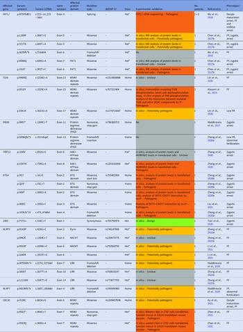
WEE2 genetic variants detected in women presenting fertilization failure or low fertilization rates after ICSI. The table indicates the specific exons and protein domains affected, the type of variant, the dbSNP nomenclature (if any), the gene dosage (Homo: homozygosity, Het: heterozygosity, Het*: compound heterozygosity) and the total number of patients in which the specific variant has been identified. The information about experimental validation for each genetic variant is also indicated, and the colour code provides information of their pathogenicity: no experimental validation reported (grey), reported as benign (green), reported as pathogenic (orange), potentially pathogenic or unclear according to existing data (yellow).

The severity of WEE2 pathogenic variants is diverse; while frameshift variants seem to produce a dramatic effect on oocyte activation ability, resulting in total FF after ICSI, in other cases residual fertilization can occur (Zhang et al., Reference Zhang, Mu, Zhao, Zhou, Chen, Wu, Yan, Wang, Zhao, Dong, Sun, Kuang, Li, Wang and Sang2019). Similarly to the outcome for PLCζ, some reports have associated the presence of variants with lower expression levels of the protein and alterations in its subcellular localization within the oocyte or in transfected cells, something that can explain the FF phenotype (Sang et al., Reference Sang, Li, Kuang, Wang, Zhang, Chen, Wu, Lyu, Fu, Yan, Mao, Xu, Mu, Li, Jin, He and Wang2018; Zhou et al., Reference Zhou, Wang, Liu, Zhang, Jin and Zhang2019).
The primary effect of WEE2 exonic variants is a dysfunction of kinase activity. WEE2 protein contains a Pkinase domain essential for this activity, a domain directly affected by 15 out of 26 (57.7%) of WEE2 variants compiled in the present review (Figure 3). Some variants (such as p.R207C) are predicted to destroy the hydrogen bonds between residues crucial for WEE2 function (Yang et al., Reference Yang, Shu, Cai, Sun, Cui and Liu2019). Concomitantly, variants such as p.E75Vfs*6, p.D234H and p.H337Yfs*24 compromise WEE2-dependent phosphorylation of Cdc2, preventing meiotic resumption and causing FF (Sang et al., Reference Sang, Li, Kuang, Wang, Zhang, Chen, Wu, Lyu, Fu, Yan, Mao, Xu, Mu, Li, Jin, He and Wang2018).
Location of genetic variants associated with female-related fertilization failure within the protein structure. The main domains and the total number of amino acids are indicated for each protein.

TUBB8: a factor not only associated with oocyte maturation defects
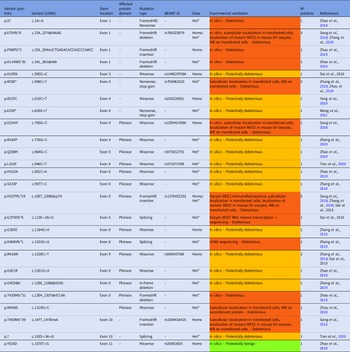
One of the genes with the most genetic variants explaining female-related zygotic problems after ICSI is TUBB8. In total, 27 TUBB8 variants present in 33 infertile women were identified in six studies (Table 4). Historically, the most frequent phenotype associated with TUBB8 sequence variants has been oocyte maturation arrest (either at the GV or MI stage) leading to the absence of MII oocytes (Feng et al., Reference Feng, Yan, Li, Yu, Sang, Tian, Xu, Chen, Qu, Sun, Sun, Jin, He, Kuang, Cowan and Wang2016; Yuan et al., Reference Yuan, Zheng, Liang, Li, Zhao, Li, Lai, Zhang and Wang2018). However, as shown in Table 4, genetic alterations in TUBB8 display a wide range of phenotypes including FF, low FR, abnormal fertilization and also zygotic arrest (PN are formed but the first mitotic division does not occur). Different studies have estimated that ∼30–40% of infertile women experiencing oocyte maturation arrest may present genetic variants in the TUBB8 gene (Feng et al., Reference Feng, Yan, Li, Yu, Sang, Tian, Xu, Chen, Qu, Sun, Sun, Jin, He, Kuang, Cowan and Wang2016; Chen et al., Reference Chen, Li, Li, Yan, Mao, Xu, Mu, Li, Jin, He, Kuang, Sang and Wang2017a; Chen et al., Reference Chen, Wang, Peng, Jiang, Zhang, Li, Li, Fu, Kuang, Sun, Wang, Zhang, Wu, Zhou, Lyu, Yan, Mao, Xu and Mu2019; Yang et al., Reference Yang, Yin, Li, Ma, Cao, Zhang, Chen and Zhao2021). Variants associated with this phenotype or embryo developmental arrest at the cellular stage were not included in this review.
TUBB8 genetic variants detected in women presenting fertilization failure, low fertilization rates abnormal fertilization and/or cleavage failure after ICSI. The table indicates the specific exons and protein domains affected, the type of variant, the dbSNP nomenclature (if any), the gene dosage (Homo: homozygosity, Het: heterozygosity, Het*: compound heterozygosity) and the total number of patients in which the specific variant has been identified. The information about experimental validation for each genetic variant is also indicated, and the colour code provides information of their pathogenicity: no experimental validation reported (grey), reported as benign (green), reported as pathogenic (orange), potentially pathogenic or unclear according to existing data (yellow). *Variants also observed in other patients exhibiting total oocyte maturation arrest or embryo developmental arrest

Microtubules, part of the cell cytoskeleton, are polymers formed by α- and β-tubulin heterodimers. These filaments constitute the meiotic spindle and are essential for oocyte maturation, but they are also involved in early fertilization events such as the extrusion of the second polar body and the first mitotic division. TUBB8 codes for β-tubulin isotype 8 (one of the nine β-tubulin isotypes), the main constituent of the oocyte (from the GV to MII stages) and embryo spindles. Therefore, genetic alterations of TUBB8 may result in irregular arrangements of the spindles and compromise the meiotic divisions in the oocyte or the first mitotic division in the zygote (Chen et al., Reference Chen, Wang, Peng, Jiang, Zhang, Li, Li, Fu, Kuang, Sun, Wang, Zhang, Wu, Zhou, Lyu, Yan, Mao, Xu and Mu2019). Indeed, 21 out of 27 variants compiled (77.8%) are considered pathogenic or potentially pathogenic (Table 4).
TUBB8 variants are associated with a wide spectrum of phenotypes, but the molecular alterations behind them are similar. TUBB8 variants can affect the folding and stability of β-tubulin, and reduce or prevent the interaction between α- and β-tubulin, causing problems in α/β heterodimer assembly and microtubule dynamics (Feng et al., Reference Feng, Yan, Li, Yu, Sang, Tian, Xu, Chen, Qu, Sun, Sun, Jin, He, Kuang, Cowan and Wang2016). These problems result in the disrupted organization of cytoplasmic microtubules and spindles with aberrant morphology, altogether compromising oocyte maturation and early mitotic divisions (Feng et al., Reference Feng, Yan, Li, Yu, Sang, Tian, Xu, Chen, Qu, Sun, Sun, Jin, He, Kuang, Cowan and Wang2016). As shown in Figure 3, most TUBB8 variants directly affect the GTPase domain and the C-terminal domain, potentially affecting protein function.
It is unclear which molecular mechanisms explain why some TUBB8 alterations are exclusively associated with total oocyte maturation arrest or embryo arrest at the cleavage stage (more than 50, data not shown), while others cause FF or zygotic arrest, and for some variants (such as p.E108K, p.T143Dfs*12, p.D295H, as some examples) the vast majority of retrieved oocytes were morphologically MII (Yuan et al., Reference Yuan, Zheng, Liang, Li, Zhao, Li, Lai, Zhang and Wang2018; Chen et al., Reference Chen, Wang, Peng, Jiang, Zhang, Li, Li, Fu, Kuang, Sun, Wang, Zhang, Wu, Zhou, Lyu, Yan, Mao, Xu and Mu2019). The phenotypic variability observed for the same TUBB8 variant could be explained by variable penetrance (Castel et al., Reference Castel, Cervera, Mohammadi, Aguet, Reverter, Wolman, Guigo, Iossifov, Vasileva and Lappalainen2018).
Additional genetic variants associated with female-related FF and zygotic arrest
In addition to WEE2 or TUBB8, other genetic variants have been identified in infertile women in nine more genes affecting early fertilization events, all of them reported in Table 5. For example, two recent studies have identified mutations affecting CDC20 in two infertile women in homozygosity or compound heterozygosity, both presenting FF or low FR after ICSI (Xu et al., Reference Xu, Zhu, Wang, Cai, Ge, Fu and Jin2021; Zhao et al., Reference Zhao, Guan, Meng, Wang, Wu, Chen, Hu, Zhu, Zhang, Mu, Chen, Sun, Wu, Wang, Zhou, Dong, Zeng, Liu and Li2021). Apart from these women, described in Table 5, CDC20 variants have been identified in patients experiencing complete oocyte maturation arrest at the MI stage or embryonic arrest (Huang et al., Reference Huang, Wang, Kong, Wang, Song, Lu, Ji, Luo and Tong2021; Zhao et al., Reference Zhao, Guan, Meng, Wang, Wu, Chen, Hu, Zhu, Zhang, Mu, Chen, Sun, Wu, Wang, Zhou, Dong, Zeng, Liu and Li2021). CDC20 is a cell division cycle protein that acts as an activator of the anaphase-promoting complex/cyclosome (APC/C), enabling the degradation of cyclin B1 and promoting the start of anaphase (Hwang et al., Reference Hwang, Lau, Smith, Mistrot, Hardwick, Hwang, Amon and Murray1998); based on that, one would expect an incomplete occurrence of meiosis in cases in which CDC20 is non-functional within the oocyte: oocyte maturation arrest or failure to resume meiosis after sperm entry, as observed in the infertile women previously mentioned. Indeed, these variants cause a reduction in CDC20 protein levels and, for the p.R322* variant, the generation of a non-functional truncated protein (Zhao et al., Reference Zhao, Guan, Meng, Wang, Wu, Chen, Hu, Zhu, Zhang, Mu, Chen, Sun, Wu, Wang, Zhou, Dong, Zeng, Liu and Li2021). All three CDC20 variants reported in this review directly affect the WD40 repeats, a region required for protein–protein interactions (Figure 3; Kraft et al., Reference Kraft, Vodermaier, Maurer-Stroh, Eisenhaber and Peters2005).
Genetic variants affecting different genes identified in patients with fertilization failure or low fertilization rates after ICSI, abnormal fertilization, oocyte degeneration or zygotic arrest at the pronuclear stage. The affected gene, specific protein domain, the phenotype/s of the patients, the gene dosage, and the experimental evidence are indicated. Het: heterozygosity, Homo: homozygosity; Het*: compound heterozygosity, FF: fertilization failure, FR: fertilization rate. The colour code provides information on the pathogenicity for each variant: no experimental validation reported (grey), reported as benign (green), reported as pathogenic (orange), potentially pathogenic or unclear according to existing data (yellow)

Another affected gene is PATL2, with six variants identified in three women, with significant phenotypic variability: oocyte maturation arrest, FF, low FR, cleavage failure, and embryo developmental arrest (Chen et al., Reference Chen, Li, Li, Yan, Mao, Xu, Mu, Li, Jin, He, Kuang, Sang and Wang2017a). PATL2 encodes for PAT1 homolog 2 (PATL2), an mRNA-binding protein that, in the oocyte, regulates the expression of specific mRNAs encoding proteins essential for oocyte maturation, meiotic progression and early embryo development (Nakamura et al., Reference Nakamura, Tanaka, Miyauchi, Huang, Tsujimoto and Matsumoto2010). Most PATL2 exonic variants reported to date are pathogenic or potentially pathogenic (Table 5).
Similarly to TUBB8 variants, the phenotypic variability observed for PATL2 variants may imply a difficult diagnosis, as some result in complete maturation arrest (in some cases, already at the GV stage), while others are associated with a high percentage of apparently normal MII oocytes, which stop their development a few hours after insemination.
PATL2 variants seem to affect protein function or reduce the gene’s expression to different extents both in oocytes and in transfected cells, therefore resulting in different phenotypes, mainly oocyte maturation arrest (either at the GV or MI stage; Wu et al., Reference Wu, Chen, Li, Song, Chen, Yan, Lyu, Wang, Kuang, Li and Sang2019). For example, ∼26–44% of patients with oocyte maturation arrested at the GV stage carry PATL2 pathogenic variants (Christou-Kent et al., Reference Christou-Kent, Kherraf, Amiri-Yekta, Le Blévec, Karaouzène, Conne, Escoffier, Assou, Guttin, Lambert, Martinez, Boguenet, Fourati Ben Mustapha, Cedrin Durnerin, Halouani, Marrakchi, Makni, Latrous and Kharouf2018; Huang et al., Reference Huang, Tong, Wang, Luo, Jin, Fu, Zhou, Li, Song, Liu and Zhu2018). However, there is no evidence that the reported PATL2 variants have affected PATL2 function as an mRNA-binding protein and its effect on mRNA translation in the oocyte, perhaps because the function of PATL2 is still largely unknown. For one splice variant identified in this gene (p.R75Vfs*21), sequencing of cDNA obtained from granulosa cells in the affected individuals showed the generation of abnormal transcripts, leading to truncated non-functional forms of the PATL2 protein (Chen et al., Reference Chen, Li, Li, Yan, Mao, Xu, Mu, Li, Jin, He, Kuang, Sang and Wang2017a).
Recently, bi-allelic pathogenic variants in TRIP13 have been identified in an infertile woman (Table 5); this woman had normal fertilization, but the zygotes failed to undergo the first mitotic division (Zhang et al., Reference Zhang, Li, Fu, Li, Diao, Li, Chen, Du, Zhou, Mu, Yan, Wu, Liu, Wang, Zhao, Dong, He, Liang and Kuang2020). TRIP13 encodes for thyroid hormone receptor interactor 13 (or Pachytene checkpoint protein 2 homolog), a factor with ATPase activity involved in meiotic recombination and essential for chromosome synapsis (Roig et al., Reference Roig, Dowdle, Toth, de Rooij, Jasin and Keeney2010).
Apart from p.I198V and p.V247M (included in the present review), other TRIP13 variants have been reported in infertile women who could produce a variable proportion of morphologically normal MII oocytes. Among the molecular alterations caused by TRIP13 variants there is a reduction of TRIP13 protein abundance, an inability to remove HORMAD2 from synapse chromosome axes (a checkpoint required for meiotic progression), and reduced ATPase activity (Zhang et al., Reference Zhang, Li, Fu, Li, Diao, Li, Chen, Du, Zhou, Mu, Yan, Wu, Liu, Wang, Zhao, Dong, He, Liang and Kuang2020). Indeed, both missense variants reported by Zhang and colleagues directly affected the AAA+ ATPase domain (Figure 3).
The four next genes implicated in FF and zygotic arrest are part of the subcortical maternal complex (SCMC). The SCMC is a complex of proteins encoded by maternal-effect genes that controls the first cellular divisions of the preimplantation embryo (Bebbere et al., Reference Bebbere, Masala, Albertini and Ledda2016). SCMC orchestrates the zygotic events occurring in parallel to oocyte activation, including the regulation of subcortical actin and mitotic spindle positioning, the regulation of the translation of maternal transcripts and the epigenetic reprogramming (Li et al., Reference Li, Baibakov and Dean2008; Bebbere et al., Reference Bebbere, Masala, Albertini and Ledda2016). Functional studies using the mouse model have shown that the knockout for these factors causes lethal phenotypes such as preimplantation embryo arrests at the cleavage stage and, in some cases, at the zygotic stage (Tashiro et al., Reference Tashiro, Kanai-Azuma, Miyazaki, Kato, Tanaka, Toyoda, Yamato, Kawakami, Miyazaki and Miyazaki2010). The first is TLE6, which encodes for transducin-like enhancer protein 6 (TLE6); three variants affecting four women with FF after ICSI have been reported (Table 5). The crucial role of SCMC is demonstrated by the phenotypes of the patients carrying the TLE6 p.S510Y variant in homozygosity, who present a dramatic phenotype: three oocytes developing to 2PN among 77 inseminated by ICSI from a total of seven cycles (3.9% FR; Alazami et al., Reference Alazami, Awad, Coskun, Al-Hassan, Hijazi, Abdulwahab, Poizat and Alkuraya2015). The molecular mechanisms behind this phenotype are explained by some authors: specific TLE6 variants can disturb TLE6 protein phosphorylation sites (as demonstrated in vitro in transfected cells) and compromise the ability of the protein to be phosphorylated by PKA, as well as inhibit its interaction with other SCMC components such as OOEP and KDHC3L, altogether preventing the meiotic resumption of the inseminated oocyte (Alazami et al., Reference Alazami, Awad, Coskun, Al-Hassan, Hijazi, Abdulwahab, Poizat and Alkuraya2015).
The second gene is PADI6, which encodes for protein arginine deiminase type-6 (PADI6); with two variants identified in two infertile women (Table 5). Its knockout in mice (Padi6−/−) caused embryo developmental arrest mainly at the 2-cell stage, the stage in which mouse embryonic genome activation (EGA) occurs (Yurttas et al., Reference Yurttas, Vitale, Fitzhenry, Cohen-Gould, Wu, Gossen and Coonrod2008). In humans, three waves of transcriptional genome activation occur at the 2-cell, 4-cell and 6- to 10-cell stages (Vassena et al., Reference Vassena, Boué, González-Roca, Aran, Auer, Veiga and Izpisua Belmonte2011) and, consistently, Xu and colleagues identified PADI6 variants in infertile women presenting embryonic arrest at these stages (Xu et al., Reference Xu, Shi, Fu, Yu, Feng, Sang, Liang, Chen, Qu, Li, Yan, Mao, Kuang, Jin, He, Sun and Wang2016). In these women, the oocytes lacked detectable PADI6 protein in the cytoplasm, and arrested embryos had reduced levels of phosphorylated RNA polymerase II, something compromising EGA (Xu et al., Reference Xu, Shi, Fu, Yu, Feng, Sang, Liang, Chen, Qu, Li, Yan, Mao, Kuang, Jin, He, Sun and Wang2016). As indicated in Table 5, in some cases all these alterations can clinically manifest already at the zygotic stage, causing poor FR, abnormal fertilization or zygotic arrest (Maddirevula et al., Reference Maddirevula, Coskun, Awartani, Alsaif, Abdulwahab and Alkuraya2017; Zheng et al., Reference Zheng, Chen, Dai, Dai, Guo, Lu, Gong, Lu and Lin2020a).
The third gene is NLRP5 (NLR Family Pyrin Domain Containing 5, also known as MATER). While some studies have reported genetic variants in infertile patients presenting embryonic arrest (Mu et al., Reference Mu, Zhang, Wu, Fu, Chen, Yan, Li, Zhou, Wang, Zhao, Dong, Kuang, Sun, He, Wang and Sang2020; Xu et al., Reference Xu, Qian, Liu, Wang, Wang, Zhou, Zhang, Pang, Ye, Xue and Sun2020), seven variants have been identified in four women experiencing FF after ICSI (Table 5). Similarly, one mutation in a fourth SCMC factor – NLRP7 (NOD-like receptor family pyrin domain containing 7) – has been recently identified in a woman with FF (Maddirevula et al., Reference Maddirevula, Awartani, Coskun, AlNaim, Ibrahim, Abdulwahab, Hashem, Alhassan and Alkuraya2020). Other components of the SCMC are KHDC3L, OOEP, ZBED3 and NLRP2, but genetic variants previously reported in these genes were associated exclusively with embryo arrest at the cleavage stage, without any apparent effect at the zygote stage (Zheng et al., Reference Zheng, Hu, Dai, Zhang, Gu, Dai, Guo, Xu, Li, Zhang, Hu, Gong, Lu and Lin2021b).
For NLRP5 and NLRP7 variants, only in silico approaches have been performed to predict their effects, so further research is required to gain insight into their specific molecular pathogenesis. However, both proteins have an N-terminal pyrin domain required for binding adaptor proteins, a central nucleotide-binding oligomerization domain (NACHT) and a C-terminal leucine-reach repeat (LRR) for ligand binding and the establishment of protein–protein interactions that regulate cellular functions (Proell et al., Reference Proell, Riedl, Fritz, Rojas and Schwarzenbacher2008). As shown in Figure 3, most variants detected in infertile patients affect these domains. In mouse oocytes and embryos, NLRP5 or MATER was involved in mitochondrial activation, endoplasmic reticulum distribution and calcium homeostasis (Fernandes et al., Reference Fernandes, Tsuda, Perumalsamy, Naranian, Chong, Acton, Tong, Nelson and Jurisicova2012; Kim et al., Reference Kim, Zhang, Kan, Cohen, Mukai, Travis and Coonrod2014). For this reason, pathogenic variants in these SCMC genes could impair protein structure and/or function, altering cellular functions during the zygote stage.
Regarding SCMC, genetic variants affecting PADI6, NLRP5 and NLRP7 (and other SCMC genes such as KHDC3L) have been associated with the occurrence of hydatidiform moles, which are aberrant pregnancies characterized by the absence of, or abnormal, embryonic development, and hyperproliferation of the trophoblast or placenta (Qian et al., Reference Qian, Nguyen, Rezaei, Huang, Tao, Zhang, Cheng, Yang, Asangla, Majewski and Slim2018; Fallahi et al., Reference Fallahi, Anvar, Razban, Momtahan, Namavar-Jahromi and Fardaei2020). The role of these genes in setting the genomic imprinting could be an explanation (Docherty et al., Reference Docherty, Rezwan, Poole, Turner, Kivuva, Maher, Smithson, Hamilton-Shield, Patalan, Gizewska, Peregud-Pogorzelski, Beygo, Buiting, Horsthemke, Soellner, Begemann, Eggermann, Baple and Mansour2015). Interestingly, the occurrence of hydatidiform moles can be male related also, associated with sperm unable to trigger the normal pattern of Ca2+ oscillations and/or the presence of pathogenic variants in PLCZ1 (Nikiforaki et al., Reference Nikiforaki, Vanden Meerschaut, De Gheselle, Qian, Van den Abbeel, De Vos, Deroo, De Sutter and Heindryckx2014; Guggilla et al., Reference Guggilla, Ferrer-Buitrago, Bonte, De Sutter, Coucke and Heindryckx2019). Altogether, these observations not only increase the spectrum of clinical consequences associated with alterations in factors involved in early fertilization, but also confirm that calcium oscillations and early fertilization events affect the future development of the embryo.
Finally, BTG4 and ZAR1 are two genes associated with female-related zygotic arrest. Five BTG4 exonic variants were identified in homozygosity in five patients, all of them presenting morphologically normal oocytes that could be successfully fertilized by ICSI (2PN formed), but embryo development was arrested at the zygotic stage (Zheng et al., Reference Zheng, Zhou, Sha, Niu, Sun, Shi, Zhao, Zhang, Dai, Cai, Meng, Hu, Gong, Li, Fu, Shi, Lu, Chen and Fan2020b). BTG4 (B cell translocation gene 4) is a key factor for maternal–zygotic transition (MZT). BTG4 forms a complex with CNOT7 and EIF4E, facilitating the decay of maternal mRNAs during the zygotic stage, an oocyte cytoplasmic maturation step required for MZT and the first mitotic division to occur (Yu et al., Reference Yu, Ji, Sha, Dang, Zhou, Zhang, Liu, Wang, Hu, Sun, Sun, Tang and Fan2016). The mechanisms by which BTG4 variants produce zygotic arrest are the reduction or absence of BTG4 (for c.1A>G and p.Q25* variants), generation of truncated non-functional protein (for p.I159Lfs*15), or an altered BGT4–CNOT7 interaction (for the p.A56T variant), problems causing a loss of maternal mRNA decay in the zygote, abnormal MZT and the subsequent cleavage failure (Zheng et al., Reference Zheng, Zhou, Sha, Niu, Sun, Shi, Zhao, Zhang, Dai, Cai, Meng, Hu, Gong, Li, Fu, Shi, Lu, Chen and Fan2020b). Three out of four variants reported are located at the N-terminal region of the protein, directly affecting the BGT domain (Figure 3).
ZAR1 encodes Zygote arrest protein 1, and studies using the murine model have demonstrated its essential role in early fertilization: Zar1 –/– female mice were infertile, and less than 20% of fertilized zygotes underwent the first mitotic division (Wu et al., Reference Wu, Viveiros, Eppig, Bai, Fitzpatrick and Matzuk2003). A recent study analyzed the ZAR1 sequence in 47 patients who presented cleavage failure after ICSI and in 188 fertile women, and one exonic variant was exclusively identified in the first group (Table 5). However, the synonymous variant in ZAR1 (c.516C>T) was not a cause of infertility, probably because there was no change in the protein sequence and variants in simple heterozygosity are not expected to cause a phenotype of zygotic arrest (Zar +/– female mice are fertile) (Wu et al., Reference Wu, Viveiros, Eppig, Bai, Fitzpatrick and Matzuk2003). Future studies may be helpful to clarify if ZAR1 variants in homozygosity can cause alterations in oocyte-to-embryo transition in humans.
Nature and inheritance of genetic variants associated with FF and zygotic arrest
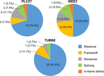
Most variants compiled in the present review are missense (n = 82; 67.8%), and the rest of the variants were frameshift (either insertion or deletions), nonsense or stop gain, splicing variants, in-frame deletions or insertions, whereas one patient carried a synonymous variant. For the three genes with a higher number of different variants identified in infertile patients (PLCZ1, WEE2, and TUBB8), the proportion of each type of variant is indicated in Figure 4. Pathogenic effects are expected especially for frameshift, nonsense, and splicing variants, as they mostly result in truncated or aberrant proteins. However, most missense variants reported here also generate dramatic effects on protein structure and/or function, characterized by diminished or lost enzymatic function, altered ability to interact with the substrate or protein partners, alteration of phosphorylation sites, or reduction of expression levels.
Proportion of each type of genetic variant for the three genes in which more genetic variants have been identified in infertile patients presenting FF or zygotic arrest after ICSI: PLCZ1, WEE2 and TUBB8.

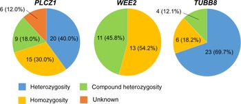
Among the 141 infertile men and women included in this review, 46 (32.6%) carried a single variant in heterozygosity, 89 carried bi-allelic variants, either in homozygosity (n = 55; 39.0%) or in compound heterozygosity (n = 34; 24.1%); while the inheritance pattern was unknown for six patients (4.3%). However, while pathogenic variants in heterozygosity have been identified in infertile patients for PLCZ1 and TUBB8, other genes (WEE2, PATL2, TLE6, PADI6, BTG4, NLRP5, NLRP7, CDC20, TRIP13, DNAH17, ACTL9 and ACTLA7) seem to cause a phenotype of infertility in a recessive manner, as both alleles are mutated in all patients identified. Moreover, for this second group of genes, many studies included the relatives of the affected patients in the analysis, demonstrating that variants in heterozygosity did not affect fertility and overall health (Alazami et al., Reference Alazami, Awad, Coskun, Al-Hassan, Hijazi, Abdulwahab, Poizat and Alkuraya2015; Chen et al., Reference Chen, Zhang, Sun, Kuang, Mao, Wang, Yan, Li, Xu, Yu, Fu, Mu, Zhou, Li, Jin, He, Sang and Wang2017b; Sang et al., Reference Sang, Li, Kuang, Wang, Zhang, Chen, Wu, Lyu, Fu, Yan, Mao, Xu, Mu, Li, Jin, He and Wang2018; Zhang et al., Reference Zhang, Li, Fu, Li, Diao, Li, Chen, Du, Zhou, Mu, Yan, Wu, Liu, Wang, Zhao, Dong, He, Liang and Kuang2020; Zheng et al., Reference Zheng, Zhou, Sha, Niu, Sun, Shi, Zhao, Zhang, Dai, Cai, Meng, Hu, Gong, Li, Fu, Shi, Lu, Chen and Fan2020b). In Figure 5, the distribution of the genetic load of the variants is represented for the three genes affecting a higher number of patients (PLCZ1, WEE2 and TUBB8).
Proportion of patients presenting genetic variants in homozygosity, heterozygosity or compound heterozygosity for the genes affecting a higher number of infertile patients: PLCZ1, WEE2 and TUBB8.

After the first reports using gene sequencing to study male-related FF, the initial hypothesis was that pathogenic PLCZ1 variants needed to be present in both maternal and paternal alleles to cause infertility (Kashir et al., Reference Kashir, Konstantinidis, Jones, Lemmon, Lee, Hamer, Heindryckx, Deane, De Sutter, Fissore, Parrington, Wells and Coward2012). However, nearly half the men with FF or low FR included in this study carried PLCZ1 in simple heterozygosity (20/46, 43.5%; Figure 5), suggesting that heterozygosity may be sufficient to cause oocyte activation deficiencies for certain variants. If this is the case, some PLCZ1 variants would be acting in a dominant negative mechanism; while this statement needs further research and confirmation, there are some facts that support it. For instance, spermatids share cytoplasmic bridges during their development, interchanging RNA transcripts, implicating that variants in heterozygosity could cause a population of testicular sperm genetically different (part of the sperm would be wild-type) but phenotypically similar (Dadoune et al., Reference Dadoune, Siffroi and Alfonsi2004). Indeed, none of the pathogenic or potentially pathogenic PLCZ1 variants indicated in Table 1 has been found in patients or donors with good FR, commonly used as controls in different studies. Second, the presence of mutated forms of PLCζ could affect the ability of the wild-type protein to trigger calcium oscillations in the oocyte, as demonstrated by co-injecting wild-type and mutant forms of human recombinant rLCζ into mice oocytes (Heytens et al., Reference Heytens, Parrington, Coward, Young, Lambrecht, Yoon, Fissore, Hamer, Deane, Ruas, Grasa, Soleimani, Cuvelier, Gerris, Dhont, Deforce, Leybaert and De Sutter2009). Third, especially for genetic variants causing a total disruption of PLCζ enzymatic activity (such as p.V189Cfs*12, p.C196* and p.V326Kfs*25), heterozygosity would either reduce the amount of wild-type protein present in all sperm or at least reduce the percentage of sperm with wild-type protein among the whole sperm population, diminishing the chances to achieve oocyte activation after ICSI, especially in oocytes with cytoplasmic immaturity. The same is true for variants in domains like C2, which may prevent PLCζ protein from reaching and attaching to the perinuclear theca, being disposed through the residual body and accumulating in abnormal areas within the oocyte after fertilization (Escoffier et al., Reference Escoffier, Lee, Yassine, Zouari, Martinez, Karaouzène, Coutton, Kherraf, Halouani, Triki, Nef, Thierry-Mieg, Savinov, Fissore, Ray and Arnoult2016). The complexity of human fertilization, and the different PLCZ1 variants can explain why the phenotype observed in the patients ranges from complete infertility to subfertility or low FR, and possibly explain why the fathers of the patients included, carrying PLCZ1 variants in heterozygosity, were able to generate offspring in vivo (Torra-Massana et al., Reference Torra-Massana, Cornet-Bartolomé, Barragán, Durban, Ferrer-Vaquer, Zambelli, Rodriguez, Oliva and Vassena2019; Yan et al., Reference Yan, Fan, Wang, Yan, Li, Ouyang, Wu, Yin, Zhao, Kuang, Li and Lyu2020). This phenotypical behaviour, which may seem contradictory, is similar in Plcz1 KO mice: while total FF with a complete absence of calcium oscillations is observed through ICSI, offspring could be produced in vivo (Nozawa et al., Reference Nozawa, Satouh, Fujimoto, Oji and Ikawa2018; Hachem et al., Reference Hachem, Godwin, Ruas, Lee, Ferrer Buitrago, Ardestani, Bassett, Fox, Navarrete, de Sutter, Heindryckx, Fissore and Parrington2017).
Regarding TUBB8, the same phenotypes are observed for both monoallelic and bi-allelic variants (Figure 5). As previously reported, TUBB8 variants can have dominant negative effects on microtubule function (Feng et al., Reference Feng, Yan, Li, Yu, Sang, Tian, Xu, Chen, Qu, Sun, Sun, Jin, He, Kuang, Cowan and Wang2016). Indeed, in affected infertile women, most TUBB8 variants are either de novo or inherited from their fathers (alterations in TUBB8 do not cause male infertility; Feng et al., Reference Feng, Yan, Li, Yu, Sang, Tian, Xu, Chen, Qu, Sun, Sun, Jin, He, Kuang, Cowan and Wang2016). Surprisingly, in some cases the variants were inherited from the mother, which could be explained by differences in the protein structure abnormalities caused by different variants, or incomplete penetrance (Chen et al., Reference Chen, Zhang, Sun, Kuang, Mao, Wang, Yan, Li, Xu, Yu, Fu, Mu, Zhou, Li, Jin, He, Sang and Wang2017b, Reference Chen, Wang, Peng, Jiang, Zhang, Li, Li, Fu, Kuang, Sun, Wang, Zhang, Wu, Zhou, Lyu, Yan, Mao, Xu and Mu2019).
In contrast, all WEE2 variants identified were either in homozygosity or compound heterozygosity (Figure 5). The inheritance of WEE2 pathogenic variants is autosomal recessive, and carriers of just one mutated allele do not display infertility (Zhao et al., Reference Zhao, Chen, Yu, Bian, Cao, Ning, Su, Zhang and Zhao2019). This inheritance pattern is shared by maternal-effect genes associated with the SCMC (TLE6, NLRP5, NLRP7 and PADI6) and by the rest of the genes compiled in the present review, either associated with female infertility (PATL2, TRIP13 and BGT4) or male infertility (ACTL9, ACTLA7 and DNAH17). For all genes with recessive inheritance, the proportion of infertile individuals is expected to be higher in families with a history of consanguinity when compared with the general population. In fact, among the 54 infertile patients carrying homozygous variants in the studies included in the present review, at least 29 (53.7%) were members of consanguineous families. For this reason, the possibility of consanguinity and the genetic information of the closest relatives should be considered when addressing the genetic analysis of male or female infertility associated with FF or zygotic arrest.
Treatment options and genetic counselling for patients carrying variants associated with FF and zygotic arrest
The treatment options for patients with FF or zygotic arrest are still scarce but, as indicated in Table 6, some experimental alternatives have been reported recently for patients affected by PLCZ1, WEE2, ACTL9, ACTLA7 and, to a lesser extent, TUBB8, CDC20 and TRIP13 pathogenic variants. In contrast, no treatment options have been attempted or proposed for patients with DNAH17, PATL2, TLE6, PADI6, NLRP5, NLRP7 and BTG4 variants, while variants identified in WBP2NL and ZAR1 are considered non-pathogenic.
Summary of treatment options for patients carrying infertility-related genetic variants causing fertilization failure or zygotic arrest after ICSI. The experimental status of these treatments and its clinical efficiency are also indicated.

Fortunately, ICSI combined with AOA using calcium ionophores has proven to be effective for infertile men carrying deleterious PLCZ1 variants (Torra-Massana et al., Reference Torra-Massana, Cornet-Bartolomé, Barragán, Durban, Ferrer-Vaquer, Zambelli, Rodriguez, Oliva and Vassena2019; Yan et al., Reference Yan, Fan, Wang, Yan, Li, Ouyang, Wu, Yin, Zhao, Kuang, Li and Lyu2020), and AOA is also effective in cases of ACTL9 and ACTLA7 variants to overcome the FF problem (Dai et al., Reference Dai, Zhang, Guo, Zhou, Gu, Zhang, Hu, Zong, Song, Zhang, Dai, Gong, Lu, Zheng and Lin2021). The use of AOA generates an increase in the oocyte cytoplasmic levels of calcium. Apparently, this signal is enough to compensate for the sperm’s inability to do so and initiate embryo development. For this reason, genetic screening of at least PLCZ1 should be encouraged in cases of OAF after ICSI of suspected male origin, and used for supporting the use of AOA in future ICSI cycles. Recently, Cheung et al. demonstrated the necessity of confirming sperm-related OAD before exposure of the gametes to the chemicals used in AOA (Cheung et al., Reference Cheung, Xie, Parrella, Keating, Rosenwaks and Palermo2020). Alternatively, the injection of PLCZ1 wild-type cRNA or recombinant PLCZ1 protein is demonstrated to trigger oocyte activation in mature oocytes, and some studies have already set the protocol conditions to use this method in humans (Yoon et al., Reference Yoon, Eum, Lee, Lee, Kim, Han, Won, Park, Shim, Lee, Fissore, Lee and Yoon2012; Nomikos et al., Reference Nomikos, Yu, Elgmati, Theodoridou, Campbell, Vassilakopoulou, Zikos, Livaniou, Amso, Nounesis, Swann and Lai2013; Yamaguchi et al., Reference Yamaguchi, Ito, Kuroda, Takeda and Tanaka2017). This method has been proposed as a better option compared with AOA, as the calcium oscillations generated are much more similar to the outcomes in an in vivo situation. Nevertheless, no evidence of a benefit in the rPLCζ–AOA protocol over the one using the Ca2+ ionophore was observed when considering preimplantation embryo development (Ferrer-Buitrago et al., Reference Ferrer-Buitrago, Tilleman, Thys, Hachem, Boel, Van Nieuwerburgh, Deforce, Leybaert, De Sutter, Parrington and Heindryckx2020).
Conversely, the FF problem originated by WEE2 and NLRP5 variants (in women) or DNAH17 alterations (in men) is not overcome by AOA using calcium ionophores (Table 6). AOA seems to enable the extrusion of the second polar body in oocytes with pathogenic WEE2 variants inseminated by ICSI, but these oocytes failed to produce pronuclei (Sang et al., Reference Sang, Li, Kuang, Wang, Zhang, Chen, Wu, Lyu, Fu, Yan, Mao, Xu, Mu, Li, Jin, He and Wang2018; Yang et al., Reference Yang, Shu, Cai, Sun, Cui and Liu2019). As suggested by other authors, the presence of non-functional WEE2 led to high levels of MPF that prevented the oocyte from completing meiotic resumption, and AOA will not benefit these patients. A correct diagnosis before treatment counselling gains relevance in this scenario, as previous studies have reported that AOA did not overcome the FF problem in some patients, especially in cases of suspected oocyte-related origin (Combelles et al., Reference Combelles, Morozumi, Yanagimachi, Zhu, Fox and Racowsky2010; Vanden Meerschaut et al., Reference Vanden Meerschaut, Nikiforaki, De Gheselle, Dullaerts, Van den Abbeel, Gerris, Heindryckx and De Sutter2012; Ferrer-Buitrago et al., Reference Ferrer-Buitrago, Dhaenens, Lu, Bonte, Vanden Meerschaut, De Sutter, Leybaert and Heindryckx2018). Recent evidence has indicated that injection of WEE2, TRIP13 or CDC20 wild-type cRNA may be a viable alternative for infertile women carrying pathogenic variants, but further research needs to be conducted to test the safety and efficiency in human oocytes and to evaluate any potential damaging effects on the developing embryo (Yang et al., Reference Yang, Shu, Cai, Sun, Cui and Liu2019; Table 6).
While AOA may be a good option for part of the cases of OAF, this is not so clear for patients successfully forming 2PN but failing to undergo the first mitotic division. As previously detailed, arrest at the pronuclear stage or cleavage failure is a phenotype observed in some patients carrying variants in TLE6, TUBB8 or PADI6, or BTG4. Preliminary studies have shown that AOA could be a good option for some patients presenting this phenotype (Ebner et al., Reference Ebner, Oppelt, Wöber, Staples, Mayer, Sonnleitner, Bulfon-Vogl, Gruber, Haid and Shebl2015), but further research is required to confirm this hypothesis (Darwish and Magdi, Reference Darwish and Magdi2015). Interestingly, a recent study in the mouse has demonstrated that the genome transfer technique could be useful in overcoming the infertility condition originated by mutations in maternal-effect genes (Bai et al., Reference Bai, Sun, Jia, Yin, Zhang, Li, Gao, Du, Li, Lin, Tu, Wang, Pan, Liang, Guo, Ruan, Kou, Zhao and Wang2020) but, currently, its application is just speculative. Oocyte spindle transfer could be another potential option for patients showing oocyte-related FF or embryo arrest (Costa-Borges et al., Reference Costa-Borges, Spath, Miguel-Escalada, Mestres, Balmaseda, Serafín, Garcia-Jiménez, Vanrell, González, Rink, Wells and Calderón2020). This technique was used for the first time in humans in 2016 to prevent the transmission of Leigh syndrome (a mitochondrial disease), and led to the birth of a healthy boy (Zhang et al., Reference Zhang, Liu, Luo, Lu, Chávez-Badiola, Liu, Yang, Merhi, Silber, Munné, Konstantinidis, Wells, Tang and Huang2017).
Despite the dramatic clinical consequences caused by the variants described in this review, patients with partial FF or zygotic arrest as well as patients in whom the infertility problem can be overcome by an alternative treatment can produce viable embryos and transmit the variants to the offspring. This risk is higher for variants presenting a dominant inheritance, when a single affected allele causes a phenotype of infertility. In these patients, preimplantation genetic testing (PGT-M) could be an option to select embryos not affected by the variants, when allowed. However, so far there has been no evidence supporting the clinical utility or appropriateness of PGT-M for genetic alterations that do not cause severe diseases or affect the general quality of life.
For all of these reasons, genetic counselling should be offered to patients carrying variants causing FF, cleavage failure or other infertility problems. These patients should be informed about the diagnostic value of genetic screening and the potential risks of transmission to the offspring, but should be offered the possibility to evaluate the family history, coordinate the specific tests needed to investigate a genetic cause for infertility and receive professional support to cope with the emotional consequences of repeat failure to achieve a pregnancy. Unfortunately, only four out of 45 (8.9%) studies included in the present review mention the need for genetic counselling for these infertile patients. As the knowledge of the genetic basis of human infertility expands, genetic counselling practice is expected to do so.
Discussion
The results of the present review demonstrated that high numbers of genetic alterations are associated with FF and zygotic arrest after ICSI, and that most of them are pathogenic (except for WBP2NL and ZAR1 genes). The high effect of these alterations on the fertilization process is exemplified by the search for WEE2 inhibitors to be used as a non-hormonal contraceptive strategy (Hanna et al., Reference Hanna, Yao, Martin, Schönbrunn, Georg, Jensen and Cuellar2019).
Lower fertilization rates or oocyte activation deficiencies are somehow expected for patients with severe sperm parameters (such as severe asthenozoospermia, cryptozoospermia or use of testicular sperm) or some oocytes abnormalities, such as the presence of cytoplasmic vacuoles (Ebner et al., Reference Ebner, Moser, Sommergruber, Gaiswinkler, Shebl, Jesacher and Tews2005). For this reason, we only focused on patients presenting morphologically normal sperm and at least some morphologically normal and mature oocytes, as these cases are often unexpected. Nevertheless, previous studies have identified specific genetic variants associated with abnormal sperm morphology and, as a consequence, compromised fertilization ability after ICSI, including mutations in DPY19L2 and SPATA16 that cause globozoospermia (Rybouchkin et al., Reference Rybouchkin, Dozortsev, Pelinck, De Sutter and Dhont1996; Dam et al., Reference Dam, Koscinski, Kremer, Moutou, Jaeger, Oudakker, Tournaye, Charlet, Lagier-Tourenne, van Bokhoven and Viville2007).
Different approaches have been tested to study gametes from patients with FF through ICSI, for example the analysis of PLCZ1 protein levels and subcellular localization, as well as diverse functional assays. For example, heterologous ICSI models, using mouse or hamster oocytes in most cases, have been proposed as a diagnostic tool for sperm-related FF (Yazawa et al., Reference Yazawa, Yanagida, Katayose, Hayashi and Sato2000; Heindryckx et al., Reference Heindryckx, Van der Elst, De Sutter and Dhont2005). These systems include the mouse oocyte activation test (MOAT) and mouse oocyte calcium analysis (MOCA; Vanden Meerschaut et al., Reference Vanden Meerschaut, Leybaert, Nikiforaki, Qian, Heindryckx and De Sutter2013); as well as its variant in human gametes, HOCA (Ferrer-Buitrago et al., Reference Ferrer-Buitrago, Dhaenens, Lu, Bonte, Vanden Meerschaut, De Sutter, Leybaert and Heindryckx2018). Similarly, heterologous models using bovine, rabbit or Xenopus oocytes have been applied to test sperm aster formation and/or microtubule enucleation (Tachibana et al., Reference Tachibana, Terada, Ogonuki, Ugajin, Ogura, Murakami, Yaegashi and Okamura2009; Amargant et al., Reference Amargant, García, Barragán, Vassena and Vernos2018). However, these methods cannot easily be introduced in routine fertility clinical practice due to technical or cost–effectiveness limitations. In contrast, gene sequencing does not provide functional insights but offers distinct advantages: it is fast, relatively cheap, and does not require complex protocols or reagents when provided by an external service; it is easy to implement in fertility clinics or associated centres, it can also be applied to the patient’s parents or other relatives to analyze the inheritance profile and the origin of each specific variant, and it can be performed on a very small amount of sample (even a single cell) or using samples other than gametes (such as blood or saliva), especially useful as mature oocytes or semen presenting severe oligozoospermia are limiting samples.
The complexity of fertilization, the high diversity of male and female factors involved, as well as the ethical, legal and technical limitations to investigate this process in humans explain why fertilization and its alterations are still not completely understood. Nevertheless, many genes can be considered potential diagnostic targets. In men, genetic alterations causing poor sperm DNA condensation or protamine deficiency, sperm proteasome function, abnormal sperm head-neck junction and altered sperm centriole structure or function may explain the additional cases of male-related FF or zygotic arrest after ICSI (Chemes et al., Reference Chemes, Puigdomenech, Carizza, Olmedo, Zanchetti and Hermes1999; Rawe et al., Reference Rawe, Terada, Nakamura, Chillik, Olmedo and Chemes2002, Reference Rawe, Díaz, Abdelmassih, Wójcik, Morales, Sutovsky and Chemes2008; Lazaros et al., Reference Lazaros, Vartholomatos, Hatzi, Kaponis, Makrydimas, Kalantaridou, Sofikitis, Stefos, Zikopoulos and Georgiou2011). Potential targets in sperm could be TSSK6, involved in histone-to-protamine transition during spermiogenesis (Jha et al., Reference Jha, Tripurani and Johnson2017), or GSTO2, encoding an enzyme located in the sperm post-acrosomal sheath that, if inhibited, causes a delay in sperm decondensation and lower rates of zygotic division (Hamilton et al., Reference Hamilton, Suzuki, Aguila, Meinsohn, Smith, Protopapas, Xu, Sutovsky and Oko2019).
In women, the potential targets to identify infertility-related genetic variants are diverse. Apart from TUBB8, genetic variants in factors involved in oocyte spindle structure and function and microtubule nucleation such as PLK1 and DCTN3 could cause failed ICSI outcomes (Fan et al., Reference Fan, Zhao, Liu, Tan, Ding, Li, Zhao, Yan, Sun, Yu and Qiao2015). Similarly, novel pathogenic variants could be found in oocyte factors involved in sperm decondensation, such as HIRA and the recently described splicing kinase SRPK1 (Gou et al., Reference Gou, Lim, Ma, Aubol, Hao, Wang, Zhao, Liang, Shao, Zhang, Meng, Li, Zhang, Xu, Li, Rosenfeld, Mellon, Adams, Liu and Fu2020), but none has been identified in humans so far. As exemplified by PATL2, genetic alterations in other genes affecting oocyte maturation can cause an inability of MII oocytes to undergo complete activation. Similarly, as found with WEE2 and CDC20, genetic alterations affecting additional factors involved in the oocyte activation signalling pathway could explain part of the FF and zygotic arrest after ICSI. For example, oocytes from mice knockout for Smc1b and Mos are arrested at meiosis II (Araki et al., Reference Araki, Naito, Haraguchi, Suzuki, Yokoyama, Inoue, Aizawa, Toyoda and Sato1996; Revenkova et al., Reference Revenkova, Eijpe, Heyting, Hodges, Hunt, Liebe, Scherthan and Jessberger2004). In humans, some genetic variants in MOS have been recently identified in infertile women experiencing preimplantation embryonic arrest and embryo fragmentation, but none has been associated with OAF or with arrest prior to the first mitotic division (Zhang et al., Reference Zhang, Zheng, Ren, Hu, Tong, Zhang, Li, Wang, Jiang, Jin, Yang, Cao, He, Ma, Zhang, Gu, Hu, Luo and Gong2021; Zeng et al., Reference Zeng, Shi, Xu, Shi, Wu, Li, Xue, Zhu, Chen, Sang and Wang2022).
Some limitations may exist in the present review. For example, there is a risk of incomplete retrieval of published articles matching the research question, a risk that was reduced by evaluating different sources and performing a manual analysis of references for each study identified. In addition, due to the multiplicity of phenotypes originating from different variants in the same gene, it is possible that the present catalogue may have skipped some variants described in patients presenting alternative phenotypes (such as complete oocyte maturation arrest or preimplantation developmental arrest at the cleavage stage) but with the potential to cause FF or zygotic arrest. Finally, as may happen in other similar reviews, not all ethnicities are represented, as most studies include patients of Asian origin. Future research is needed to overcome this limitation, by providing an independent verification on additional families and ethnic groups.
In summary, according to the existing evidence, 14 out of 16 genes included in the present review are potential targets for genetic screening of variants related to FF or zygotic arrest after ICSI: PLCZ1, DNAH17, ACTL9 and ACTLA7 (in infertile men), and WEE2, TUBB8, PATL2, TLE6, PADI6, TRIP13, BTG4, CDC20, NLRP5, and NLRP7 (in infertile women). Pathogenic variants in some of these genes (such as PLCZ1 and WEE2) can explain a large proportion of unexpected and often repetitive FF cases. Many more targets and novel variants are expected to appear in the near future not only related to FF, but also to all types of infertility, allowing the application of what is known as genome-based reproductive medicine or preconception genomic medicine (Capalbo et al., Reference Capalbo, Poli, Riera-Escamilla, Shukla, Kudo Høffding, Krausz, Hoffmann and Simon2021).
Supplementary material
For supplementary material accompanying this paper visit https://doi.org/10.1017/S096719942300014X
Financial support
This work was supported by intramural funding from Eugin.
Competing interest
The authors report no financial or commercial conflicts of interest.