INTRODUCTION
Amnestic mild cognitive impairment (MCI)—by many regarded as the prodromal stage of Alzheimer’s disease (AD)—has been spotlighted in clinical research in recent years. MCI is characterized by a selective impairment of episodic memory exceeding the decline observed in normal aging (Petersen et al., Reference Petersen, Stevens, Ganguli, Tangalos, Cummings and DeKosky2001). A large proportion of patients with MCI, particularly of the amnesic type, show typical physiopathological features of AD, especially in the hippocampal formation (Petersen et al., Reference Petersen, Parisi and Dickson2000; Price & Morris, Reference Price and Morris1999).
The study of memory in AD patients has recently been expanded from the investigation of veridical (correct) recognition and recall (i.e., memory for information that was presented) to the investigation of false memory (i.e., false memory for information that was not presented). A common research tool to test false memories is the Deese–Roediger–McDermott (DRM)-paradigm. It was developed by Deese (Reference Deese1959) and rediscovered by Roediger and McDermott (Reference Roediger and McDermott1995). The DRM-paradigm elicits a high number of false recognition responses in healthy adults. In the basic procedure, lists of words (e.g., hill, climb, valley, summit, top, molehill, peak, plain, glacier, goat, bike, climber, range, steep) are consecutively presented to the participant. Each word on these lists is semantically related to a nonpresented theme word or a so-called critical lure in memory tests (here mountain). Healthy individuals frequently reproduce the theme word on free recall tests or erroneously “recognize” the critical lure in recognition tests (Roediger & McDermott, Reference Roediger and McDermott1995). Unlike memory errors in other paradigms, false memories in the DRM-paradigm are typically recognized with high confidence and remembered vividly by participants.
Using the DRM-paradigm, it has been demonstrated that patients with very mild and mild AD show a decrease in both veridical and false recognition compared to age-matched healthy controls. In contrast, the AD individuals are more susceptible to falsely recognize unrelated lures (false alarm baseline; Balota et al., Reference Balota, Cortese, Duchek, Adams, Roediger, McDermott and Yerys1999; Budson et al., Reference Budson, Daffner, Desikan and Schacter2000). Furthermore, AD patients typically show a significant reduction of their corrected veridical and false recognition (corrected recognition refers to the proportion of “old” responses to studied items and related lures minus the proportion of “old” responses to unrelated lures) compared to healthy age-matched controls (Budson et al., Reference Budson, Daffner, Desikan and Schacter2000, Reference Budson, Desikan, Daffner and Schacter2001, Reference Budson, Michalska, Sullivan, Rentz, Daffner and Schacter2003a, Reference Budson, Sullivan, Daffner and Schacter2003b).
In a modification of the DRM-task, it was shown that false recognition increased in AD patients across five trials but decreased in controls (Budson et al., Reference Budson, Daffner, Desikan and Schacter2000). Healthy controls were able to use repeated trials to determine that the lures were not presented and consequently rejected them as nonstudied. This indicates that AD patients show more gist-based recollection, whereas healthy participants, in turn, adopt item-specific recollection (Budson et al., Reference Budson, Daffner, Desikan and Schacter2000).
The present study extends the scope of the existing literature on false memory in AD patients by comparing AD and amnesic MCI patients. We hypothesized that MCI patients would show impairments in the DRM-task that are qualitatively similar to those found in AD patients. Similar to AD patients, we predicted that MCI patients show a reduced corrected veridical and false recognition rate and an enhanced false alarm baseline.
Furthermore, the present study employed an emotional variant of the DRM-paradigm. While emotional material is usually better retained than neutral information (emotional memory effect; Medford et al., Reference Medford, Phillips, Brierley, Brammer, Bullmore and David2005), it is also more prone to false memories (e.g., Moritz et al., Reference Moritz, Glascher and Brassen2005). However, the latter point is still subject to an ongoing debate. In contrast to Moritz et al., a study by Kensinger and Corkin (Reference Kensinger and Corkin2004) found that (negative) emotional lures are less often falsely recalled or recognized than neutral lures, and recent studies report that individuals remember negative information more vividly and with more detail than they remember positive information (e.g., Denburg et al., Reference Denburg, Buchanan, Tranel and Adolphs2003; Ochsner, Reference Ochsner2000). Healthy older adults show a normal emotional memory effect (Denburg et al., Reference Denburg, Buchanan, Tranel and Adolphs2003; Kensinger et al., Reference Kensinger, Anderson, Growdon and Corkin2004). In comparison, AD patients do not benefit from emotional memory according to some (Adolphs et al., Reference Adolphs, Tranel and Buchanan2005; Budson et al., Reference Budson, Todman, Chong, Adams, Kensinger, Krangel and Wright2006; Kensinger et al., Reference Kensinger, Anderson, Growdon and Corkin2004; Mori et al., Reference Mori, Ikeda, Hirono, Kitagaki, Imamura and Shimomura1999) but not all studies (Boller et al., Reference Boller, El Massioui, Devouche, Traykov, Pomati and Starkstein2002; Kazui et al., Reference Kazui, Mori, Hashimoto, Hirono, Imamura, Tanimukai and Hanihara2000; Moayeri et al., Reference Moayeri, Cahill, Jin and Potkin2000). A recent review concludes that AD patients partly suffer from a reduced emotional memory effect (Blessing et al., Reference Blessing, Martin, Wenz and Zöllig2006). An influence of emotional valence (positive vs. negative) on emotional memory in AD has been put forward, but results are inconsistent (Fleming et al., Reference Fleming, Kim, Doo, Maguire and Potkin2003; Hamann et al., Reference Hamann, Monarch and Goldstein2000). So far, no study exists exploring emotional memory in MCI.
To summarize, we try to shed further light on three important questions in MCI research. First, we expect that MCI patients show comparable impairments in false recognition relative to those with AD. Second, we examine whether patient groups differ in their veridical recognition for emotional versus neutral stimuli. Third, we investigate whether emotionally false memories in both patient groups are governed by different processes (item-specific vs. gist-based memory).
METHODS
Participants
A total of 83 participants were recruited from the participant pool at the Memory Clinic of the University Hospital Hamburg. The entire sample consisted of three subgroups. The first group was composed of 20 healthy older adults [control group (CG): 8 males and 12 females] with a mean age of 69 years (SD = 7.0) and a range from 60 to 83 years. The second group was composed of 27 participants with an MCI (12 males and 15 females) with a mean age of 70 years (SD = 6.1) and a range from 61 to 86 years. The third group was composed of 36 participants with manifest AD (16 males and 20 females) with an average age of 72 years (SD = 7.0) and a range from 59 to 86 years. All samples did not significantly differ on sociodemographic background characteristics (education, gender, and age; all comparisons, p > .05).
Participants were screened to eliminate those with a history of substance abuse, severe heart disease, possible reversible dementias, and other disorders that could affect cognitive performance (significant neurological, psychiatric, and medical disease). No participants fulfilled criteria for major depression—all samples did not differ on the 15-item Geriatric Depression Scale (Sheikh & Yesavage, Reference Sheikh and Yesavage1986; all comparisons, p > .05). Besides neuropsychological examination (see Cognitive Assessment described below), all patients underwent in-depth physical, neurological, and psychiatric evaluations [AD patients inclusive blood and liquor analyses, magnetic resonance imaging (MRI), computed tomography (CT), or positron emission tomography (PET)]. Medical history was taken from both patients and relatives. The MCI and AD individuals were diagnosed by a multidisciplinary team of physicians and psychologists based on the criteria set of the International Statistical Classification of Diseases and Related Health Problems (ICD-10; World Health Organization, 2000). MCI patients were diagnosed following the criteria proposed by Petersen et al. (Reference Petersen, Stevens, Ganguli, Tangalos, Cummings and DeKosky2001): subjective memory complaint (preferably validated by relatives), normal general cognitive functioning, intact activities of daily living, objective memory impairment (1.5 SD below age-matched controls in any memory test) but no dementia. Because of the challenging level of difficulty of some tasks, only those AD individuals with mild or moderate dementia were included in the study [Mini Mental State Examination (MMSE) score mean = 24.0, range 13–29, and clinical estimation in accordance to ICD-10 diagnostic criteria for research; World Health Organization, 1993: The overall severity of the dementia depends on the level of memory or intellectual impairment, whichever is the more severe].
No invasive procedures were administered, and all instruments represented standard assessment in memory clinics. Subjects who were not willing to participate or withdraw consent received the same treatment and care as study participants. All experiments were performed in compliance with the corresponding laws and institutional guidelines.
Cognitive Assessment
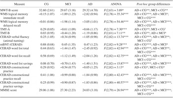
All participants underwent a neuropsychological examination including the battery developed by the “Consortium to Establish a Registry for Alzheimer’s Disease” (CERAD; Thalmann et al., Reference Thalmann, Monsch, Schneitter, Bernasconi, Aebi, Camachova-Davet and Staehelin2000) with measurements of verbal and nonverbal memory functions, visuospatial functions, and verbal functions like verbal fluency and naming. This also included a measurement of general intellectual function (MMSE; Folstein et al., Reference Folstein, Folstein and McHugh1975), a test of memory [the Logical Memory subtest of the revised Wechsler Memory Scale (WMS-R); Härting et al., Reference Härting, Markowitsch, Neufeld, Calabrese, Deisinger and Kessler2000], a test of visual–conceptual and visual–motor tracking including motor speed and attention functions (Trail Making Test; Reitan, Reference Reitan1992), and a multiple-choice word test as an estimate for premorbid intelligence [Mehrfach–Wortwahl Test Version B; Lehrl, Reference Lehrl1995]. The mean psychometric performance of all groups is listed in Table 1.
Psychometric performance as a function of group expressed as standard z scores (except for MWT-B and MMSE score)

† p < .1.
* p < .05.
** p < .01.
*** p ≤ .001.
Note. Standard deviations are in parentheses. MWT-B, Mehrfach–Wortwahl Test Version B; TMT, Trail Making Test; mBNT, modified Boston Naming Test.
False Memory Paradigm
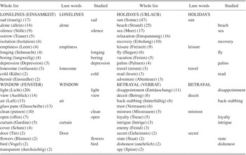
An emotional variant of the DRM-paradigm (Roediger & McDermott, Reference Roediger and McDermott1995) was administered that was based on a previously employed paradigm (Moritz et al., Reference Moritz, Glascher and Brassen2005) deemed feasible for a geriatric population. The test was composed of four thematic word lists. Each of the lists was created by choosing associates for a critical word (critical lure: cl). The thematic words were depression-relevant (cl: loneliness), delusion-relevant (cl: betrayal), positive (cl: holidays), and neutral words (cl: window). Since AD is also associated with psychiatric symptoms like depression, social isolation, hallucinations, and delusions, we included two negative psychological states (depression- and delusion-relevant) in our study paradigm. The items of the study lists (nine items per list) were sorted in descending order according to the word frequencies in the association study (strongly, medium, and weakly related; for details, see Moritz et al., Reference Moritz, Glascher and Brassen2005), that is, the first word in a learning list was the word most often produced in the study of Moritz et al. (Reference Moritz, Glascher and Brassen2005) (see Appendix for the lists used). Items from the four lists did not differ in word length, word frequency, and associative strength.
The recognition list consisted of 24 words (“old” words, comprising two per word list of the following types: strongly, medium, and weakly related words of all four lists) and 24 “new” words [one per word list of the following types: critical lure and strongly, medium, and weakly related lure words as well as eight unrelated words (depression-relevant: powerless, tiredness; delusion-relevant: oppression, devil; positive: gift, refreshment; neutral: girl, chair)]. Words from each item type were pseudorandomized within the recognition list, whereby the same order was used for all participants.
Procedure
Participants were tested individually as part of a larger cognitive investigation (Table 1). Participants were instructed that words from four lists would be read to them (approximately 2 s per word). They were further instructed that they had to recognize studied words among nonpresented items subsequently. The four lists were then read consecutively to the participant, whereby list order was randomized between participants to avoid recency or primacy effects. Each list was announced as list 1, list 2, and so on. To elicit the false memory effect, it is crucial that the items are presented list-wise.
Subsequent to the presentation of the learning lists, the recognition list was administered without delay. After each item of the recognition list, participants were required to state whether it was old or new (old–new). Responses were recorded by the experimenter.
Data Analysis
With regard to the conventional neuropsychological test battery, dependent variables were first entered in a multivariate analysis of variance (ANOVA) (MANOVA) model. In case of a significant overall effect, planned ANOVAs with subsequent Bonferroni-adjusted single comparisons (in case of absent homogeneity of variance, the alpha-adjusted Tamhane-T2 procedure was adopted) followed for all cognitive measurements. For the DRM-task-measures, a four-way mixed ANOVA was calculated with group (CG, MCI, AD) as the between-subjects factor and recognition type (veridical and false recognition), relatedness (weakly related or unrelated, medium related, strongly related, or critical lure), and word list (neutral, positive, depression-relevant, or delusion-relevant) as the within-subjects factors. Exposition of results is confined to main effects and to effects involving group as a factor. To explore whether groups display an emotional memory effect, we conducted planned t-test comparisons for each group separately for veridical and false recognition for emotional versus neutral words. Pearson correlation coefficients were computed between true and false recognition to analyze recognition strategy (i.e., gist-based vs. item-specific memory). Differences between correlation coefficients were computed by Fisher’s z. The alpha level was set at p < .05 (two-tailed).
RESULTS
Neuropsychological Performance
The MANOVA [Pillai Trace = 1.16, F(32,66) = 2.84, p < .001] indicated that neuropsychological performance was compromised as a function of increasing disease severity (Table 1). Subsequent univariate ANOVAs revealed that the AD group was the most impaired. MCI patients scored below age-matched healthy adults but still performed in the normal range according to normative data (standard z score −1 to +1) except for some memory scores (e.g., WMS Logical Memory).
Recognition Performance
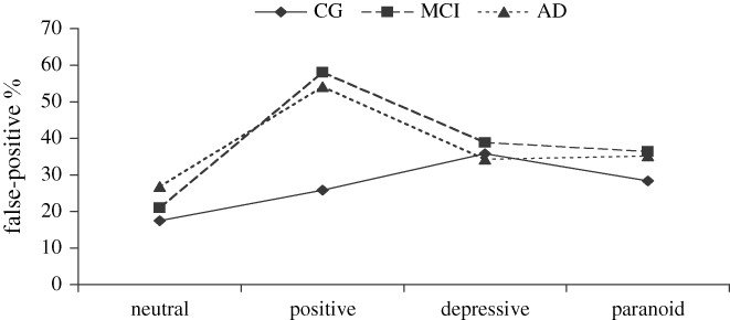
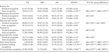
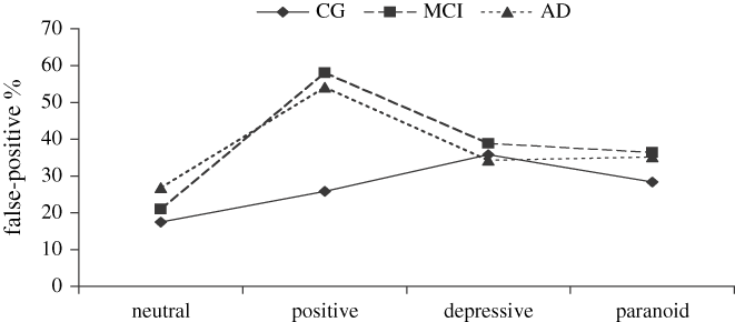
A mixed ANOVA was conducted with group (CG, MCI, and AD) as the between-subject factor and word lists, relatedness, and recognition type as the within-subject factors. Percentage of recognition served as dependent variable. A main effect for word lists emerged, F(3,240) = 24.46, p < .001. As can be seen in Table 2, recognition rate for neutral words was markedly lower than that for the emotional words. The main effect for response type also achieved significance, F(1,80) = 272.06, p < .001, reflecting more veridical than false recognition of words. The group effect did not yield significance, F(2,80) = 0.31, p > .7. A significant interaction emerged for group and relatedness, F(4,160) = 4.32, p = .002. The effect will be explained further below (see the following paragraph). The interaction for group and word list reached significance, F(6,240) = 3.99, p = .001. As can be seen in Table 2, this was because both patient groups showed higher recognition rates for the positive list compared to healthy controls. This was further qualified by a three-way interaction of group, word type, and relatedness which tended toward significance, F(6,240) = 2.07, p = .06, showing that both MCI and AD patient groups had an excess of false recognition for positive words (please see Table 2 and Figure 1). We will deal with the underpinnings of the two-way interaction of group and word type in more detail below (see the Emotional Memory Effect section).
False recognition rate as a function of group and emotional valence. Group differences occur only in the positive valence. Both patient groups show significant higher false alarm rates than healthy controls.
Mean proportion (in %) of veridical recognition rate, false recognition rate, noncritical false alarm baseline, corrected veridical recognition rate, corrected false recognition rate, and veridical and false recognition rates for the four word lists as a function of groups

† p < .1.
* p < .05.
** p < .01.
*** p ≤ .001.
Note. Standard deviations are in parentheses. Corrected veridical recognition = veridical recognition minus false alarm baseline; corrected false recognition = false recognition minus false alarm baseline.
To further explore the effect of group and relatedness, we adopted the procedure of Balota et al. (Reference Balota, Cortese, Duchek, Adams, Roediger, McDermott and Yerys1999). They found that AD individuals are more susceptible to falsely recognize unrelated lures. Balota et al. (Reference Balota, Cortese, Duchek, Adams, Roediger, McDermott and Yerys1999) assumed that veridical recognition may reflect true remembering of the specific study items versus a bias to say “old” (false alarm baseline). False recognition can also be thought of remembering the gist of the study list (semantic false memory) or again the tendency to say “old.” Table 2 shows also the false alarm baseline, the corrected veridical recognition rate (veridical recognition minus false alarm baseline), and the corrected false recognition rate (false recognition minus false alarm baseline) as a function of groups. Analyses of corrected recognition rates revealed that both AD and MCI patients exhibited significantly reduced levels of corrected veridical recognition compared to healthy older adults, F(2,80) = 21.36, p < .001. There was no significant difference between the MCI and AD patients on this variable. With regard to the corrected false recognition rate, AD but not MCI patients made significantly fewer “old” responses to related lures than did healthy controls, F(2,80) = 9.86, p < .001. There was no significant difference between the MCI patients and the healthy older adults on this variable.
Relationship Between Veridical and False Recognition
To analyze response strategy, we correlated the number of correctly recognized items with the number of falsely recognized items (only related lures; unrelated lures were dropped). Whereas no correspondence emerged between veridical and false recognition for healthy older participants, r = .04, p > .8, a strong relationship between both indices emerged for MCI patients, r = .51, p = .006, and an even stronger correlation emerged for AD patients, r = .78, p < .001. This indicates that healthy older adults pursue an item-specific response strategy, and both MCI and AD patients employed a gist-based recognition strategy. While this was more prominent for AD than for MCI patients, the differences failed to reach statistical significance, Fisher’s z = −0.5, t = −1.86, p < .1.
Emotional Memory Effect
To look at emotional memory more closely, we contrasted the emotional items (i.e., positive, depression-relevant, delusion-relevant) with the neutral ones. A set of t tests indicated that both healthy older adults, t(19) = −2.01, mD = −10.83, p = .06, and MCI patients, t(26) = −4.81, mD = −24.69, p < .001, showed enhanced veridical recognition for emotional relative to neutral items indicating an emotional memory effect. The AD group, in contrast, displayed no emotional memory effect and gave an equal percentage of veridical “old” responses to emotional and neutral studied items, t(35) = −0.78, mD = −3.24, p > .4. When the data were analyzed separately for positive, depressive-relevant, paranoid-relevant, and neutral items, healthy controls showed better veridical recognition for positive, mD = −13.3, p < .03, and delusion-relevant, mD = −11.7, p = .06, items compared to neutral items. The MCI group remembered all emotional valenced lists better than the neutral one, positive: mD = −27.2, p < .001; depression-relevant: mD = −22.2, p = .001; delusion-relevant: mD = −24.7, p < .001. AD patients only remembered positive valenced items better than neutral ones, mD = −13.4, p < .02.
Multiple group-wise paired t tests conducted for false recognition (emotional vs. neutral) showed that all groups falsely recognize emotional valent stimuli more often than neutral ones, CG: t(19) = −5.51, mD = −24.83, p < .001; MCI: t(26) = −7.61, mD = −34.26, p < .001; AD: t(35) = −5.91, mD = −20.14, p < .001. When the data were analyzed separately for positive, depression-relevant, delusion-relevant, and neutral items, healthy controls showed more false recognition for depression-relevant, mD = −18.3, p < .001, and delusion-relevant, mD = −10.8, p = .006, words compared to nonemotional words. The MCI group showed more false recognition on all emotional valenced lists than in the neutral one, positive: mD = −37.0, p < .001; depression-relevant: mD = −17.9, p = .001; delusion-relevant: mD = −24.7, p < .001. The AD group also showed a higher false recognition rate on all emotional valenced lists than in the neutral one—even when in the depression-relevant list only trend level was achieved, positive: mD = −27.3, p < .001; depression-relevant: mD = −7.4, p = .06; delusion-relevant: mD = −8.3, p < .05.
DISCUSSION
The present study is the first that investigated similarities between MCI and AD patients on an emotional variant of the DRM-paradigm. In line with previous studies (e.g., Balota et al., Reference Balota, Cortese, Duchek, Adams, Roediger, McDermott and Yerys1999; Budson et al., Reference Budson, Daffner, Desikan and Schacter2000, Reference Budson, Desikan, Daffner and Schacter2001, Reference Budson, Michalska, Sullivan, Rentz, Daffner and Schacter2003a, Reference Budson, Sullivan, Daffner and Schacter2003b; Pierce et al., Reference Pierce, Sullivan, Schacter and Budson2005; Watson et al., Reference Watson, Balota and Sergent-Marshall2001), AD patients showed impaired baseline-corrected veridical recognition, a decrease in corrected false recognition and increased false recognition of unrelated lures (false alarm baseline) in comparison to age-matched healthy participants. For AD patients, a lower recognition rate and a raised false alarm baseline is well established from conventional memory tests (Calabrese, Reference Calabrese, Calabrese and Förstl2000) and reflects neurodegeneration and atrophic processes in the medial temporal cortex (Schacter et al., Reference Schacter, Verfaellie, Anes and Racine1998).
On conventional cognitive tests, MCI patients often performed in between healthy older adults and AD patients but still were in the age-corrected normal range on most tests. In contrast, MCI patients in our study were equally impaired as AD patients on some indices of the DRM-task. They displayed a dramatically raised false alarm baseline and a diminished corrected veridical recognition rate like the AD group. In contrast to the AD group, reports of raised false memory baseline in MCI patients are rare. Only one study found enhanced intrusions in MCI patients (Greenaway et al., Reference Greenaway, Lacritz, Binegar, Weiner, Lipton and Munro Cullum2006). On closer examination, it is obvious that both MCI and AD patient groups had an excess of false recognition for positive words. The implication is that these findings occur relatively early in the course of incipient AD. Furthermore, the MCI group in our study was indistinguishable from the healthy group on their corrected false recognition rate. This result is in line with studies employing conventional memory tests in which MCI patients had a lower tendency to intrusions and confabulations than AD patients (e.g., Schreiber et al., Reference Schreiber, Ackl, Sonntag and Zihl2005; Welsh et al., Reference Welsh, Butters, Hughes, Mohs and Heymann1991). However, our study showed that MCI patients exhibited an increased false alarm baseline in the DRM-task. Differences between our results and the results of other studies may be accommodated by differences in study material or the emotional valence of the material. Studies on the emotional memory effect in AD patients so far only used negatively valenced information. In the present study, we employed both negative and positive stimuli to examine the emotional memory effect. Both healthy older adults and MCI patients showed better memory for the emotional word lists compared to the neutral list. Only the AD group did not show the usual memory benefit. This result is in accordance with other studies reporting a reduced emotional memory effect in AD patients (Budson et al., Reference Budson, Todman, Chong, Adams, Kensinger, Krangel and Wright2006; Kensinger et al., Reference Kensinger, Anderson, Growdon and Corkin2004), which could be caused by the atrophy and dysfunction in limbic structures including the amygdala (Kensinger et al., Reference Kensinger, Brierley, Medford, Growdon and Corkin2002; Mizuno et al., Reference Mizuno, Wakai, Takeda and Sobue2000). Although AD patients did not gain from emotional valence regarding the overall emotional effect, they still benefited from the positive valence of the information because only for this type they displayed a better veridical memory compared to the neutral valence, which, however, was more pronounced for false recognition. However, a preserved emotional memory effect for positively valenced information in AD patients has also been reported by other studies (Hamann et al., Reference Hamann, Monarch and Goldstein2000; Kensinger et al., Reference Kensinger, Brierley, Medford, Growdon and Corkin2002). Perhaps AD patients are especially attracted by words with a positive valence because of an arousal-based response bias. The positive words may have a stronger stimulative nature for AD patients than negative words, the latter prompting disapproval or avoidance. This could be an explanation for an at least preserved emotional memory effect for positively valenced items in AD. Whether AD patients show a preserved or an impaired emotional memory effect and to what extent the emotional valence plays a role are still under debate (compare Budson et al., Reference Budson, Todman, Chong, Adams, Kensinger, Krangel and Wright2006; Fleming et al., Reference Fleming, Kim, Doo, Maguire and Potkin2003; Hamann et al., Reference Hamann, Monarch and Goldstein2000; Kazui et al., Reference Kazui, Mori, Hashimoto, Hirono, Imamura, Tanimukai and Hanihara2000; Kensinger et al., Reference Kensinger, Brierley, Medford, Growdon and Corkin2002; Moayeri et al., Reference Moayeri, Cahill, Jin and Potkin2000). Like healthy controls, MCI patients in our study remembered emotionally valent words better than neutral words. This is remarkable because recent studies with MCI patients described significant atrophy not only in hippocampus but also in amygdala (Bottino et al., Reference Bottino, Castro, Gomes, Buchpiguel, Marchetti and Neto2002; den Heijer et al., Reference den Heijer, Geerlings, Hoebeek, Hofman, Koudstaal and Breteler2006; Mega et al., Reference Mega, Small, Xu, Felix, Manese, Tran, Dailey, Ercoli, Bookheimer and Toga2002).
In addition to the emotional memory effect as evidenced by raised veridical recognition, emotional valence of item lists also leads to an increase of false memories. Emotionally valent lures were more often falsely recognized than nonemotional lures. This result contradicts a recent study in which emotional lures were less often falsely recalled or recognized than neutral lures (Kensinger & Corkin, Reference Kensinger and Corkin2004). In contrast to the present study, the aforementioned paper used an ordinary word list recognition paradigm and employed lures of strong negative valence (e.g., rape, slut, hell). Furthermore, in this paper the studied words did not have the same emotional valence as the critical lures, so perhaps the lure words had a stronger pop-out effect thus counteracting the false memory effect. We infer that emotional words lead to a stronger perception of familiarity than nonemotional words because of the enhanced false recognition of emotional compared to nonemotional words that we found. On closer examination, healthy controls showed this effect with negatively valenced items only. This result is in accordance with recent studies reporting that individuals remember negative information more vividly and with more detail than they do remember positive information (e.g., Denburg et al., Reference Denburg, Buchanan, Tranel and Adolphs2003; Ochsner, Reference Ochsner2000).
Whereas healthy older adults followed an item-specific strategy during recognition, we found that both the AD and MCI groups employed a gist-based recognition strategy: This was more pronounced for AD than for MCI patients. For the AD patients, this result is in line with previous studies that found reliance on gist-based recognition in patients with very mild dementia (Balota et al., Reference Balota, Cortese, Duchek, Adams, Roediger, McDermott and Yerys1999; Budson et al., Reference Budson, Sullivan, Daffner and Schacter2003b). In contrast, more severely affected AD patients cannot rely on gist-based recognition anymore as even the overall theme of a list cannot be properly retrieved (Balota et al., Reference Balota, Cortese, Duchek, Adams, Roediger, McDermott and Yerys1999; Budson et al., Reference Budson, Sullivan, Daffner and Schacter2003b). Following from this, we assume that our AD group was less affected by dementia than the more severely affected patients in the studies above mentioned. The gist-based recognition strategy that we found in our AD group presumably depends on dysfunctions of the frontal network leading to a disinhibition of gist information during recognition. This interpretation is indeed backed-up by studies reporting frontal pathology in AD patients (de Leeuw et al., Reference de Leeuw, Barkhof and Scheltens2004; Esiri et al., Reference Esiri, Lee and Trojanowski2004; Jellinger, Reference Jellinger, Iqbal and Winblad2003). But it is not clear whether this explanation for gist-based recognition applies to our MCI patients, because MCI patients do not always exhibit neuropathological frontal changes (den Heijer et al., Reference den Heijer, Geerlings, Hoebeek, Hofman, Koudstaal and Breteler2006).
To conclude, the results of the present study indicate that MCI patients, like AD patients, are more susceptible to falsely recognize unrelated and emotional positive valent lures, as compared to healthy older adults. On the other hand, MCI patients are similar to healthy older adults in that both benefit from emotional memories in veridical recognition. Finally, the data presented might be helpful for understanding the breakdown of emotional memories along the continuum between normal aging and AD. Further experiments will be needed to replicate our findings and to clarify whether positive and negative valent words are processed in the same way.
Since we have already employed and validated these word lists and the procedure in a prior study (Moritz et al., Reference Moritz, Glascher and Brassen2005), it is unlikely that group differences in the present study reflect idiosyncratic differences among word lists (younger depressive patients of the prior study and our healthy older adults performed entirely different to our MCI and AD patients). A limitation of the present paradigm is that we collected lure words around single constructs (e.g., loneliness). Moreover, although the lists did not differ regarding associative strength, frequency, and word length, it cannot be ruled out that the lists differed on other aspects like familiarity, vividness, or concreteness. Furthermore, it is not clear if the perceived associative strength for a control person would necessarily be the same for an AD or MCI patient. This concern could be eliminated with the administration of multiple lists per condition or further questions on emotional valence and salience, although any prolongation of the encoding phase may easily frustrate and exhaust older and cognitive impaired MCI and even more AD patients. While we acknowledge this, in our view these limitations cannot fully account for word list–unspecific findings, particularly higher false recognition of unrelated items.
ACKNOWLEDGMENTS
This study was conducted as part of the dissertation thesis of K.B. The study was supported by the University of Hamburg (Ph.D. Grants at the University of Hamburg). We are grateful for the logistic support provided by Dr. H. Jahn (University Hospital Hamburg Eppendorf) and Dr. S. Stodieck (Epilepsy Center Hamburg) and would like to thank Dr. B. Bengner and U. Schwarze (Epilepsy Center Hamburg) for their helpful advice on a previous version of this manuscript.
Word lists. The original German items are set in parentheses followed by the number of naming frequency in the norming study. In case associative words have the same frequency, the order was determined by the first author of the association study (Moritz et al., Reference Moritz, Glascher and Brassen2005). Critical lure words (i.e., the thematic words for the association study) are set in uppercase. Lure words are the distractor words in the recognition test. Studied are the “old” words in the recognition test.