Introduction
Dictyocaulosis is a parasitic disease caused by nematodes of the genus Dictyocaulus, which can infect the trachea, bronchi and bronchioles of cattle, sheep, goats and horses (Urquhart et al., Reference Urquhart, Armour, Ducan, Dunn and Jennings1998; Fortes, Reference Fortes2004). Although Dictyocaulus viviparus (Nematoda: Trichostrongyloidea) occurs worldwide, it causes problems mainly in moist regions (Ploeger, Reference Ploeger2002). Adult worms lay their eggs in the trachea and bronchi, being expelled by coughing or swallowing. The eggs may hatch in the airways or in the digestive tract, and the larvae are eliminated in the faecal material. Humidity is essential for larval survival and development, and moderate temperatures, around 18–21°C, allow their complete development to the infective stage (L3) after 3–7 days. Infection occurs when cattle ingest larvae while grazing. Ingested larvae may cross the intestinal wall and migrate to the mesenteric lymph nodes, reaching first the vena cava through lymphatic pathways, and finally the lungs via the bloodstream (Urquhart et al., Reference Urquhart, Armour, Ducan, Dunn and Jennings1998; Fortes, Reference Fortes2004).
Outbreaks of dictyocaulosis occur sporadically, usually during summer or autumn, affecting mainly calves (Bateman et al., Reference Bateman, Baird, Slocombe, Leslie, Curtis and Menzies1986; Radostits et al., Reference Radostits, Gay, Blood and Hinchcliff2002; Da Silva et al., Reference Da Silva, De Barros and Graça2005; Konradt et al., Reference Konradt, Bianchi, Fredo, Leite Filho, Dalto and Pavarini2015). The epidemiology of the disease is strongly related to the number of infective larvae on pasture (Radostits et al., Reference Radostits, Gay, Blood and Hinchcliff2002). An epidemiological investigation on 25 herds infected by D. viviparus in The Netherlands suggested that light pasture contamination, followed by auto-infections with heavy pasture contamination by carriers, is the most prominent cause of lungworm disease. Infections due to overwintered pasture seem to play a minor role (Saatkamp et al., Reference Saatkamp, Eysker and Verhoeff1994). There are recent reports of dictyocaulosis outbreaks in cattle in southern Brazil. The affected animals were between 5 and 12 months of age and the main clinical signs reported were anorexia, low body weight, cough, abdominal breathing and serous nasal discharge (Da Silva et al., Reference Da Silva, De Barros and Graça2005; Konradt et al., Reference Konradt, Bianchi, Fredo, Leite Filho, Dalto and Pavarini2015). In Europe, outbreaks of this disease in young animals have become less common in recent decades, mainly due to the use of a vaccine against the parasite (Ploeger, Reference Ploeger2002). However, adult animals have been more frequently affected, contributing to losses in milk production (Ploeger, Reference Ploeger2002; Radostits et al., Reference Radostits, Gay, Blood and Hinchcliff2002). In North America, although less frequent, dictyocaulosis outbreaks in dairy cows have been reported as a cause of a drastic reduction in productivity and, eventually, animal death (Bateman, Reference Bateman, Baird, Slocombe, Leslie, Curtis and Menzies1986; Wapenaar, Reference Wapenaar, Barkema, Eysker and O'Handley2007).
Numerous studies have been performed to clarify the role of free radicals on inflammatory responses to injuries (Reuter et al., Reference Reuter, Gupta, Chaturvedi and Aggarwal2010). Free radicals are powerful oxidizing agents that react with organic molecules. An excess of oxidizing agents and/or deficiency of the protective system leads to an imbalance that characterizes oxidative stress (Ferreira & Matsubara, Reference Ferreira and Matsubara1997). Oxidative stress occurs when the production of reactive oxygen species (ROS) is higher than the anti-oxidant mechanism, which contributes to lesions of biological molecules (Xing et al., Reference Xing, Li, Wang, Gao, Xu and Wang2012). To minimize ROS production, organisms activate anti-oxidant mechanisms, as well as anti-oxidant defences (Pamplona & Costantini, Reference Pamplona and Costantini2011). Catalase (CAT) and superoxide dismutase (SOD) are important anti-oxidant enzymes of the anti-oxidant system (Constantini, Reference Costantini2008). Also, studies have linked oxidative stress to clinical pathological features and the pathogenesis of several parasitic diseases (Karsen et al., Reference Karsen, Sunnetcioglu, Ceylan, Bayraktar, Taskin, Aksoy and Erten2011; Machado et al., Reference Machado, Da Silva, Schafer, Aires, Tonin, Oliveira, Hermes, Almeida, Moresco, Stefani, Lopes, Mendes, Schwertz and Leal2014; Bottari et al., Reference Bottari, Mendes, Lucca, Schwertz, Henker, Olsson, Piva, Sangoi, Campos, Moresco, Jaques and Da Silva2015).
Parasitic infections can induce oxidative stress and increase ROS levels. The latter can trigger lipid peroxidation, causing damage to the host. Anti-oxidant enzymes, such SOD and CAT, protect against oxidative stress and scavenge the excessive generation of ROS (Martins et al., Reference Martins, Pereira and Baptista2014). Therefore, the aim of this study was to evaluate the oxidative stress, pre- and post-treatment with eprinomectin, of cows naturally infected by D. viviparus and showing mild or severe clinical forms of the disease.
Materials and methods
Collection and examination of dairy cows
The dairy cows used in this study were obtained from a farm located in Arabutã (52°13′463ʺW; 27°05′983ʺS), Santa Catarina State, southern Brazil, where an outbreak of verminous pneumonia caused by D. viviparus was reported. The age of cows ranged between 3 and 9 years, although variation in age occurred at different stages of lactation (first, second and third phase) and reproduction (pregnant and non-pregnant). At the time of the outbreak, 23 cows were in the semi-intensive system, with access to oats (Avena sativa), ryegrass (Lolium multiflorum), corn silage and concentrate. The first cow showing clinical signs of respiratory disease was subjected to euthanasia in extremis due to the severity of the disease and its poor prognosis.
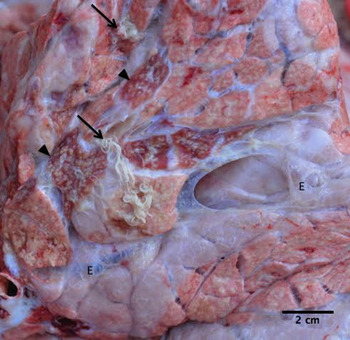
According to the farmer, cows that had access to pasture showed behavioural changes such as low head and apathy, as well as reduced appetite and rumination. Also, some cows started to show nasal secretion and reduced milk production (50% reduction in total production of the herd). Necropsy of the first animal confirmed the cause of the disease, since there was a heavy infection caused by D. viviparus inside the bronchi, associated with moderate multifocal to coalescing pneumonia and moderate multifocal interlobular emphysema (fig. 1).
The lung of a dairy cow naturally infected with Dictyocaulus viviparus (arrow), associated with moderate, multifocal, lobular atelectasis (arrowhead) and moderate, multifocal, interlobular emphysema (E).

As a result, all the remaining cows in the semi-intensive system were submitted to clinical examination and faecal sampling to confirm the infection. Out of 22 dairy cows that showed respiratory clinical signs (cough and nasal secretion), ten were classified as mild (n = 10) and 12 as severe (n = 12); in addition, three showed progressive weight loss (n = 3), three had abdominal breathing (n = 3) and one aborted (n = 1). Infected cows also showed high heart (HR) and respiratory rates (RR) (average: HR, 85.4 beats per minute (bpm); RR, 52.0 mpm). The average body temperature of infected cows was 38.6°C.
Faecal examination was carried out for parasite eggs (McMaster technique) and larvae (Baermann technique) as described in detail by Monteiro (Reference Monteiro2010). These tests were performed pre- and post-treatment, as described below.
Treatment
Levamisole was the drug commonly used on the farm for prophylaxis and parasitic treatment before the outbreak. Levamisole acts as an agonist on nicotinic acetylcholine receptors (nAChRs) at the nematode neuromuscular junction, leading to sustained neuromuscular depolarization and spastic paralysis (Martin & Robertson, Reference Martin and Robertson2007). Levamisole is the main antiparasitic drug used in dairy cows in Brazil. However, based on the diagnosis of dictyocaulosis and the possibility of parasitic drug resistance to levamisole, animals were treated with a single dose of eprinomectin (360 μg/kg; pour on).
Experimental design and sampling
Infected cows were divided into two groups according to the severity of the disease: one group of 10 animals showing mild clinical signs, and a second group of 12 cows with severe clinical signs of dictyocaulosis. For oxidative and anti-oxidant profile analyses, blood samples were collected with and without sodium citrate on day 0 (the day of the diagnosis) and 10 days post-treatment. Blood samples without anticoagulant were centrifuged at 3500 rpm for 10 min to obtain serum samples, which were collected and stored in Eppendorf tubes at −20°C until analysed for ROS and thiobarbituric acid-reactive substances (TBARS). Blood samples with anticoagulant were homogenized and frozen at −20°C for CAT and SOD analyses. TBARS levels were performed on the same sampling day, while the other analyses were performed up to 1 week after collection.
Oxidative stress variables
Lipid peroxidation was determined based on the levels of TBARS in serum samples, as described by Jentzsch et al. (Reference Jentzsch, Bachmann, Furst and Biesalski1996). The results were obtained by spectrophotometry (535 nm) and expressed as nmol of malondialdehyde per millilitre.
Serum samples were prepared as described by Ali et al. (Reference Ali, Lebe and Bondy1992) in order to determine ROS levels. Briefly, serum samples were diluted (1:10) with 10 mm Tris (pH 7.4) and loaded with 5 μl of 2′,7′-dichlorofluorescin diacetate (DCFH-DA) in methanol at 37°C in 5% CO2 for 15 min, enough time for esterase to form a non-fluorescent compound (2′,7′-dichlorofluorescin (DCFH)) (Bass et al., Reference Bass, Parce, Dechatelet, Szejda, Seeds and Thomas1983). Fluorescence was measured before and after an additional 60 min of incubation. The production of the fluorescent oxidized derivative of DCFH, 2′,7′-dichlorofluorescein (DCF), was measured using a spectrophotofluorometer (Perkin-Elmer LS-50; Perkin-Elmer, Waltham, Massachusetts, USA) at an excitation wavelength of 488 nm and an emission wavelength of 525 nm. The cuvette holder was thermostatically maintained at 37°C. Autofluorescence was checked by the inclusion of blank samples (unloaded tissue fraction) and their readings were always less than 10% of the total fluorescence. ROS formation was quantified from a standard curve of DCF in methanol (0.05–1.0 μm).
Determination of CAT activity was carried out according to a modified method of Nelson & Kiesow (Reference Nelson and Kiesow1972). This assay takes into account absorbance changes at 240 nm due to CAT-dependent decomposition of hydrogen peroxide. An aliquot (0.02 ml) of blood (diluted 1:10 with saline solution) was homogenized in 0.910 ml of potassium phosphate buffer 50 mm, pH 7.0. The spectrophotometric determination started with the addition of 0.07 ml of 0.3 mol/l hydrogen peroxide (H2O2). The change in absorbance at 240 nm was measured for 2 min. CAT activity was calculated using the molar extinction coefficient (0.0436 cm2/lmol) and the results were expressed as nmol of CAT per mg of protein.
SOD activity was measured based on the inhibition of radical superoxide reaction with adrenaline, as described by McCord & Fridovich (Reference McCord and Fridovich1969). In this method, SOD competes with the detection system for radical superoxide. One unit of SOD is defined as the amount of enzyme that inhibits 50% of the speed of adrenaline oxidation, which leads to the formation of a red-coloured product, adrenochrome, that can be detected by spectrophotometry. SOD activity is determined by measuring the speed of adrenochrome production, observed at 480 nm, in a reaction medium containing glycine–NaOH (50 mm, pH 10) and adrenaline (1 mm). The results were expressed as UI SOD per mg of protein.
Data analysis
Experimental data were first analysed descriptively; measures of central tendency (median) and data dispersion (range, which stands for the interval between the minimum and maximum values in the data) were computed. Further, all variables were submitted to the Shapiro–Wilk W test for normal distribution verification. Since none of the variables met the assumption of parametric testing, a Mann–Whitney U test was applied to the data of both groups of cows (showing either mild or severe infections) for the analysis of TBARS, ROS, SOD and CAT. Secondly, we checked the effect of eprinomectin (pre-treatment versus post-treatment) on TBARS, ROS, SOD and CAT. In addition, a special correlation (Polyserial) was computed for the number of larvae per gram of faeces (LPG) separated into two degrees of infection (1: <25 LPG; and 2, ≥25 LPG), biochemical variables (TBARS, ROS, SOD and CAT) and the degree of the disease (mild or severe). Results were considered statistically different at P values <0.05. The statistical process followed R-language, v.2.15.2 (R Development Core Team, 2012).
Results
Cows that showed severe clinical signs of the disease on day 0 (pre-treatment) had 1–448 larvae on faecal examination. On day 10 post-treatment, cows showed no clinical signs of respiratory disease and milk production increased by up to 50%. In addition, faecal examination was negative for nematode larvae in all cows. Treated cows showed normal HR and RR (average: HR, 58.2 bpm; RR, 26.3 mpm). The average body temperature of infected cows was 38.6°C.
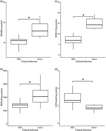
Degree of clinical infection between the mild and severe showed a significant difference for TBARS, ROS, SOD and CAT (fig. 2). Cows with severe clinical disease showed increased TBARS (P < 0.001) and ROS (P = 0.002) levels, as well as SOD activity (P < 0.001), compared to those with mild disease. CAT activity was significantly (P < 0.001) reduced in cows with severe disease compared to those with mild disease.
Blood and serum samples from dairy cows naturally infected with Dictyocaulus viviparus, showing mild and severe clinical signs of the disease, regarding their concentrations of (a) serum thiobarbituric acid reactive substances (TBARS: mmol/ml), (b) serum reactive oxygen species (ROS: uf/mg protein), (c) superoxide dismutase in blood (SOD: UI/mg protein), and (d) catalase in blood (CAT: mmol/mg protein). *Level of significance with P < 0.05.

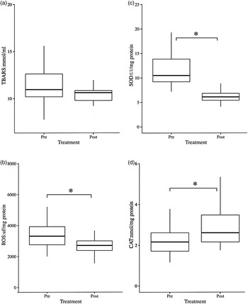
Eprinomectin treatment affected all variables, except TBARS (fig. 3). After treatment (day 10), a reduction of ROS levels (P = 0.006) and SOD activity (P < 0.001) were observed, as well as an increase of CAT activity (P = 0.05) compared to day 0 (pre-treatment).
Blood and serum samples from dairy cows naturally infected with Dictyocaulus viviparus pre- and post-treatment with epinomectin, to show concentrations of (a) serum thiobarbituric acid reactive substances (TBARS: mmol/ml); (b) serum reactive oxygen species (ROS: uf/mg protein); (c) superoxide dismutase in blood (SOD: UI/mg protein) and (d) catalase in blood (CAT: mmol/mg protein). *Level of significance with P < 0.05.

A significant positive correlation was found between LPG and TBARS levels (r = 0.1976, P < 0.001); LPG and ROS levels (r = 0.2933, P < 0.001); and LPG and SOD activity (r = 0.4147, P < 0.001). A negative correlation between LPG and CAT activity (r = −0.1146, P < 0.001) was observed. A positive correlation was also found between LPG and severe clinical signs of the disease (r = 0.6230, P < 0.001).
Discussion
Many studies have reported that D. viviparus may activate a variety of inflammatory cells, which may lead to the production of reactive species as a strategy to kill the parasite (Johnson et al., Reference Johnson, Sales and Matthews2005). However, there are few studies on dictyocaulosis regarding its effect on reactive species. On the other hand, the relation between ROS and oxidative damage on DNA was evaluated by Gao et al. (Reference Gao, Wang Di Zhang, Xu, Yang, Yan, Liu, Luo, Wu, Yao, Yan and Liu2015), and the consequent negative effect on the host was already described in studies with other nematodes, such as Haemonchus contortus (Pivoto et al., Reference Pivoto, Torbitz, Aires, da Rocha, Severo, Grando, Peiter, Moresco, da Rocha and Leal2015). In this study, levels of ROS, lipid peroxidation and the anti-oxidant status were measured in cows naturally infected by D. viviparus, and the oxidative profile in sera of these animals showed an increase in variables of the oxidative damage. These results corroborate the findings of Nanev et al. (Reference Nanev, Gabrashanska, Vladov and Gunchev2011), who demonstrated that oxidative stress is a significant feature of dictyocaulosis in mouflons, and provided evidence of the production of oxidative stress detected by the increase of malondialdehyde (MDA) levels in the lung, which indicates lipid peroxidation. According to Rosenfeldt et al. (Reference Rosenfeldt, Wilson, Lee, Kure, Ou, Braun and de Hann.2013), ROS acts as a defence mechanism against parasites, but it may also damage various cells and tissues, such as those observed in this study. Anti-oxidant enzymes are altered in an attempt to modulate the oxidative damage present in other parasitic infections (Baldissera et al., Reference Baldissera, Oliveira, Rech, Rezer, Sagrillo, Alves, Da Silva, Leal, Boligon, Athayde, Da Silva, Mendes and Monteiro2014).
According to researchers, CAT and SOD are the most important anti-oxidant enzymes (Ranjithkumar et al., Reference Ranjithkumar, Kamili, Saxena, Dan, Dey and Raut2011), and their measurement is the proper indirect way to assess the status of the anti-oxidant defence (Dimri et al., Reference Dimri, Sharma, Yamdagni, Ranjan and Zama2010). In the present study, a significant decrease in CAT and increase in SOD activities were observed in sera from cows with a severe clinical form of the disease. Nanev et al. (Reference Nanev, Gabrashanska, Vladov and Gunchev2011) found a significant decrease in CAT and SOD activities in mouflons naturally infected by D. viviparus. The literature reports that nematode parasites cause inflammation and generate ROS (Siwela et al., Reference Siwela, Mambvura, Masanganise and Dube2010), which induce oxidative damage of macromolecules such as polyunsaturated fatty acids in membrane lipids (Beyer et al., Reference Beyer, Segura-Aguilar, Di Bernardo, Cavazzoni, Fato, Fiorentini, Galli, Setti, Landi and Lenaz1996). This study observed an increase in MDA content that caused deleterious effects on cells, followed by cell death (Beyer et al., Reference Beyer, Segura-Aguilar, Di Bernardo, Cavazzoni, Fato, Fiorentini, Galli, Setti, Landi and Lenaz1996), which contributed to the severity of the disease. Catalase enzyme is responsible for the removal of hydrogen peroxide; therefore, the reduction in CAT activity in cows infected by D. viviparus (severe compared to mild disease) suggests an increase in hydrogen peroxide concentration, such as those observed in other parasitic infections (Nanev et al., Reference Nanev, Gabrashanska, Vladov and Gunchev2011; Baldissera et al., Reference Baldissera, Oliveira, Rech, Rezer, Sagrillo, Alves, Da Silva, Leal, Boligon, Athayde, Da Silva, Mendes and Monteiro2014).
Higher levels of ROS would increase the overall activity of the first line of defence as an adaptive response, an effect demonstrated in our SOD results. It is possible that up-regulation or increased activity of SOD might be a result of an adaptive response, triggered by increased production of H2O2 caused by D. viviparus infection. This adaptation, in which cells respond to oxidative stress by inducing SOD activity after infection, has the intention to counteract the toxic effects of H2O2 caused by decreasing CAT activity (De Domínguez et al., Reference De Domínguez, Charris, Domínguez, Monasterios, Angel and Rodrigues2015).
A relationship between oxidative stress and pathophysiology of nematode parasites was observed by Machado et al. (Reference Machado, Da Silva, Schafer, Aires, Tonin, Oliveira, Hermes, Almeida, Moresco, Stefani, Lopes, Mendes, Schwertz and Leal2014) in lambs infected by H. contortus, agreeing with this present study. A positive correlation between LPG and ROS, TBARS and SOD was observed in the current study, as well as a negative correlation between LPG and CAT activity. This infers that the parasite burden directly contributes to oxidative stress, which may also contribute to the pathophysiology of the disease, because the oxidative imbalance has been associated with severity of pathological conditions due to cell damage caused by free radicals (Wolkmer et al., Reference Wolkmer, Da Silva, Traesel, Paim, Cargnelutti, Pagnoncelli, Picada, Monteiro and Lopes2009). It should be noted that our study did not quantify the number of adult parasites, only larvae in the faeces. Therefore, it is impossible to assert that these animals with a high number of larvae in their faeces also had high numbers of adult parasites. However, there was a positive correlation between LPG and severe degree of clinical disease, that is, cows with a higher number of larvae in faecal examinations also had severe clinical disease. So, indirectly, it is possible to suggest that the parasitic load is higher in these cows, and that there is a relationship between parasite load by D. viviparus and oxidative stress, as described in other parasitic diseases (Wolkmer et al., Reference Wolkmer, Da Silva, Traesel, Paim, Cargnelutti, Pagnoncelli, Picada, Monteiro and Lopes2009; Machado et al., Reference Machado, Da Silva, Schafer, Aires, Tonin, Oliveira, Hermes, Almeida, Moresco, Stefani, Lopes, Mendes, Schwertz and Leal2014).
In this study, post-treatment analyses (day 10) showed reduced ROS levels and SOD activity, as well as increased CAT activity. Alterations in biochemical variables post-treatment were not related to any direct effect of eprinomectin treatment, but a consequence of the elimination of the infection caused by D. viviparus (death of the parasite) and, thus, animal recovery. Note that on day 10 post-treatment, no clinical signs (mostly nasal secretion and cough) observed during the diagnosis (day 0) were observed in cows.
In summary, the intense production of ROS, aiming to kill the parasite, also caused lipid peroxidation, which altered CAT and SOD activities, as an adaptive response against D. viviparus infection in dairy cows. This fact may contribute to the pathophysiology and severity of disease clinical signs. It is possible to conclude that oxidative stress is exacerbated by the degree of clinical disease, that is, the severe clinical signs lead to higher oxidant levels. It was also found that treatment with eprinomectin was efficient, since it eliminated the parasite and, thus, minimized the oxidative stress in dairy cows.
Financial support
This research received no specific grant from any funding agency, commercial or not-for-profit sectors.
Conflict of interest
None.
Ethical standards
The present study was approved by the Ethics Committee for Use of Animals (CEUA) of Instituto Federal Catarinense under protocol number 17/2015.