1. INTRODUCTION
Imaging small features of transient systems has found applications in many fields, from the study of dynamic material properties such as phase transitions and mechanical failure modes at high density, pressure, and strain rates to studying the processes important to inertial confinement fusion.
There are two primary means of measuring these transient properties within an opaque material. The most common is radiography, where an external source directs the probe particles through the object. These probes interact with the object material, encoding the information of the internal structure and carrying this information to the detection system. For X-ray radiography, this information is encoded through absorption and scattering processes and the X-ray intensity leaving the object carries the information of the integrated mass along the X-ray paths through the object. The second imaging technique utilizes the generation of probes within the object itself. As these probe particles exit the object, they carry information about both the internal source distribution, which generated the probe particles, as well as the material surrounding this source. Other techniques exist such as magnetic resonance imaging, but these techniques tend to require the features to persist longer than practical for these types of dynamic measurements.
Since the discovery of X rays in the late 1800s, X-ray radiographic imaging has become ubiquitous in the study of structure within otherwise opaque materials. Today X-ray radiography is applied to a wide range of applications from medical investigations with low-energy bremsstrahlung radiation to the study of exotic states of matter that survive for only a small fraction of a second with X rays generated at free-electron lasers. Through the development of new technologies, however, new probes exist for applications where X-ray radiography is difficult or impossible.
For example, in the past two decades proton radiography has been developed, providing flash radiography measurements for the study of thick dynamic systems. Several facilities have recently been commissioned at existing proton accelerators (King et al., Reference King, Ables, Adams, Alrick, Amann, Balzar, Barnes, Crow, Cushing, Eddleman, Fife, Flores, Fujino, Gallegos, Gray, Hartouni, Hogan, Holmes, Jaramillo, Knudsson, London, Lopez, McDonald, McClelland, Merrill, Morley, Morris, Naivar, Parker, Park, Pazuchanics, Pillai, Riedel, Sarracino, Shelley, Stacy, Takala, Thompson, Tucker, Yates, Ziock and Zumbro1999; Burtsev et al., Reference Burtsev, Lebedev, Mikhailov, Ogorodnikov, Oreshkov, Panov, Rudnev, Svirskii, Syrunin, Trutnev and Khramov2011; Morris et al., Reference Morris, Ables, Alrick, Aufderheide, Barnes, Buescher, Cagliostro, Clark, Clark, Espinoza, Ferm, Gallegos, Gardner, Gomez, Greene, Hanson, Hartouni, Hogan, King, Kwiatkowski, Liljestrand, Mariam, Merrill, Morgan, Morley, Mottershead, Murray, Pazuchanics, Pearson, Sarracino, Saunders, Scaduto, Schach von Wittenau, Soltz, Sterbenz, Thompson, Vixie, Wilke, Wright and Zumbro2011), which are now performing flash proton radiography measurements and microscope systems have been commissioned to improve the resolution of these systems (Mottershead et al., Reference Mottershead, Barlow, Blind, Hogan, Jason, Memll, Morley, Moms, Saunders and Valdiviez2003; Merrill et al., Reference Merrill, Campos, Espinoza, Hogan, Hollander, Lopez, Mariam, Morley, Morris, Murray, Saunders, Schwartz and Thompson2011). In the past decade, low-energy electron radiography has been demonstrated for the radiographic study of thin systems (Merrill et al., Reference Merrill, Morris, Folkman, Harmon, Hunt and King2005; Reference Merrill, Harmon, Hunt, Mariam, Morley, Morris, Saunders and Schwartz2007), showing promise for future measurements. Theoretical studies of high-energy electron radiography, described below, show promise for providing even higher resolution imaging through thicker systems.
Because of these new developments it is important to understand the benefits and draw backs of each probe when choosing the tools required for diagnosing a new system. A method for comparing these probes is described in Section 2, the capabilities of each of these techniques are described in Sections 3–6 and in Section 7 the capabilities of these techniques are compared.
2. METHOD FOR COMPARISON
There are three primary metrics for the comparison of the imaging probes discussed here: penetration capability, spatial resolution, and time structure. The fundamental resolution of the probes described are all very small (the Debroglie wavelengths are all <10 −14 m). Therefore, the resolution limitation comes from the practical limits of the source characteristics and detector. For many applications :10 μm is sufficient resolution and has been demonstrated for many of the probes considered here. There is also essentially no fundamental limitation to the time structure of generating the probes discussed. The generation of these probes in the required time structure can be difficult and costly, and therefore becomes a consideration in the probe choice.
The penetration capability of each of these probes, however, does have fundamental limitations. For example, charged particles cannot be used to radiograph objects that are thicker than their range in these objects. Because of these fundamental limitations we will focus on comparing the penetration capability of each of these probes. In fact, most probes do not provide useful images when used at the limit of their penetration capability. For example, probes that have been substantially attenuated can suffer from significant background contamination, resulting in potentially large systematic errors in the density reconstructions.
Therefore, it is most useful to compare the object thickness where each probe provides the maximum information per particle within the images being formed. To find this optimum we must look at the interaction processes which lead to contrast generation for each probe in various materials. This optimum can be found by looking for the largest contrast to noise ratio, which is defined as the change in transmission divided by the noise of the measurement, for a given fractional change in object thickness. In the ideal measurement system the noise level will be limited by counting statistics and the noise can be approximated as the square root of the total particles transmitted. A useful radiographic figure of merit can be formed by dividing the contrast to noise ratio by the fractional change in object thickness. Equation (1) shows the formula for this radiographic figure of merit, f, for the known transmission T through the object thickness L.
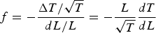 $$f = - \displaystyle{{\Delta T/\sqrt T} \over {dL/L}} = - \displaystyle{L \over {\sqrt T}} \displaystyle{{dT} \over {dL}} $$
This figure of merit can then be determined for each probe and the maximum of this figure of merit is the optimum object thickness to be radiographed with that probe. Of course, thicker or thinner objects may successfully be imaged, but the location of this maximum provides a guide for choosing the appropriate probe for the object to be imaged.
3. X-RAY RADIOGRAPHY
X-ray radiography provides a good example to demonstrate this method of identifying the optimum object thickness for a given imaging probe. The X-ray transmission fraction through an object is relatively simple and is governed by Beer's law (Beer, Reference Beer1852), shown in Eq. (2), where λ is the mass attenuation coefficient and L is the object thickness. The derivative of X-ray transmission fraction can be substituted into Eq. (1) to provide a figure of merit for X-ray radiography, shown in Eq. (3).
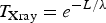 $${T_{{\rm X ray}}} = {e^{ - L/\lambda}} $$
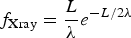 $${\,f_{{\rm X ray}}} = \displaystyle{L \over \lambda} {e^{ - L/2\lambda}} $$
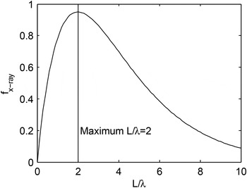
The figure of merit shown in Eq. (3) is plotted in Figure 1, where the maximum is found when L = 2λ. This result shows that an X-ray source with a mass attenuation coefficient λ is best suited to image through an object thickness of 2λ, resulting in a transmission of ≈14%.
The figure of merit shown in Eq. (3) is plotted as a function L/λ. The maximum of this function is shown at L/λ= 2. At this object thickness the contrast generated by a small change in object thickness is largest relative to the counting statistics noise of any measurement with this probe.

In the description above the incident particle flux, N 0 is not included in the figure of merit. Equation (4) shows how the incident flux can be taken into account in this figure of merit. This modification is a simple multiplier, which does not affect the location of the maximum. Equation (4) illustrates how a probe can be used to image through an object far from the ideal thickness, the figure of merit, f, can be recovered by increasing the particle flux. This approach is often used, but can result in difficulties when pushed to the extreme. For example, if one chooses an X-ray energy which is far from optimal, either the contrast becomes small for thin systems (L ≪ 2λ) or the transmission is low for thick systems (L ≫ 2λ). In these cases the contrast to noise ratio becomes small and it is easy for systematic errors to dominate, making the desired measurement difficult or impossible.
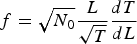 $$f = \sqrt {{N_0}} \displaystyle{L \over {\sqrt T}} \displaystyle{{dT} \over {dL}} $$
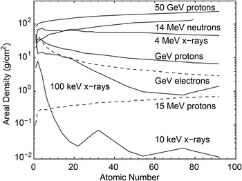
Figure 5 summarizes and compares the penetration power of the probes considered in the following sections, starting with the optimal object thickness for three X-ray energies (10 keV, 100 keV, and 4 MeV) as a function of atomic number of the radiographed material. Because the interaction length is maximum for 4 MeV X rays, this is the most penetrating electromagnetic radiation and 100 and 10 keV X rays are best suited to imaging thin systems (~1 g/cm2 and ~10 mg/cm2, respectively).
In the following sections this figure of merit is studied for various probes, determining the optimum object thickness for each probe, assuming the same incident flux. Because the interaction processes are typically dependent on object material, the location of this optimum will be considered as a function of material.
4. NEUTRON RADIOGRAPHY
Because neutrons only interact through nuclear forces, they are a very penetrating probe. These interactions tend to scatter the neutrons to large angles, resulting in an exponential attenuation process following Beer's law, similar to X-ray interactions. In this analytic form for attenuation the nuclear interaction cross-section is used to determine the mean free path of the neutrons in the material.
14 MeV neutrons generated through deuterium–tritium fusion processes are a common neutron imaging probe. Therefore, the nuclear cross-sections for 14 MeV neutrons are well known and this information can be used to determine the ideal object thickness for these measurements.
Of course, many other neutron energies are available with a wide range of nuclear cross-sections, allowing one to choose the best neutron energy for a specific imaging task. However, the process of generating a particular energy neutron can be difficult, as neutrons are typically generated through fission and fusion reactions or spallation processes. To generate neutrons of lower energies, a thermalization process is required, where the neutrons scatter multiple times within a moderator material losing energy in each scatter until reaching the energies of interest. These thermalized neutrons are not typically reasonable for imaging fast processes, as the moderation times (>100 ns) can be too large when compared with the time scale of the fast (~1 ns) dynamic processes being imaged. New laser driven neutron sources show promise for future applications of flash neutron radiography (Roth et al., Reference Roth, Jung, Falk, Guler, Deppert, Devlin, Favalli, Fernandez, Gautier, Geissel, Haight, Hamilton, Hegelich, Johnson, Merrill, Schaumann, Schoenberg, Schollmeier, Shimada, Taddeucci, Tybo, Wagner, Wender, Wilde and Wurden2013).
For self-emission imaging, such as the neutron pinhole imaging techniques common in the fusion sciences (Merrill et al., Reference Merrill, Bower, Buckles, Clark, Danly, Drury, Dzenitis, Fatherley, Fittinghoff, Gallegos, Grim, Guler, Loomis, Lutz, Malone, Martinson, Mares, Morley, Morgan, Oertel, Tregillis, Volegov, Weiss, Wilde and Wilson2012), however, the small interaction cross-section is ideal. This allows the neutron to escape the burning plasma without interaction in the surrounding material, even allowing the beam to escape the containment vessel and through vacuum system boundaries. With self-emission, however, the time structure of the imaging measurement matches the fusion burn history of the fusion process, providing a time averaged image.
5. PROTON RADIOGRAPHY
In the past 20 years two forms of proton radiography have been developed, both generating new and interesting measurement capabilities. The first was the development of high-energy proton radiography at Los Alamos National Laboratory (King et al., Reference King, Ables, Adams, Alrick, Amann, Balzar, Barnes, Crow, Cushing, Eddleman, Fife, Flores, Fujino, Gallegos, Gray, Hartouni, Hogan, Holmes, Jaramillo, Knudsson, London, Lopez, McDonald, McClelland, Merrill, Morley, Morris, Naivar, Parker, Park, Pazuchanics, Pillai, Riedel, Sarracino, Shelley, Stacy, Takala, Thompson, Tucker, Yates, Ziock and Zumbro1999). The introduction of a magnetic imaging lens (Mottershead & Zumbro, Reference Mottershead and Zumbro1997) between the object being studied and the detection system, revolutionized a decades old idea of using high-energy protons as direct radiographic probes for thick object radiography. Second, 15 MeV protons from (d,3He) fusion have been used to study the laser-driven implosion processes at the Laboratory for Laser Energetics (LLE's) Omega laser facility (Li et al., Reference Li, Seguin, Rygg, Frenje, Manuel, Petrasso, Betti, Delettrez, Knauer, Marshall, Meyerhofer, Shvarts, Smalyuk, Stoeckl, Landen, Town, Back and Kilkenny2008; Rygg et al., Reference Rygg, Séguin, Li, Frenje, Manuel, Petrasso, Betti, Delettrez, Gotchev, Knauer, Meyerhofer, Marshall, Stoeckl and Theobald2008).
Protons interact with materials through three primary processes: Coulomb interactions with the electrons, Coulomb interactions with the nuclear charge, and nuclear interactions with the nucleus. Because the proton mass is ~2000 times the mas of the electron, collisions with the electrons results in minimal deflection of the high-energy protons, but results in energy loss of the protons as it traverses the material, often referred to as dE/dx. Coulomb interactions between the protons and nucleus are also common, resulting in many small deflections of the incident protons, often referred to as Multiple Coulomb Scattering (MCS). The third process is mediated through strong force interactions and is therefore more rare than either ionization processes or MCS. Because of the larger nuclear mass, the protons tend to be scattered to large angles through these nuclear interactions, removing the protons from the angular acceptance of the proton radiography imaging system. Coulomb interactions are more important for low-energy proton radiography (~1 GeV), but become less important for high-energy proton radiography (>10 GeV). Equation (5) shows an analytic form for the calculation of proton transmission for high-energy proton radiography.
 $${T_{{\rm pRad}}} = {e^{ - L/{L_c}}}(1 - {e^{ - {\rm \theta} _{\rm c}^2 /2({{\rm \phi} ^2} + {\rm \theta} _0^2 )}}) $$
The first exponential term in Eq. (5) is Beers law capturing the loss of protons through nuclear processes, where L c is the nuclear collision length. The second term in Eq. (5) characterizes the removal term due to Coulomb interactions where θc is the angular acceptance of the imaging lens, ϕ is the natural angular spread of the proton beam, and θ0 is the additional spread in the angular distribution introduced through MCS in the object.
The derivative of the transmission shown in Eq. (5) can be substituted into Eq. (1). The resulting maximum of the figure of merit is plotted for 50 and 1 GeV protons in Figure 5 below. The 1 GeV optimal areal density curve was generated assuming the parameters of the imaging lenses described in Merrill et al. (Reference Merrill, Barlow, Espinoza, Hollander, Kwiatkowski, Lopez, Mariam, Morley, Morris, Nedrow, Saunders, Tainter, Tupa and Tybo2014) and the 50 GeV curve was plotted assuming an imaging system similar to that described in Morris et al. (Reference Morris, King, Kwiatkowski, Mariam, Merrill and Saunders2013).
Unlike neutrons or X rays, the mechanism for generating contrast in GeV proton radiographs, where MCS is the dominant removal process, can be adjusted without modifying the energy of the incident proton beam. This can be seen in the MCS removal term in Eq. (5) where θc can be adjusted by introducing a smaller collimator, which reduces the angular acceptance of the lens system. This results in an increase in the derivative of transmission for thinner systems, improving the sensitivity to thin systems. With the small emittance of linac generated beams (ϕ is small), this optimization can allow the radiographic sensitivity to be uniform through three orders of magnitude below the maximum optimal thickness shown in Figure 5.
Proton radiography with 15 MeV protons is best suited to very thin objects, as the range of 15 MeV protons is relatively small, fundamentally limiting the applications of this probe to imaging objects thinner than the range of 15 MeV protons. The upper limit for the application of this probe is also shown in Figure 5.
6. ELECTRON RADIOGRAPHY
Electrons undergo some of the same processes as protons as they pass through materials; Coulomb interactions with electrons and Coulomb interactions with the nuclear charge. Electrons with energies lower than ~30 MeV are similar to high-energy protons. For electron energies greater than ~30 MeV the interactions with the large nuclear electric field are not just small deflections, but stimulate the production of bremsstrahlung radiation, where the electrons emit photons as they are accelerated in the electric field of the nucleus. This process results in a new energy loss mechanism for electrons, which are not present for protons. These bremsstrahlung interactions can result in the production of high-energy X rays, removing substantial energy from the incident electron in a few interactions.
A linear, magnetic charged particle imaging lens can be set to focus only one energy of electrons. When lower energy electrons, such as those generated through the production of bremsstrahlung radiation, pass through the lens they are not properly focused at the detector plane. This blurring effect is often referred to as chromatic aberrations. Through these chromatic aberrations in the magnetic imaging lens this large energy loss can result in a significant blur that increases the width of the point spread function. In fact, this processes results in the practical limit of the penetration capability of high-energy electrons. Unfortunately, an analytic form describing this process is difficult and it is more convenient to utilize Monte Carlo techniques to simulate the penetrating power of high-energy electrons as they pass through the materials of interest.
To investigate these effects, EGS5 (Hirayama et al., Reference Hirayama, Namito, Nelson, Bielajew and Wilderman2005) was used to simulate 1 GeV electrons passing through varying thicknesses of iron plates. The electrons exiting the iron in this simulation were then transported through the magnetic imaging optics of a standard charged particle imaging lens composed of four quadrupole magnets similar to the system described in Merrill et al. (Reference Merrill, Harmon, Hunt, Mariam, Morley, Morris, Saunders and Schwartz2007). This simulated lens system had a magnification of 5 and was limited to a 3 mrad angular acceptance with a collimator at the Fourier plane of the imaging optics which was located just upstream of the third quadrupole magnet. This collimator removed electrons scattered out of the angular acceptance of the imaging lens. The electrons within the angular acceptance were transported to the image plane, where the root-mean-square width of the point spread function was calculated as a metric of resolution. As the object thickness is increased the probability for a large energy loss increases, resulting in a significant energy tail that blurs the image. When the object thickness reaches a full radiation length nearly all the electrons have suffered large energy loss interactions and this probe appears to become unusable for high-resolution image formation.
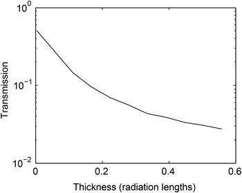
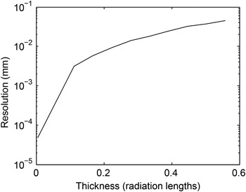
Figure 2 shows the transmission of a 1 GeV electron beam through the magnetic imaging lens after passing through an iron object of various thicknesses, which are measured in units of radiation lengths. This simulation shows that the transmission reaches the percent level when the object thickness reaches ~1/2 of a radiation length. Unlike protons the removal process is not simple scattering of the electrons out of the angular acceptance of the imaging lens, but a result of the large energy loss combined with the magnetic lens transport to the collimator position. When the low-energy electrons are transported through the first two magnets of the imaging system, only a small fraction pass through the collimation, but these off-energy electrons can reach the image plane and degrade the resolution through chromatic aberrations. Figure 3 shows the root mean square width of the system point spread function versus the thickness of the iron object in units of radiation lengths, showing that the resolution is remarkably good (<1 μm) for thin objects, but is degraded by nearly three orders of magnitude as the object thickness approaches ~1/2 a radiation length.
Transmission of 1 GeV electrons through a × 5 magnifying magnetic lens with an angular acceptance of 3 mrad after passing through various thicknesses of an iron sample.

Root mean square width of the point spread function versus object thickness for 1 GeV electrons penetrating a range of iron object thicknesses. The fast degradation of resolution is due to the large energy loss through bremsstrahlung processes. This large energy loss results in blur through chromatic aberrations introduced by the magnetic imaging lens.

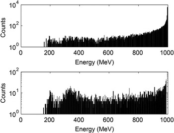
Figure 4 shows the distribution of electron energies reaching the detector plane after passing through 1 mm (~1/10 radiation length) and 9 mm (~2/3 radiation length) of iron. For objects 1/10 of a radiation length thick the image is formed mainly from 1 GeV electrons, but as the object thickness increases, the low-energy electrons begin to dominate the image formation, which results in significant image blur. From this simulation one can conclude that high-energy electron radiography is fundamentally limited to the study of objects thinner than a radiation length and ideal for objects between 1/10 and 1/3 of a radiation length. The curve for1 GeV electrons in Figure 5 plots the optimal thickness as 1/4 of a radiation length. A similar independent analysis has drawn similar conclusions (Wei et al., Reference Wei, Qiu and Jing2014).
Energy spectrum from an EGS5 simulation of electrons passing through 1 mm of iron (~1/10 radiation length) (top) and 9 mm of iron (~2/3 radiation length) (bottom). The electrons exiting the iron object in the simulation were transported through a magnetic imaging lens, with a collimator located at the Fourier plane of the imaging system. The energy of the electrons arriving at the image location was histogrammed to form the two graphs in this figure.

Summary of penetration capability for several common radiographic probes. The solid lines represent the location of the optimal thickness for the given probe and the dashed lines show the upper limit for the given probe. As discussed in the text, a probe can be used away from the optimal object thickness by increasing the incident flux, but as the probe is pushed far from the optimal range systematic errors will begin to dominate the measurements.

7. COMPARISON OF PROBES
Because of the differences in interaction processes each of the probes discussed above provide unique measurement capabilities, covering a wide range of areal densities. Figure 5 shows the optimal areal density as a function of material atomic number for a sample of the radiographic probes discussed above with energies common for applications in imaging small features of dynamic systems.
For proton radiography of thick objects, the energies of the probe must be large to reduce the effect of Coulomb interactions, leaving the nuclear interactions as the primary interaction mechanism. Thus, 50 GeV protons are well suited for radiography of the thickest objects (250 g/cm2 of material with high atomic number). 14 MeV neutrons and 4 MeV X rays, the most penetrating electromagnetic radiation, do not match the penetration capability of 50 GeV protons, but are well suited to thick objects (150 and 50 g/cm2 of material with high atomic number, respectively). Due to the impact of Coulomb interactions, 1 GeV proton radiography is best suited for the radiography of ~10 g/cm2 of high Z material.
Because of Bremsstrahlung interactions, high-energy electron radiography is limited to relatively thin system imaging (2 g/cm2 of high Z material), but the ease of generating electron beams in the required temporal structure and the potential for very high-resolution imaging make this a desirable probe for applications of thin object radiography. The 100 keV X rays approach the capabilities of 1 GeV electrons and higher energy X rays could match the penetration of 1 GeV electron radiography, but it is difficult to generate high fluxes of mono-energetic X rays at these energies with the source characteristics to provide high resolution radiography. Low-energy X rays and protons are a sensitive probe for radiography of the thinnest objects, capable of imaging systems down at the mg/cm2 level.
As mentioned above, with proper design and careful measurements, each probe can be used to radiograph objects away from the optimal areal densities shown in Figure 5. However, the natural interaction processes of each probe should be considered and matched to the object to be radiographed when designing a new radiographic capability. The simple figure of merit, based on the contrast to noise ratio, provides a useful tool to compare the penetration capability of the various probe options.