Introduction
Cladia is a genus of lichenized fungi in the family Cladoniaceae, mostly distributed in the Southern Hemisphere (Ahti Reference Ahti2000). In modern circumscription the genus comprises 24 species, including the former genera Heterodea and Ramalinora, 15 of which belong to the Cladia aggregata complex (Lumbsch et al. Reference Lumbsch, Parnmen, Rangsiruji and Elix2010; Parnmen et al. Reference Parnmen, Rangsiruji, Mongkolsuk, Boonpragob, Elix and Lumbsch2010, Reference Parnmen, Rangsiruji, Mongkolsuk, Boonpragob, Nutakki and Lumbsch2012, Reference Parnmen, Leavitt, Rangsiruji and Lumbsch2013). Cladia aggregata, the type of the genus, is the most widely distributed species, occurring from South America, South Africa, Australasia and South-East Asia to southern Japan and India. In spite of important recent contributions to the knowledge of lichenicolous fungi in the Southern Hemisphere (e.g., Aptroot Reference Aptroot2002; Etayo Reference Etayo2002; Etayo & van den Boom Reference Etayo and van den Boom2006; Etayo & Sancho Reference Etayo and Sancho2008; Flakus et al. Reference Flakus, Ahti, Kukwa and Wilk2008; Flakus & Kukwa Reference Flakus and Kukwa2012), the diversity of these fungi in this part of the world is still far from well known. To date, just four species of lichenicolous fungi have been reported from Cladia species, viz. Echinothecium cladoniae Keissl. nom. nud. (on C. aggregata from Columbia; Etayo Reference Etayo2002), Lichenoconium echinosporum D. Hawksw. (on C. muelleri from Australia; Hawksworth Reference Hawksworth1977), Roselliniella heterodeae Matzer & Hafellner (on C. muelleri from Australia; Matzer & Hafellner Reference Matzer and Hafellner1990) and Pyrenidium actinellum Nyl. agg. (on C. aggregata from Columbia; Etayo Reference Etayo2002). In the course of a revision of Cladia specimens in the herbarium of the University of Helsinki, a few new species of lichenicolous fungi were found. The new data, along with a key to the lichenicolous fungi occurring on Cladia, are presented here.
Materials and Methods
This study is based on 14 specimens of lichenicolous fungi found among 257 viewed specimens of Cladia species. Most of the specimens examined were collected by Professor Soili Stenroos and Dr Filip Högnabba during their field trip to New Zealand in 2010. The material was examined and photographed using Zeiss microscopes Stemi 2000-CS and Axio Imager A1 equipped with Nomarski differential interference contrast optics. Microscopic examination was carried out with material mounted in water, 10% KOH (K), Lugol’s iodine, directly (I) or after a KOH pre-treatment (K/I), or phloxin; considerable variation in the size of the ascospores and conidia in these media was not observed. The length, breadth and length/breadth ratio (l/b) of asci, ascospores and conidia are given (when n>10) as: (min−)(
 ${\bar x} $
−SD)−(
 ${\bar x} $
+SD)(−max), where min and max are the extreme values,
 ${\bar x} $
the arithmetic mean, and SD the corresponding standard deviation. Specimens examined are housed in the herbaria H or LE.
The Taxa
Endococcus cladiae Zhurb. & Pino-Bodas sp. nov.
MycoBank No.: MB 813735
Lichenicolous fungus. Ascomata perithecioid, (75–)85–100(–115) µm diam., protruding up to finally semi-immersed. Hamathecium of aseptate, brown external periphyses, 8–16×2·0–3·5 µm. Asci 32–50×7·5–11·0 µm, 8-spored. Ascospores evenly light brown, very narrowly obovate, occasionally almost fusiform or clavate with thinner lower cell, (9·9–)11·5–14·1(–15·7)×(2·7–)3·1–3·9(–4·6) µm, 1-septate, smooth-walled, non-halonate, diagonally uni- to biseriate in an ascus. Similar to Endococcus parentus, which mainly differs in its host selection (Pseudocyphellaria vs. Cladia), and in having (0–)1-septate, on average slightly shorter, ascospores.
Type: New Zealand, Stewart Island, Port Pegasus, North Arm, 47°10'S, 167°41'E, track margin at a secondary bog on a burnt area in lowland rainforest, on Cladia sullivanii (podetia), 24 March 1927, G. E. & G. Du Rietz 2205:3a (H—holotype).
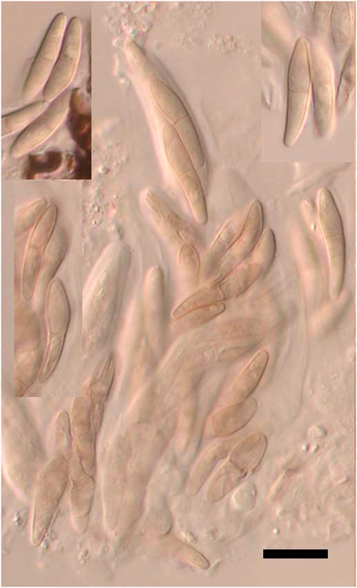
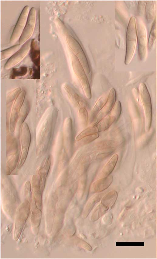
(Fig. 1)
Endococcus cladiae (holotype). Ascospores in K. Scale=10 µm. In colour online.
Vegetative hyphae scarce, immersed, pale brown, 1·1–2·5 µm diam.
Ascomata perithecioid, black, glossy, subglobose to pyriform, often conical above, (75–)85–100(–115) µm diam. (n=33), with pore 10–15 µm across, erumpent, protruding in the upper part to finally semi-immersed, dispersed; wall dark reddish brown throughout, pigmentation even, amorphous, K−, in surface view of textura angularis, composed of cells 3·3–8·8×2·8–6·5 µm, in section 10–15 µm thick, composed of 3–4 layers of radially compressed thick-walled cells. Interascal gel I+ red, K/I+ blue. Hamathecium not observed except aseptate brown external filaments 8–16×2·0–3·5 µm around the ostiole, which correspond to external periphyses sensu Etayo & Sancho (Reference Etayo and Sancho2008: 88, 90). Asci subcylindrical to narrowly obclavate, wall apically sometimes markedly thickened, occasionally with small indentation c. 1 µm tall, 32–50×7·5–11·0 µm (n=8, in I), 8-spored. Ascospores evenly light brown, very narrowly obovate, occasionally almost fusiform or clavate (narrowly obskittle-shaped) with thinner, occasionally markedly thinner, lower cell, straight to slightly curved, ends rounded to occasionally rather acute, (9·9–)11·5–14·1(–15·7)×(2·7–)3·1–3·9(–4·6) µm, l/b=(2·2–)3·2–4·2(–5·0) (n=88, in water, I or K/I), 1-septate, not or sometimes constricted at the septum, smooth-walled, non-halonate, usually with 2(–3) large guttules in each cell, diagonally uni- to biseriate in an ascus.
Conidiomata not observed.
Notes
The new species fits the Endococcus concept sensu Kainz & Triebel (Reference Kainz and Triebel2004), though the presence of brown external periphyses was not mentioned by these authors. However, such periphyses are described, for example, in Endococcus pallidosporus Etayo and E. parentus Etayo (Etayo & Sancho Reference Etayo and Sancho2008). Among the other species of the genus, Endococcus cladiae is most similar to E. alectoriae (D. Hawksw.) D. Hawksw. (on Alectoria), E. pallidosporus (on Pseudocyphellaria), E. parentus (on Pseudocyphellaria) and E. ramalinarius (Linds.) D. Hawksw. (on Ramalina). Whereas almost all species of Endococcus are confined to a specific host genus (Lawrey & Diederich Reference Lawrey and Diederich2015), none of them have been reported from members of Cladoniaceae, where Cladia belongs. Additionally, Endococcus alectoriae differs in having comparatively much wider ascospores (Hawksworth Reference Hawksworth1971: Fig. 1), E. pallidosporus has K/I+ red interascal gel and shorter ascospores, (8·0–)9·0–11·0(–12·5)×3·0–3·5(–4·0) µm (Etayo & Sancho Reference Etayo and Sancho2008), E. parentus has on average slightly shorter ascospores, (8·5–)9·0–14·0(–15·0)×3–4 µm (Etayo & Sancho Reference Etayo and Sancho2008), and E. ramalinarius has slightly shorter and wider ascospores, 10–13×4·0–4·5 µm (Hawksworth Reference Hawksworth1976: Fig. 3C). Among the pyrenocarpous cladoniicolous fungi, the most similar to Endococcus cladiae is Stigmidium cladoniicola Zhurb. & Diederich, which also has narrowly obskittle-shaped ascospores of similar size, (9·0–)11·5–15·0(–16·5)×3.0–4.0(–5.0) μm. However, it clearly differs from the latter in often having sessile and smaller ascomata 30–80 μm diam., I−, K/I− hymenial gel and colourless ascospores (Zhurbenko & Diederich Reference Zhurbenko and Diederich2008).
Distribution and hosts
Known from three localities in New Zealand, growing on healthy-looking and darkened parts of Cladia retipora and C. sullivanii podetia. An adverse effect on the host was not observed.
Additional specimens examined (both on podetia of Cladia retipora).
New Zealand: South Island: west-facing roadside near Ferguson Science Reserve, Highway 6 south of Westport, 42°58·9'S, 42°41·4'E, 0–20 m, 1999, S. Hammer 7524a (H); Southern Lakes, secus viam 9 km ad australem versus ad Te Anau, 45°37'06·0"S, 167°35'48·0"E, 210 m, 27 i 1984, M. E. Hale (H).
Lichenopeltella soiliae Zhurb. & Pino-Bodas sp. nov.
MycoBank No.: MB 813736
Lichenicolous fungus. Ascomata catathecioid, broadly conical, sometimes truncated above, ostiolar region often raised and forming a collar, without setae, (30–)46–62(–70) µm diam. Asci ellipsoid, ovoid to narrowly pyriform, (18·8–)20·6–26·2(–30·0)×(9·0–)9·7–11·7(–12·5) µm, 4-spored. Ascospores oblong, fusiform or very narrowly clavate, (11·2–)12·0–13·4(–14·5)×(2·8–)2·9–3·5(–4·0) µm, 1-septate, usually with 4 large guttules, rarely with 3 pairs of setulae. Similar to Lichenopeltella minuta, which mainly differs in its host selection (Omphalina vs. Cladia) and in having ascospores permanently without setulae.
Type: New Zealand, South Island, Southland, Wilderness Scientific Reserve, 6 km E of crossing to Manapouri on State Highway 94, 45°31'55·2"S, 167°51'21·6"E, open shrubland with Halocarpus bidwillii, on Cladia aggregata s. lat. (podetia), 6 May 2010, S. Stenroos 5778a (H—holotype; LE 264438—isotype; accompanied by Niesslia cladoniicola).
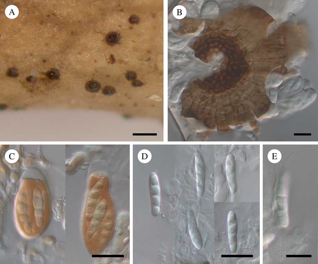
(Fig. 2)
Lichenopeltella soiliae (holotype). A, habitus; B, squashed ascoma in water (surface view); C, asci in K/I; D, ascospores in K; E, ascospore with 3 pairs of setulae in water. Scales: A=100 µm; B–D=10 µm; E=5 µm. In colour online.
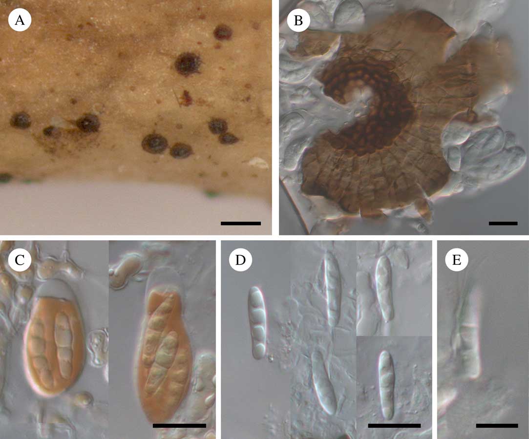
Ascomata catathecioid, medium to dark brown, usually much darker (often almost black) around the ostiole (better seen in wet state), glossy, broadly conical, sometimes truncated above, with central ostiole mainly c. 10 µm diam., sometimes gaping up to 30 µm across, ostiolar region often raised and forming a collar, without setae, (30–)46–62(–70) µm diam. (n=75), ascomatal margin entire, somewhat sinuate; upper wall medium to dark brown around the ostiole, pale to medium brown at the periphery, K+ grey-olive, one cell thick, composed of radially-arranged more or less rectangular or trapezoidal cells which are smaller, darker, more equilateral, and thick-walled around the ostiole, sometimes associated with brown branched hyphae 3–5 µm thick; lower wall similar in structure to the upper wall; ascomata usually slightly immersed in the cortex of the host thallus, dispersed, but occasionally adjacent. Paraphyses not observed. Asci ellipsoid, ovoid to narrowly pyriform, apical cap conspicuously thickened, foot not seen, (18·8–)20·6–26·2(–30·0)×(9·0–)9·7–11·7(–12·5) µm (n=18, in water, K or K/I), I−, K/I−, 4-spored. Ascospores hyaline, oblong, fusiform or very narrowly clavate (slightly wider above), rounded at the apices, (11·2–)12·0–13·4(–14·5)×(2·8–)2·9–3·5(–4·0) µm, l/b=(3·3–)3·6–4·2(–4·8) (n=43, in water, K or K/I), 1-septate, homopolar, occasionally slightly constricted at the septum, smooth-walled, non-halonate, usually with 4 large guttules, rarely with 3 pairs of setulae (seen in 5% of ascospores).
Etymology
The species is named after the Finnish lichenologist Professor Soili Stenroos who collected the type.
Notes
Most species of Lichenopeltella are known from a specific host lichen genus, and none of them have been reported on Cladia. Three species of the genus have been reported from members of Cladoniaceae, viz. L. cladoniarum E. S. Hansen & Alstrup, L. rangiferina Brackel and L. uncialicola Brackel, all believed to be confined to species of Cladonia. All of these clearly differ from Lichenopeltella soiliae: L. cladoniarum has 8-spored asci and larger ascospores, 18–21×5–6 µm, without setulae (Hansen & Alstrup Reference Hansen and Alstrup1995); L. rangiferina has ostiolar setae and (1–)3-septate, slightly longer ascospores, (13·0–)13·9–15·5(–16·0)×(3·0–)3·3–4·0 µm (Brackel Reference Brackel2011); L. uncialicola has ostiolar setae and ascospores without setulae (Brackel Reference Brackel2010). Among the other species of the genus, Lichenopeltella soiliae seems to be morpho-anatomically closest to L. minuta R. Sant., found on the basidiolichen Marchandiomphalina foliacea in South America (Santesson Reference Santesson1989). It differs from the latter by the ascospores which occasionally bear setulae and by a different host selection.
Distribution and host
Known from five localities in Australia and New Zealand, growing on healthy-looking and also darkened parts of Cladia aggregata s. lat. podetia. An adverse effect on the host not observed, even with abundant development of the fungus.
Additional specimens examined (all on podetia of Cladia aggregata s. lat.).
Australia: New South Wales: Great Dividing Range, 10 km ESE of Michelago, S. Tinderry Pic, 35°45'S, 148°18'E, 1250 m, dry sclerophyll forest, 1998, S. Hammer 7008a (H).—New Zealand: South Island: Southland, Wilderness Scientific Reserve, 6 km E of crossing to Manapouri on State Highway 94, 45°31'55·2"S, 167°51'21·6"E, open shrubland with Halocarpus bidwillii, 2010, S. Stenroos 5772a, 5774a, 5776a (all in H). North Island: Wellington, Tararau Forest Park, track from Kiriwhakapapa entrance to Blue Range Hut, before crossing of Cow Creek Hut and Blue Range Hut tracks, 40°47'52·8"–40°48'28·8"S, 175°32'45·6"E, 310–930 m, old-growth broadleaf forest, 2010, F. Högnabba 1864a (H, LE 264268); Manawatu-Wanganui, Tongariro National Park, Mt. Ruapehu, E of Whakapapa Village, Upper Taranaki Fall Track and Tama Lakes Tramping Track, about 500 m E of Taranaki Falls, 39°11'52·8"–39°12'00·0"S, 175°32'34·8"–175°34'12·0"E, 1140–1240 m, shrubland, 2010, F. Högnabba 1932a (H; accompanied by Lichenosticta hoegnabbae).
Lichenosticta hoegnabbae Zhurb. & Pino-Bodas sp. nov.
MycoBank No.: MB 813737
Lichenicolous fungus. Conidiomata pycnidial, blackish (dry) or brown with dark rim around ostiole (wet), 100–150(–200) µm diam., first immersed then up to half-erumpent. Conidiophores multi-septate, flexuous, branched, c. 2·0–2·5 µm diam. Conidiogenous cells lageniform or subcylindrical, mainly 6·5–11·5×1·5–2·5 µm, distinct collarette and proliferations not observed. Conidia bacilliform to fusiform, straight to slightly curved, rounded at the apex and attenuated and truncated at the base, (7·8–)9·1–10·9(–12·6)×(1·9–)2·1–2·3(–2·7) µm. Similar to Lichenosticta alcicorniaria, which mainly differs in its host selection (Cladonia vs. Cladia), and in having lacriform and broader conidia of (6·0–)6·5–10·0(–11·0) × (2·0–)3·0–4·5(–6·0) µm.
Type: New Zealand, North Island, Manawatu-Wanganui, Tongariro National Park, Mt. Ruapehu, E of Whakapapa Village, Upper Taranaki Fall Track and Tama Lakes Tramping Track to c. 500 m E of Taranaki Falls, 39°11'52·8"–39°12'00·0"S, 175°32'34·8"–175°34'12·0"E, c. 1200 m elev., shrubland, on Cladia aggregata s. lat. (podetia), 21 May 2010, F. Högnabba 1932a (H—holotype; accompanied by Lichenopeltella soiliae).
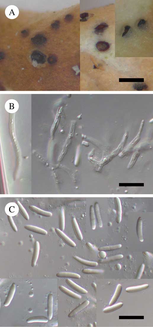
(Fig. 3)
Lichenosticta hoegnabbae (holotype). A, habitus; B, conidiophores and conidiogenous cells in water; C, conidia in water. Scales: A=200 µm; B & C=10 µm. In colour online.
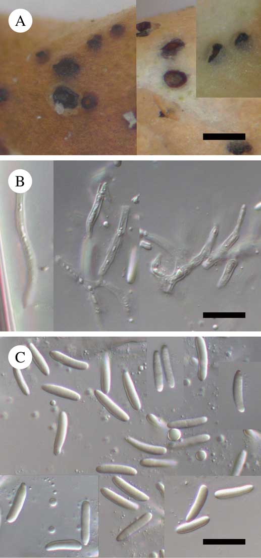
Conidiomata pycnidial, blackish (dry) or brown with dark rim around ostiole (wet), subglobose to broadly pyriform, sometimes indistinctly papillate, 100–150(–200) µm diam., ostiolate, sometimes opening by irregular aperture up to 80 µm across, first immersed then up to half-erumpent, arising singly; wall dark brown around the ostiole, otherwise medium brown throughout, K+ olive-brown, in surface view of textura epidermoidea or partly of textura angularis. Conidiophores hyaline, multi-septate, flexuous, branched, c. 2·0–2·5 µm diam., lining the pycnidial cavity. Conidiogenous cells hyaline, enteroblastic, phialidic, integrated into chains, lageniform or subcylindrical, mainly 6·5–11·5 × 1·5–2·5 µm, distinct collarette and proliferations not observed. Conidia hyaline, acro-pleurogenous, bacilliform to fusiform, straight to slightly curved, rounded at the apex and attenuated and truncated at the base, (7·8–)9·1–10·9(–12·6)×(1·9–)2·1–2·3(–2·7) µm, l/b=(3·4–)4·1–5·1(–6·0) (n=122, in water, K or phloxin), aseptate, smooth-walled, usually with 2–6 guttules clearly seen in K, excretion in a gelatinous drop not observed.
Etymology
The species is named after the Finnish lichenologist Dr Filip Högnabba who collected the holotype.
Notes
The coelomycetous lichenicolous genus Lichenosticta Zopf so far includes four species, viz. Lichenosticta alcicorniaria (Linds.) D. Hawksw. (on species of Cladonia), L. dombrovskae Zhurb. (on Stereocaulon), L. jurgae Kukwa & Flakus (on Lecanora), and L. lecanorae (Vouaux) Brackel & Zhurb. comb. ined. (on Lecanora). All of these clearly differ from L. hoegnabbae in the shape and size of conidia, which are lacriform, (6·0–)6·5–10·0(–11·0)×(2·0–)3·0–4·5(–6·0) µm in L. alcicorniaria (Hawksworth Reference Hawksworth1981); narrowly to broadly ellipsoid, occasionally almost oblong or circular, (4·5–)6·5–8·5(–11·0)×(3·0–)4·0–4·5(–5·5) μm in L. dombrovskae (Zhurbenko Reference Zhurbenko2010); broadly ellipsoid to oblong, rarely lacriform or pyriform, (4–)5–6(–8)×(2·0–)2·5–3·0(–3·5) μm in L. jurgae (Flakus & Kukwa Reference Flakus and Kukwa2012); ellipsoid to oblong, (2·7–)3·1–4·3(–5·5)×(1·5–)1·6–2·0(–2·2) µm in L. lecanorae (M. P. Zhurbenko, A. Frisch. Y. Ohmura & G. Thor, unpublished data).
Distribution and host
Known from two localities in New Zealand, growing on podetia of Cladia aggregata s. lat. An adverse effect on the host not observed.
Additional specimen examined
New Zealand: South Island: Wilderness Scientific Reserve, 6 km E of crossing to Manapouri on State Highway 94, open shrubland with Halocarpus bidwillii, 45°31'55·2"S, 167°51'21·6"E, on Cladia aggregata s. lat. (podetia), 2010, S. Stenroos 5774b (H).
Niesslia cladoniicola D. Hawksw. & W. Gams
Notes
New to Australasian region. Cladia is a new host genus.
Specimens examined (both on podetia of Cladia aggregata s. lat.). New Zealand: South Island: Southland, Kepler Track, outside of the Dock Bay entry to Fiordland National Park, 45°26'31·2"S, 167°41'16·8"E, open mossy and grassy seepage, 2010, S. Stenroos 5802a (H).; Wilderness Scientific Reserve, 6 km E of crossing to Manapouri on State Highway 94, 45°31'55·2"S, 167°51'21·6"E, open shrubland with Halocarpus bidwillii, 2010, S. Stenroos 5778a (H; accompanied by Lichenopeltella soiliae).
Discussion
A total of eight species of lichenicolous fungi are currently known to grow on species of Cladia. Four of them have also been reported on other host genera: Echinothecium cladoniae, Lichenoconium echinosporum and Niesslia cladoniicola grow on species of Cladonia in the same family Cladoniaceae (Hawksworth Reference Hawksworth1975; Suija et al. Reference Suija, Czarnota, Himelbrant, Kowalewska, Kukwa, Kuznetsova, Leppik, Motiejūnaitė, Piterāns and Schiefelbein2010; Brackel Reference Brackel2014), and Pyrenidium actinellum agg., probably representing a group of species (P. Diederich, pers. comm.), is known on many non-related host genera.
The Department of Conservation of New Zealand is thanked for the collection permits granted to Prof. S. Stenroos and Dr F. Högnabba in 2010. We are indebted to Paul Diederich and Teuvo Ahti for their valuable comments on the manuscript, to Rod Seppelt for providing details of collecting localities in New Zealand and to Paul Lockley who kindly revised the English. Mikhail P. Zhurbenko carried out this research project no. 01201255604 funded by Komarov Botanical Institute Russian Academy of Sciences. Raquel Pino-Bodas received funding from the European Union's Seventh Framework Programme (FP7/2007-2013) under grant agreement no. PIEF-GA-2013-625653.