I. INTRODUCTION
Light-emitting diodes (LEDs) have gained increased attention in daily life because of their superior lifetime, efficiency, reliability, and low pollution effect. Reference Chiu, Huang, Lee, Liu, Yeh, Jang and Liu1–Reference Li, van Steen, van Krevel, Botty, Delsing, DiSalvo, de With and Hintzen3 There are two fundamental ways to generate white light. Both methods require suitable phosphors, which refers to their ability to down-convert LED light. Reference Schlotter, Schmidt and Schneider4,Reference Mueller-Mach, Mueller, Krames and Trottier5 In recent years, newly developed (oxy)nitride phosphors have attracted the main focus of research in this field, because of their high efficiency, longer excitation and emission wavelengths, as well as their remarkable chemical and thermal stabilities. Reference Fukuda, Okada and Albessard6–Reference Yang, Song, Bian and Liu8
Aluminum nitride is a typical binary nitride with a wurtzite structure. It has been widely studied for electronic packing applications because of its high electrical insulation, high thermal conductivity, low thermal expansion coefficient, and high chemical stability. Reference Hagen, Yu, Grande, Hoier and Einarsrud9,Reference Jackson, Virkar, More, Dinwiddie and Cutler10 Aluminum nitride has been also proposed as a potential candidate for photoluminescence and electroluminescence devices due to its wide band gap (6.2 eV). Reference Taniyasu, Kasu and Makimoto11 Thus, there are many studies on AlN thin films or phosphors doped with rare-earth or transition metals. Reference Richardson, Van Patten, Richardson and Kordesch12–Reference Yin, Zhu, Yu, Hao, Xu, Hu and Lee17
The development of phosphors with controlled morphology and nanosized particles, which will make possible the achievement of high packing density and a low light scattering coefficient, is a great challenge in modern technology because it will lead to improvements in the brightness and resolution of displays. Thus, nano-oxynitride phosphors can be considered as candidates in the field of luminescent labeling. Oxynitride phosphors are superior to oxides and sulfurs because they have longer excitation wavelengths, higher emission intensity, and excellent stability.
Oxynitride phosphors are ordinarily prepared by a solid state reaction, carbothermal reduction, or gas reduction–nitridation. Reference Yin, Xu, Yu, Yang, Yang, Yang, Hao and Liu16,Reference Inoue, Hirosaki, Xie and Takeda18–Reference Hirosaki, Xie, Inoue, Sekiguchi, Dierre and Tamura20 The involvement of high temperatures, which is an inherently inevitable condition in these methods, leads to abnormal grain growth and the formation of hard agglomerates. Thus, many researchers have focused on the synthesis of binary or ternary transition metal nitrides using chemically complex precursors, Reference Kim and Kumta21–Reference Bem, Gibson and Loye24 expecting lower temperatures for the formation of crystalline phases as well as the suppression of grain growth, thereby promoting the formation of nanocrystals.
This paper presents the successful production of nanosized AlN:Eu phosphors with a homogeneous size distribution using a metal-organic precursor method. The precursors were produced by a reaction between metal chlorides and triethylamine. Triethylamine acts as a nitride former through a coordination reaction in the initial stages of the formation of the precursor.
II. MATERIALS AND EXPERIMENTAL PROCEDURE
The EuCl3 powder was obtained by the reaction between a fine powder of Eu2O3 (Sinopharm Chemical Reagent Co. Ltd, Shanghai, China, 99.99%) and 6 mol/L HCl solution (AR, referring to Analytical Grade), followed by drying with reflowing at 70 °C in the presence of thionyl chloride (SOCl2, AR). The produced powder of EuCl3 was stored in a glove box. AlCl3 (Sinopharm Chemical Reagent Co. Ltd, Shanghai, China, 99.99%), acetonitrile (CH3CN, liquid, AR), and triethylamine [N(C2H5)3, liquid, AR] were also used. All the glassware was oven dried prior to use.
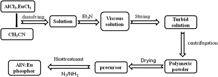
The experimental procedure is schematically outlined in Fig. 1. In a glove box, filled with N2 gas (O2 and H2O ≤ 5 ppm), the powders of EuCl3 and AlCl3were precisely weighed to satisfy the stoichiometric molar ratio of AlN:Eu. Then, these powders were put into a 250 mL glass conical flask together with a Teflon-coated magnetic stir bar. Then, about 50 mL of acetonitrile was added to completely dissolve the chloride powders. Then, triethylamine was carefully added, resulting in a yellowish solution under vigorous stirring. At a certain moment, the color of the solution changed instantly from white to yellowish. Under continuous stirring, the solution became very viscous and finally turbid due to the precipitation of the solid precursor. The solid precipitate was separated from the liquid by centrifugation and then dried under vacuum at 80 °C overnight. The obtained precursor powder was highly reactive toward oxidation and hydrolysis. Therefore, extreme precautions were taken to prevent any exposure of this powder precursor to air or moisture.
Outline of the synthesis process of nanosized AlN:Eu phosphors.

The precursor powder was put into boron nitride (BN) crucibles. The crucibles were placed in a quartz tube furnace (note that everything was inside the glove box). The back end of the tube furnace was connected to a flow meter (which was meant to count the bubbles of the gas flow through mineral oil). The front end of the tube furnace was connected to the gas line. The samples were heated at different temperatures between 500 and 1200 °C for 6 h under a gas flow of NH3 (flow rate was 300 mL/min). For comparison purposes, the same experiments were also carried out with firing under an N2 flow. The heating and the cooling rates were 3 K/min.
The crystalline phases that developed in the final products were identified by x-ray diffraction analysis (XRD, Philips PW 1700, Eindhoven, Netherlands, using Cu Kα1 radiation, λ = 1.54056 Å, at 40 kV and 100 mA with a graphite monochromator). The diffractograms were recorded in the range of 2θ between 10 and 80° with a scanning step of 2°/min. The samples were also analyzed by Fourier-transform infrared spectroscopy (FTIR, Perkin-Elmer 580B, Norwalk, CT, IR spectrophotometer using the KBr pellet technique).
The emission and excitation spectra were recorded at room temperature with a fluorescent spectrophotometer (F-4600, Hitachi, Chiyoda, Tokyo, Japan) with a 200 W Xe-lamp as an excitation source, corrected for the spectral response of the monochromator and Hamamatsu R928P photomultiplier tube (Hamamatsu Photonics K.K., Hamamatsu, Japan) by a light diffuser and tungsten lamp (Noma Electric Corp., NY; 10 V, 4 A) for the spectral distribution of the Xe lamp intensity by measuring Rhodamine-B as the reference.
The morphology of the powders was observed using a field emission scanning electron microscope (FE-SEM, JEOL-6390LA, JEOL, Tokyo, Japan), which was equipped with an electron dispersive spectroscopy (EDS) apparatus for chemical analysis. The inner structure of the produced powders was investigated using bright field and high-resolution transmission electron microscopy (BF-TEM and HR-TEM, respectively), equipped with selected area electron diffraction (SAED) analysis (JEOL-2010, JEOL, Tokyo, Japan). The valence of Eu was measured by an x-ray fluorescence spectrometer (XRF-1800, Shimadzu Corporation, Hadano, Japan). The carbon content was measured by a high frequency infrared carbon sulfur analyzer (CSC600 LECO, St. Joseph, MI).
III. RESULTS
The experimental results include those referring to the synthesis process and those referring to the characterization of the produced phosphors. With regard to the former, as reported in the experimental procedure section, the change in the color of the solution from white to yellowish (it specifically resembled the color of chrome-containing compounds), as well as changes in the viscosity of the solution, occurred after the addition of triethylamine under continuous vigorous stirring. Then, a precipitate gradually appeared. The precipitate of the solid precursor, separated from the remnant liquid by centrifugation, had a yellow color.
After heat treatment at 800 °C in an NH3 atmosphere, the powder of the precursor became dark gray. The elemental analysis (by EDS) revealed the existence of Al, Eu, N, and C in this powder. At the end of the heat treatment, a white powder was found deposited onto the walls of the quartz tube, which was identified as ammonium chloride, suggesting a reaction between HCl and NH3. The calcination of this dark gray powder at a temperature higher than 1100 °C resulted in a change in the color to white.
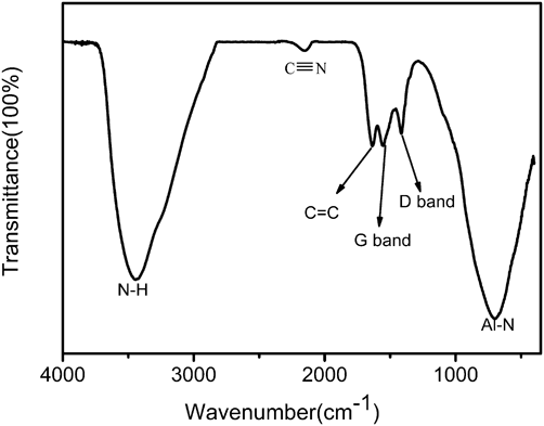
The FTIR spectrum of the powder of the precursor calcined in NH3 at 800 °C is shown in Fig. 2. The assignments of the peaks are also shown. In particular, the strong adsorption peak at about 706 cm−1 is attributed to the stretching vibration of the Al–N bond. Reference Kim, Sriram, McMichael, Kumta, Phillips and Risbud25,Reference Li, Liu, Yu, Dong, Wang, Hao and Cui26 The adsorption peaks at 1560 and 1410 cm−1 are the well-known graphite G band and the disorder-induced D band, respectively, Reference Maya, Cole and Hagaman27 whereas the peak at about 1634 cm−1 is attributed to C=C stretching vibrations. Reference McKenzie, McPhedran, Savvides and Cockayne28 The weak adsorption peak at 2160 cm−1 is attributed to the stretching vibrations of C≡N bonds. Reference Han and Feldman29 The vibration at 3440 cm−1 is assigned to the stretching vibrations of the N–H bonds, Reference Sacconi and Sabatini30 which likely occurred due to the adsorption of NH3 onto the surface of the powder during heat treatment in the NH3 atmosphere.
FTIR spectrum of the as-prepared precursor powder calcined in NH3 at 800 °C for 6 h.

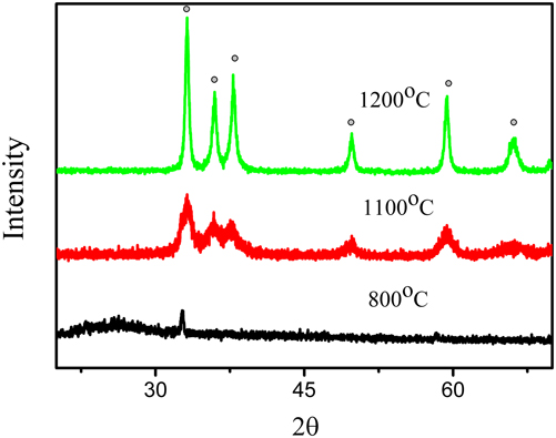
The x-ray diffractograms of the powders of the precursor calcined in NH3 for 6 h at different temperatures are plotted in Fig. 3. The precursor powder calcined at 800 °C was largely amorphous. However, the weak peak of the strongest diffraction peak of AlN at ∼32° is evidence of the onset of crystallization of AlN at this temperature. The crystallinity of AlN markedly improved with increasing calcination temperature, and well-defined patterns of AlN were clearly seen in the diffractogram at 1200 °C. There was no evidence of the formation of other phases. The carbon content of the AlN phosphor calcined at 1200 °C is less than 0.17 wt%, indicating that most of the carbon had been removed.
X-ray diffractograms of the precursor powders calcined in NH3 for 6 h at different temperatures. The circular markers indicate the peaks assigned to AlN (JCPDS card No. 00-025-1133).
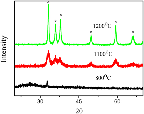
The broad XRD patterns of the sample calcined at 1200 °C (Fig. 3) suggest a significant refinement in the grain size. The calculated crystallite size of the produced AlN:Eu2+ crystals (using the Scherrer formula Reference Friedman and Briks31 ) was 16 nm.
Similar results, i.e., the formation of a single phase of AlN, were also obtained when calcination took place under N2 flow. However, it should be noted that there was no color change in the samples calcined under an N2 atmosphere at 1100 °C and the color of the precursor powder remained dark gray.
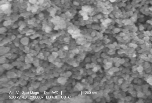
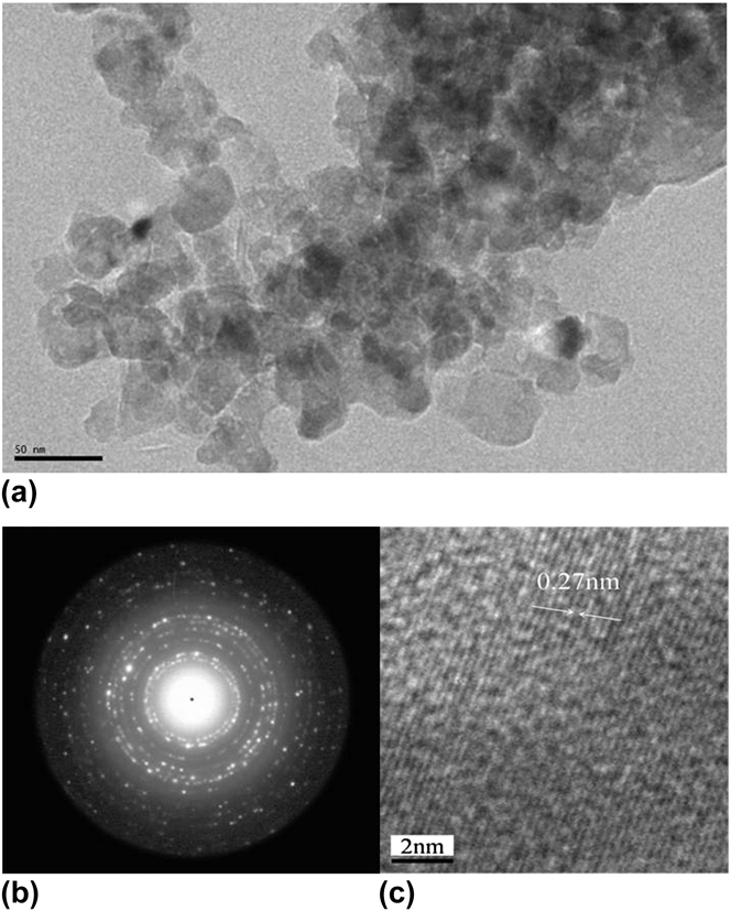
The morphology of the produced powder after calcination of the precursor in NH3 at 1200 °C is shown in the SEM image in Fig. 4. A fine powder with an apparently homogeneous particle size distribution and nanoparticles with a size of ∼10–20 nm was observed. These features were also confirmed in the BF-TEM image shown in Fig. 5(a).
FE-SEM image of the powder of nano-AlN:Eu2+ phosphor calcined in NH3 at 1200 °C for 6 h.

(a)BF-TEM image, (b) SAED pattern, and (c) HR-TEM image of nano-AlN:Eu2+ phosphor powder calcined in NH3 at 1200 °C for 6 h.

The diffraction pattern, obtained from the SAED analysis [Fig. 5(b)], consisted of rings corresponding to the polycrystalline hexagonal AlN. This finding agrees fairly well with the results of the XRD analysis (Fig. 3). To identify the exposed facets of a nanoparticle, HR-TEM was employed. According to the image in Fig. 5(c), the lattice fringe clearly reveals the presence of the (100) plane with a lattice space of 0.27 nm, which agrees with the data of the hexagonal lattice of AlN (JCPDS card No. 00-025-1133).
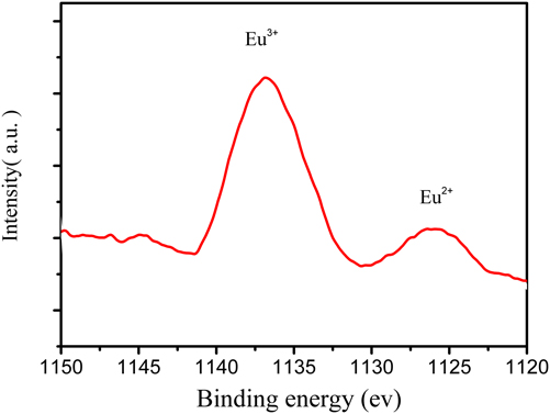
XPS was employed to identify the chemical state of the Eu, as shown in Fig. 6. A peak with a binding energy of around 1125 eV, attributed to divalent Eu, and a peak with a binding energy of around 1136 eV, attributed to trivalent Eu, are clearly seen, indicating that Eu ions exist in both the trivalent and the divalent states on the surface of the nanosized AlN:Eu2+ phosphor, which is in good agreement with our previous research. Reference Yin, Zhu, Yu, Hao, Xu, Hu and Lee17 It deserves to be pointed out that the actual Eu3+/Eu2+ ratio in the AlN:Eu2+ phosphor should not be so high, because the measurement of XPS is limited to the surface with a very small depth. Using XPS measurements, Eu ions also exist mainly in the trivalent state in the two famous blue phosphors (NaBaPO4:Eu2+ and BaMgAl10O17:Eu2+) Reference Wanjun and Donghua32,Reference Lacanilao, Wallez, Mazerolles, Dubot, Binet, Pavageau, Servant, Buissette and Le Mercier33 and in the Ca2Si5N8: Eu2+red phosphor. Reference Chung and Chou34
XPS spectra showing peaks of the AlN:Eu2+ phosphors.

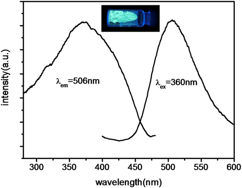
The photoluminescence spectra of the obtained AlN:Eu nanophosphor are plotted in Fig. 7. A broad green emission band at about 506 nm under excitation at 360 nm was recorded and was ascribed to the 5d–4f transition of Eu2+. Even with a much smaller grain size, the emission intensity of the obtained AlN:Eu nanophosphor can reach as high as 70% of that of the microsized AlN:Eu phosphor obtained by the carbothermal reduction route reported previously. Reference Yin, Xu, Yu, Yang, Yang, Yang, Hao and Liu16 This could be attributed to the homogeneous distribution of Eu in the nanosized AlN crystal. The line emission in the spectral region from 550 to 630 nm, corresponding to the5D0–7F1 and 5D0–7F2 transitions of the Eu3+ ions, has not been detected due to the forbidden f–f transitions.
Excitation and emission spectra of the AlN:Eu2+nanophosphor powder calcined in NH3 at 1200 °C for 6 h.

IV. DISCUSSION
The experimental results show that the proposed synthesis method resulted in fine powders of AlN:Eu2+ comprising well-crystallized nanoparticles with a spherical shape and a narrow particle size distribution between ∼10–20 nm. It is worth noting that the particle size of AlN:Eu2+ powders produced by solid state reaction methods is 3–5 μm. Reference Inoue, Hirosaki, Xie and Takeda18
The finding that a single phase of AlN was formed after calcination regardless of the atmosphere, whether NH3 or N2, suggests that the obtained AlN is not the result of a reaction between NH3 and the precursors, but rather a result of the decomposition of the precursor. Thus, this section aims at discussing the mechanism of the synthesis process in the light of the experimental findings of the characterization of the final product of the AlN:Eu2+ phosphor nanopowder.
It is well known that many metal chlorides behave as Lewis acids and tend to form adducts with electron-donating bases, such as hydrazine and triethylamine. With the term adduct, we define the product of an addition reaction, a new chemical species, where each molecular entity is formed by the direct combination of two separate molecular entities participating in such a way that there is a change in connectivity, but no loss of atoms within the two moieties. The interaction between the transition metal halides and nitriles has been well documented and reviewed in the literature. Organonitriles can coordinate with metal ions. Moreover, such coordinated nitriles can be easily replaced by other ligands. Reference Storhoff and Lewis35–Reference Sriram, Kumta and Ko37
According to the general scheme described above, it is suggested that in the present synthesis process, the chlorides first transform into adducts that can be designated by the formula CH3CN→MCl3 (where M denotes the metals, Al and Eu). After the addition of triethylamine into the solution, the color changed instantly from white to (chrome-like) yellowish and the solution became more viscous. All these features provide evidence for a chelation reaction, which means the formation of coordinate bonds between a multiple bonded ligand and a single central atom. The ligand exchange reaction between the adduct and triethylamine can be described with the following chemical equation:
 $${\rm{E}}{{\rm{t}}_3}{\rm{N}} + {\rm{C}}{{\rm{H}}_3}{\rm{CN}} \to {\rm{Al}}\left( {{\rm{Eu}}} \right){\rm{C}}{{\rm{l}}_3} \to {\rm{E}}{{\rm{t}}_3}{\rm{N}} \to {\rm{Al}}\left( {{\rm{Eu}}} \right){\rm{C}}{{\rm{l}}_3} + {\rm{C}}{{\rm{H}}_3}{\rm{CN}}\quad {\rm{.}}$$
The FTIR spectrum of the precursor powder showed weak evidence of bonds between C and N (adsorption peak at about 2160, Fig. 2). This suggests that the C≡N groups were coordinated to Al(Eu) and seemingly occurred at the stage of the reaction of Eq. (1).
The solid precursor is likely the result of condensation and polymerization reactions, summarized in Eqs. (2) and (3):
 $${\rm{E}}{{\rm{t}}_3}{\rm{N}} \to {\rm{Al}}\left( {{\rm{Eu}}} \right){\rm{C}}{{\rm{l}}_3} \to {\rm{E}}{{\rm{t}}_2}{\rm{N}}-{\rm{Al}}\left( {{\rm{Eu}}} \right){\rm{C}}{{\rm{l}}_2} + {\rm{EtCl}}\quad ,$$
 $$\eqalignb{ & {\rm{2E}}{{\rm{t}}_2}{\rm{N}}-{\rm{Al}}\left( {{\rm{Eu}}} \right){\rm{C}}{{\rm{l}}_2} \to {\rm{E}}{{\rm{t}}_2}{\rm{N}}-{\rm{Al}}\left( {{\rm{Eu}}} \right){\rm{C}}{{\rm{l}}_2} \cr & \downarrow \cr & {\rm{E}}{{\rm{t}}_2}{\rm{N}}-{\rm{Al}}\left( {{\rm{Eu}}} \right){\rm{C}}{{\rm{l}}_2} \cr}$$
The product of Eq. (2), Et2N–Al(Eu)Cl2, is obviously both a Lewis acid and a Lewis base (because there is an unoccupied orbital in the electron valance shell of Al and a lone pair of electrons in N, respectively). This dual (acid–base) feature of Et2N–Al(Eu)Cl2 seemingly favors the chemical reaction of Eq. (3). The dimer product of Eq. (3) can form oligomers whose growth gradually results in the formation of an inorganic three-dimensional Al–N network. When the network reaches a sufficient size, precipitation occurs. Since the polymerization process is a rather slow stage, control of the grain growth of the precursor powder seems to be possible.
The direct coordination of nitrogen to metallic ions during the precipitation of precursors was confirmed by the FTIR spectrum (adsorption peak at about 706 cm−1, attributed to the stretching vibration of the Al–N bond, Fig. 2). The establishment of Al(Eu)–N bonds in the as-prepared precursor formed due to the homogeneous reaction of Al(Eu)Cl3 with triethylamine, and should result in the homogeneous distribution of AlN:Eu2+ nanocrystallized particles, shown in the BF-TEM image [Fig. 5(a)].
The experimental results show that the precursor powder contained carbon in the form of amorphous graphite (peaks at 1560 and 1410 cm−1, Fig. 2). In an atmosphere of NH3, this carbon should react according to the chemical equation Reference Kim and Kumta21,Reference Kim and Kumta38,Reference Colque and Grange39 :
 $${\rm{C}} + {\rm{N}}{{\rm{H}}_3} \to {\rm{HCN}} + {{\rm{H}}_2}\quad ,$$
causing a change in the color of the precursor after calcination from dark gray (after calcination at 800 °C) to white (after calcination at 1100 °C). Equation (4) is confirmed because there was no color change in the samples calcined under an N2 atmosphere. The experimental results of the present study do not allow us to propose anything about the effect of carbon. However, in the literature, it has been stated that the residual carbon could suppress grain growth. Reference Weil and Kumta23 Thus, in the present study, residual carbon might also prevent the grain growth of nanosized AlN:Eu2+ phosphor particles.
With regard to the potential applications of the produced phosphor, the relatively long emission wavelength (with a peak at about 506 nm, Fig. 7) might be attributed to the high N content Reference Yin, Yu, Xu, Hao and Simeon15 or to the high Eu concentration. The solubility of Eu2+in AlN is quite low (∼0.3 mol%), Reference Yin, Zhu, Yu, Hao, Xu, Hu and Lee17,Reference Inoue, Hirosaki, Xie and Takeda18 but the Eu concentration in nanosized phosphors pronouncedly increases up to 2 mol%. Apparently, the homogeneous mixing of Eu and Al ions in the solution as well as crystal distortion in the nanophosphor can affect the solubility of Eu2+ ions in AlN. A higher Eu concentration should enhance the energy transfer among Eu2+ ions, Reference Colque and Grange39,Reference Sakuma, Hirosaki and Xie40 leading to a red shift in the emission wavelength.
V. CONCLUSIONS
Nanophosphors of AlN:Eu2+ with good photoluminescence properties, supporting their use as alternative high efficiency emitting phosphors, were synthesized using a metal-organic method. Compared to the powders and processing involved in conventional methods, such as the solid state reaction, the method applied in this study resulted in the establishment of direct Al(Eu)–N bonds in the initial stage of the formation of the precursor. This is very important for the production of fine and homogenous AlN:Eu2+ nanopowders (with a particle size of about 15 nm) at moderately low temperatures.
ACKNOWLEDGMENTS
This research was supported by the National Science Foundation of China (Grant Nos. 51072191, 11179037), the National Basic Research Program of China (2012CB922004), the USTC-NSRL association funding (KY2060140005), and the Chinese Academy of Sciences under the Bairen Program.