Introduction
The ‘lambari-do-rabo-amarelo’, Astyanax bimaculatus (Linnaeus, 1758), is a small-sized species that is widely distributed, being found in several South American basins (Lima et al., Reference Lima, Malabarba, Buckup, Pezzi da Silva, Vari, Harold, Reis, Kullander and Ferraris2003). This species does not migrate for reproduction, it reproduces both in lentic and lotic water environments and has a long reproductive season and multiple spawning (Godinho et al., Reference Godinho, Lamas and Godinho2010). The genus Astyanax previously belonged to the Tetragonopterinae sub-family but is currently included in the incertae sedis group in the Characidae family (Lima et al., Reference Lima, Malabarba, Buckup, Pezzi da Silva, Vari, Harold, Reis, Kullander and Ferraris2003). This situation is due to a lack of monophyly among the sub-families that was evident from phylogenetic studies using osteologic, biometric and meristic parameters (Weitzman & Malabarba et al., Reference Weitzman, Malabarba, Malabarba, Reis, Vari, Lucena and Lucena1998). The species of the incertae sedis group are small in size and are very important for energy flow along the food chain as they are food for piscivore species (Martins et al., Reference Martins, Arantes, Sato, Enemir, Rizzo and Bazzoli2011).
Eggs of different fish species may be distinct in size, shape, colour, specific density and degree of adhesiveness (Rizzo et al., Reference Rizzo, Sato and Godinho2002) and may be classified as adhesive, semi-adhesive or non-adhesive. However, the presence of a jelly coat is not directly related to egg adhesiveness as the jelly coat is present frequently in Siluriformes eggs, but not all fish of this order have adhesive eggs (Rizzo et al., Reference Rizzo, Sato and Godinho2002). Some species of Characiformes, such as Schizodon knerii, Triportheus guentheri, Bryconops affinis, Hoplias spp., Hoplerythrinus unitaeniatus, Acestrorhynchus spp. and Curimatella lepidura present adhesive eggs but do not have a jelly coat (Rizzo et al., Reference Rizzo, Sato and Godinho2002; Gomes et al., Reference Gomes, Scarpelli, Arantes, Sato, Bazzoli and Rizzo2007). In some fish groups, adhesion of the egg to the substrate is accomplished via an adhesive disc around the micropyle (Honorato-Sampaio et al., Reference Honorato-Sampaio, Santos, Bazzoli and Rizzo2009; Perini et al., Reference Perini, Sato, Rizzo and Bazzoli2010). Although the jelly coat is a feature observed commonly in Siluriformes eggs, to our knowledge the occurrence of this coat in Characiformes eggs has not been reported previously.
Embryonic development of fish consists of a series of morphological events and cellular movements that starts with the penetration of the spermatozoid into the oocyte through the micropyle and culminates with the hatching of the larvae to the external environment (Solnika-Krezel, Reference Solnica-Krezel2005). After fertilisation, the perivitelline space is formed by the oocyte hydration. Pelagic eggs have a large perivitelline space and greater buoyancy, whereas demersal eggs have a small perivitelline space and less buoyancy when compared in a water column (Lubzens et al., Reference Lubzens, Young, Bobe and Cerdà2010). Knowledge of the initial development may be useful towards a better understanding of the reproductive strategies of the species, as well as for ex situ species preservation experiments, besides being a tool for environmental quality assessment (Ninhaus-Silveira et al., Reference Ninhaus-Silveira, Foresti and Azevedo2006). Water temperature is a parameter that directly affects embryo development time and also the fertilisation rate (Hansen & Falk-Petersen, Reference Hansen and Falk-Petersen2002). Low temperatures delay embryonic development, whereas very high temperatures may cause morphological alterations and decrease larvae survival (Sund & Falk-Petersen, Reference Sund and Falk-Petersen2005).
Research on fish oocyte morphology and embryonic development is important to provide information for future studies on reproduction, phylogeny and taxonomy. This study aims to analyse the morphology of recently released vitellogenic oocytes using histological and histochemical techniques and on the embryonic development of A. bimaculatus specimens that were subjected to artificial reproduction.
Materials and methods
Specimen collection and fertilization
Astyanax bimaculatus specimens, 10 males each with a total length of 8.6 ± 0.29 cm and body weight of 9.16 ± 0.60 g and 10 females each with a total length of 9.3 ± 0.83 cm and body weight of 14.09 ± 3.73 were captured in the São Francisco River and kept in 200 m2 aquaculture tanks with an average depth of 1 m at the Três Marias Hydrobiology and Pisciculture Station, CODEVASF (18°11′58″S, 45°15′07″W). Fish that presented external morphological characteristics of gonadal maturation, such as swollen abdomen, red urogenital papilla and spenting males, were selected and transferred to reproduction tanks with a water temperature of 26°C and underwent induced reproduction. Both males and females received a single dose (6 mg kg−1) of crude common carp pituitary extract (Cyprinus carpio) injected into the coelomic cavity. Oocyte extrusion was performed manually and fertilisation was carried out using the dry method, i.e. by mixing semen with oocytes followed by hydration. The fertilised eggs were kept in funnel-shaped incubators (20 l) with constant circulation of water at 24°C. A camera was attached to a stereoscopic microscope and egg samples were collected every 10 min in order to monitor the embryonic development.
Histological analysis
For recently ovulated oocytes, samples were fixed in Bouin's fluid for 12 h for the histological analysis and subjected to routine histological techniques: paraffin embedding, 5-μm thick microtomy sections, and hematoxylin–eosin stain (HE). The following histochemical techniques were used to determine the histochemical content of the oocyte structures: periodic acid–Schiff (PAS) stain, Alcian blue (AB) stain pH 2.5 and Alcian blue (AB) stain pH 0.5.
Measurement of oocyte diameter
Oocyte diameter, perivitelline space and jelly coat thickness were determined by measuring 25 fresh oocytes from each female using a micrometric ocular attached to a light microscope.
Results
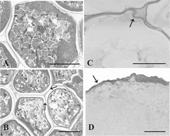
Recently ovulated vitellogenic oocytes of Astyanax bimaculatus were found to be spherical, yellowish, and covered by a thin, slightly adhesive jelly coat. The jelly coat of the recently ovulated oocytes (Fig. 1A,B) reacted positively to PAS stain (Fig. 1C) and AB stain pH 2.5 (Fig. 1D) histochemical techniques, and neutral glycoproteins and carboxylated acid glycoconjugates, forming mucosubstances, were detected on this jelly coat. The mean non-hydrated A. bimaculatus oocyte diameter was 1.03 ± 0.03 mm, the perivitelline space was 0.21 ± 0.02 mm and the thickness of the jelly coat was 0.04 ± 0.01 mm.
Recently ovulated oocytes of Astyanax bimaculatus. (A) Eggs after extrusion, hematoxylin–eosin stain (HE). (B) Oocytes agglutinated by jelly coat, HE. (C) Positive reaction to periodic acid–Schiff (PAS) stain on the jelly coat. (D) Positive reaction to Alcian blue (AB) stain pH 2.5. Scale bars = 200 μm in (A) and (B); 80 μm in (C) and (D); arrows indicate the jelly coat.

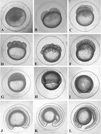
During embryonic development, the following stages were registered, with their respective post-fertilisation times:
• Zygote (20 min): formation of the unicellular blastodisc, evident perivitelline space, abundant yolk (Fig. 2A).
• Cleavage (40 min): successive mitotic divisions of the blastodisc. Discoidal meroblastic cleavage. Blastomeres spaced apart from each other (Fig. 2B–F).
• Blastula (2 h 45 min): blastodisc stratification and formation of a protuberance of cells characterising the high blastula. The space between blastomeres decrease. The embryo's spherical shape returns due to the blastomeres’ reorganisation (Fig. 2G).
• Gastrula (3 h 45 min): cells from the blastodisc start to migrate towards the vegetative pole characterising the epiboly movement. Together with this movement, the invagination movement starts, forming the epiblast and hypoblast (Fig. 2H).
• Blastopore closure (5 h): end of the epiboly movement culminating with the blastopore closure. Yolk completely surrounded by the blastodisc, forming the yolk sac (Fig. 2I).
• Differentiation of embryonic layers (7 h 15 min): the epiblast forms the ectoderm and the notochord and hypoblast form the mesoderm and endoderm. Cranial and caudal portions are distinct (Fig. 2J).
• Tail release (10 h 30 min): successive contractions of the myotomes cause the tail to separate from the rest of the body (Fig. 2K,L).
• Hatching (17 h): vigorous muscle contractions cause the chorion rupture and the release of the larvae into the external environment.
Embryonic development stages of Astyanax bimaculatus. (A) Zygote. (B–F) Segmentation. (G) Blastula. (H) Gastrula. (I) Blastopore closure. (J) Differentiation of embryonic layers. (K) Start of the tail release. (L) Completely free tail, pre-eclosion.

Discussion
This study analysed the morphology of vitellogenic oocytes and examined the main stages of embryonic development of A. bimaculatus, as well as finding a jelly coat in Characiformes species for the first time.
The oocyte colouration is directly related to diet and it represents an important parameter in studies in which the identification of eggs collected in nature is required (Blount & Houston, Reference Blount and Houston2000). The yellow colour is associated with the presence of carotenoids, but little information is known regarding its importance for the oocyte (Lubzens et al., Reference Lubzens, Lissauer, Leavavi-Sivan, Avarre and Sammar2003). The difference of oocyte colour among species or among fish of the same species may also be related to diet and habitat segregation (Arcifa et al., Reference Arcifa, Northcote and Froelich1991).
Teleostei eggs have different diameters, i.e. they are smaller in species that have reproductive migration and larger in non-migratory species (Suzuki et al., Reference Suzuki, Agostinho and Winemiller2000; Sato et al., Reference Sato, Fenerich-Verani, Nuñer, Godinho, Verani, Godinho and Godinho2003). These differences may be related to the reproductive strategies of each species (Lubzens et al., Reference Lubzens, Young, Bobe and Cerdà2010). Astyanax bimaculatus eggs are small with a diameter of 1.03 ± 0.03 mm, however it is not a species that migrates to reproduce, therefore their small egg size may be related to body size as observed for other small-sized Characiformes species (Sato et al., Reference Sato, Fenerich-Verani, Nuñer, Godinho, Verani, Godinho and Godinho2003). The neutral glycoproteins and carboxylated acid glycoconjugates on the jelly coat of A. bimaculatus oocytes form mucosubstances that may be synthetized by the follicular cells and transferred to the surface of the zona radiata in order to form the jelly coat, as recorded for other neotropical teleost species (Andrade et al., Reference Andrade, Bazzoli, Rizzo and Sato2001; Santos et al., Reference Santos, Padilha, Bomcompagni-Júnior, Santos, Rizzo and Bazzoli2006; Martins et al., Reference Martins, Arantes, Sato, Enemir, Rizzo and Bazzoli2011). In some species of the Erythrinidae family that have adhesive eggs, lectin was found in the follicular cells and on the zona radiata's surface, but without formation of a jelly coat (Gomes et al., Reference Gomes, Scarpelli, Arantes, Sato, Bazzoli and Rizzo2007). These substances may thus be related to adhesion of the egg to the spawning substrate. Adhesive eggs are related to parental care (Kolm & Ahnesjö, Reference Kolm and Ahnesjö2005), however, there is no study that claims that this is a reproductive strategy in A. bimaculatus.
The A. bimaculatus jelly coat is narrow (0.04 ± 0.01 mm), compared with the coat found in Siluriformes eggs, which can reach 0.51 mm in Rhamdia quelen (Sato et al., Reference Sato, Fenerich-Verani, Nuñer, Godinho, Verani, Godinho and Godinho2003). In teleostei, the thickness of the jelly coat is directly related to the location of spawning (Riehl, Reference Riehl1996). Sedentary species that leave eggs in rocky or sandy areas and inside nests have a thick jelly coat for protection against mechanical shocks and to support the weight of the material that is deposited over them (Groot & Alderdice, Reference Groot and Alderdice1985). However, species that lay their eggs near the surface, such as the aquatic macrophytes or marginal vegetation, have a thin coat to facilitate gas exchange (Riehl, Reference Riehl1996). The jelly coat is a characteristic of several other groups of fish and is present in the eggs of Siluriformes, Perciformes, Cypriniformes, Cyprinodontiformes, Acipenseriformes and Dipnoi (Kemp, Reference Kemp1994; Riehl & Patzner, Reference Riehl and Patzner1998; Rizzo et al., Reference Rizzo, Sato and Godinho2002; Zelazowska, Reference Zelazowska2010). Usually, species with adhesive eggs have prismatic follicular cells and a large number of organelles for mucosubstance synthesis. A. bimaculatus, however, has squamous follicular cells and a smooth zona radiata with pore-canals (Rizzo et al., Reference Rizzo, Sato and Godinho2002; Martins et al., Reference Martins, Arantes, Sato, Enemir, Rizzo and Bazzoli2011), a finding that could explain the thin jelly coat and the weak adhesiveness of A. bimaculatus eggs.
The perivitelline space in A. bimaculatus (0.21 ± 0.02 mm) was smaller than that in migrating Characiformes, such as Brycon orthotaenia, Prochilodus spp. and Salminus franciscanus, which have a space of 0.96–1.41 mm (Sato et al., Reference Sato, Fenerich-Verani, Nuñer, Godinho, Verani, Godinho and Godinho2003). Fish that have a migratory reproductive strategy and usually leave their eggs in rivers with strong flow have eggs with a larger perivitelline space to protect them against this abrasive force (Alexandre et al., Reference Alexandre, Ninhaus-Silveira, Veríssimo-Silveira, Buzollo, Senhorini and Chaguri2010). Astyanax bimaculatus, however, leave their eggs in lotic or lentic environments, where flow is not very strong, so that the eggs are not subjected to a severe force. For this reason, their perivitelline space is smaller when compared with migratory species.
The stages of A. bimaculatus embryonic development were similar to those described for other species of neotropical Teleostei (Gomes et al., Reference Gomes, Scarpelli, Arantes, Sato, Bazzoli and Rizzo2007; Amorim et al., Reference Amorim, Gomes, Martins, Sato, Rizzo and Bazzoli2009; Alexandre et al., Reference Alexandre, Ninhaus-Silveira, Veríssimo-Silveira, Buzollo, Senhorini and Chaguri2010; Perini et al., Reference Perini, Sato, Rizzo and Bazzoli2010; Gomes et al., Reference Gomes, Sato, Rizzo and Bazzoli2011). The pattern of egg segmentation in vertebrates depends on the quantity and distribution of yolk and its proportion in relation to the blastodisc (Solnika-Krezel, Reference Solnica-Krezel2005). Fish eggs are usually classified as telolecithal due to the large quantity of yolk concentrated at the vegetative pole (Ninhaus-Silveira et al., Reference Ninhaus-Silveira, Foresti and Azevedo2006). Cleavage in A. bimaculatus was meroblastic, the same pattern found in the majority of teleost species (Meijide & Guerreiro, Reference Meijide and Guerreiro2000; Gomes et al., Reference Gomes, Scarpelli, Arantes, Sato, Bazzoli and Rizzo2007; Perini et al., Reference Perini, Sato, Rizzo and Bazzoli2010). Blastopore closure occurred 5 h after oocyte fertilisation. This stage is an important indicator that fertilisation was successful because the unfertilised eggs are only able to divide parthenogenetically up to this stage (Perini et al., Reference Perini, Sato, Rizzo and Bazzoli2010). Usually, the duration of embryogenesis is associated with the migration patterns (Rizzo et al., Reference Rizzo, Sato and Godinho2002). Migratory species presents a short embryonic development and lack of adhesive eggs, while non-migratory species present a long embryonic development and some degree of egg adhesiveness (Ninhaus-Silveira et al., Reference Ninhaus-Silveira, Foresti and Azevedo2006; Gomes et al., Reference Gomes, Scarpelli, Arantes, Sato, Bazzoli and Rizzo2007; Perini et al., Reference Perini, Sato, Rizzo and Bazzoli2010). Despite the fact that A. bimaculatus do not proceed with reproductive migration, they have slightly adhesive eggs, a rapid embryogenesis compared with other sedentary species and a long reproductive period (Godinho et al., Reference Godinho, Lamas and Godinho2010). These characteristics place this species as one with an opportunistic reproductive strategy (Winemiller & Rose, Reference Winemiller and Rose1992).
This study registered the presence of a jelly coat in a Characiformes species, and provided information on the embryonic development of A. bimaculatus. Results such as oocyte morphology, presence of a jelly coat and embryonic development are essential to the biological understanding of the species being studied, and provide important information for future studies on the phylogeny of the incertae sedis group.
Acknowledgments
The authors wish to thank the CEMIG-GT/CODEVASF partnership, the Brazilian agencies – FAPEMIG and CNPq – for their financial support and also Dr Stephen Latham for his review of the English language in this document.