




No CrossRef data available.
Article contents
Regional and global right ventricular dysfunction in asymptomatic or minimally symptomatic patients with congenitally corrected transposition
Published online by Cambridge University Press: 20 January 2005
Abstract
Patients with congenitally corrected transposition are at risk of right ventricular dysfunction and failure. With this in mind, we examined 13 patients with congenitally corrected transposition, 7 not having undergone surgery, and 6 after physiological repair, comparing them with 6 healthy subjects matched for age and sex, using cardiac magnetic resonance imaging, at rest and during dobutamine stress, in order to determine regional and global right ventricular response to stress.
At rest, the patients had significantly decreased overall wall motion compared to their healthy peers (7.2 ± 0.5, versus 9.8 ± 0.4 mm). During infusion of dobutamine, overall wall motion increased to 12.8 ± 0.4 mm in the healthy subjects, versus 8.8 ± 1.0 mm in patients. At the regional level, significant differences in mural motion were found between patients and controls in the anterior (9.5 ± 1.1, versus 13.2 ± 0.6 mm), posterior (10.2 ± 1.6, versus 13.2 ± 0.8 mm), and septal segments (5.0 ± 0.8, versus 11.2 ± 0.6 mm).
At rest, overall mural thickening in patients was similar to that of controls, but significantly less in patients during stress. During dobutamine stress, patients showed significantly less regional wall thickening than controls, particularly in the septal (2.7 ± 0.6, versus 6.0 ± 0.4 mm, respectively) and in the anterior segments (4.2 ± 0.6, versus 7.8 ± 0.6 mm, respectively). Right ventricular ejection fraction strongly correlated with mural motion and thickening, both at rest and during stress.
Abnormal regional function in the systemic morphologically right ventricle may occur in patients with congenitally corrected transposition, which strongly correlates with right ventricular ejection fraction. Our findings support the hypothesis that, in patients with congenitally corrected transposition, ischemia of the right ventricular myocardium contributes to the development of right ventricular dysfunction.
Information
- Type
- Original Article
- Information
- Copyright
- © 2004 Cambridge University Press
Congenitally corrected transposition, the combination of discordant connections at both atrioventricular and ventriculoarterial junctions, accounts for about 0.5% of all forms of congenital cardiac disease.1 In patients with this defect, it is the morphologically right ventricle and the tricuspid valve that support the systemic circulation. In the course of time, systemic ventricular dysfunction and failure are known to develop.1, 2 The factors contributing to this problem are poorly understood, but it has been hypothesized that myocardial ischemia may be one reason for right ventricular dysfunction.3, 4 Chronic systemic pressure overload increases wall stress in the morphologically right ventricle, resulting in hypertrophy and myocardial ischemia, and causing abnormalities in regional myocardial function.5, 6 Abnormalities of ventricular mural dynamics, such as motion and thickening, at rest and during stress, as determined by magnetic resonance imaging, are sensitive markers of myocardial ischemia.7–9
The aims of our study were:
- To quantify regional mural motion and thickening, both at rest and during stress, as possible parameters for early detection of right ventricular dysfunction in patients with congenitally corrected transposition.
- To examine the correlation between regional myocardial function as shown by mural motion and thickening, and global right ventricular function as demonstrated by ejection fraction.
Materials and methods
Patients
We included 13 asymptomatic or minimally symptomatic adults with congenitally corrected transposition, with a mean age of 28.7 ± 3.3 years, and 6 aged-matched healthy volunteers. Of the patients, 6 had undergone multiple operations between 1978 and 1985 along the pathway leading to physiologic repair. Preoperatively, 2 patients had presented with cyanosis, and 4 patients had been in intractable heart failure. All patients preoperatively had been in stages III or IV of the classification of the New York Heart Association. Palliative procedures were performed in 3 patients at a mean age of 1.1 years, with a range from 0.4 to 2 years, including 2 who had banding of the pulmonary trunk, and one construction of a modified left Blalock-Taussig shunt. Intracardiac physiologic repair was performed at a mean age of 15.8 years, with a range from 3 to 61 years, and included closure of a ventricular septal defect in three patients, closure of an atrial septal defect in three patients, replacement of the tricuspid valve in two patients, repair of the tricuspid valve in one patient, pulmonary valvar comissurotomy in one patient, and insertion of a valved homograft conduit from the left ventricle to the pulmonary arteries in another patient. The three patients requiring interventions on the tricuspid valve had a moderate form of Ebstein's malformation. There was no operative mortality, and no incidence of postoperative complete heart block. At the time of the magnetic resonance imaging, mean age was 29.5 ± 18 years, with a range from 17 to 65 years.
We also studied 7 asymptomatic adults with unoperated congenitally corrected transposition, having a mean age of 26.7 years, with a range from 22 to 35 years. Spontaneous complete atrioventricular block had developed in 1 patient, who did not require pacing, while the other 6 patients were in regular sinus rhythm. Associated intracardiac anomalies were found in 5, including an atrial septal defect in 2 patients, a ventricular septal defect in 3 patients, pulmonary valvar stenosis in 2 patients, and Ebstein’s malformation in 2 patients. We submitted 6 aged-matched healthy adult volunteers, with a mean age 26.1 ± 1.5 years, to the same study protocol. We compared the function of the morphologically right ventricle in the patients with congenitally corrected transposition, unoperated or after physiologic repair, to the function of the systemic morphologically left ventricle of the control subjects.
Magnetic resonance imaging
We placed the subjects supine in a 1.5 Tesla magnetic resonance imaging scanner with high power gradients (Vision, Siemens, Erlangen Germany). Acquisition of images involved a standardized protocol. Imaging sessions were initiated with scout images to determine the position of the heart in the thoracic cavity. Based on these images, an electrocardiogram-triggered T1-weighted turbo spin echo series of axial images was acquired. A gradient-echo cine sequence was then performed in a plane bisecting the systemic atrioventricular valvar orifice and passing through the apex, visualizing the long-axis view in order to localize the plane of the atrioventricular valve. Then an electrocardiogram-triggered, ultrafast, breath-hold gradient-echo cine sequence with repetition time equal to the R-R interval, time of 4.8 ms, slice thickness at 10 mm, imaging matrix of 256 by 256, field of view of 350 mm, and flip angle of 20° was used. With this sequence, images were acquired in the short axis plane, in contiguous 10 mm slices, encompassing the heart from the valvar plane to the apex. End-systolic and end-diastolic volumes were calculated from this set of multisliced and multiphased images.
Dobutamine infusion
Dobutamine was administered by a digital infusion pump, which was placed outside the scanner. After the acquisition of images at rest, dobutamine was infused with an initial dose of 5 μg/kg/min. After 3 min, the rate was increased by 5 μg every 3 min to a maximum of 15 mg/kg/min. The protocol of imaging during the dobutamine study started 3 min after the maximum dose. We monitored the heart rate and electrocardiogram during each examination, and measured systolic and diastolic blood pressures every 3 min using a sphygmomanometer cuff on the brachial artery.
We discontinued the study in case of ventricular tachycardias, an increase greater than 50%, or a decrease of more than 20% in systolic blood pressure from the resting state, an increase in heart rate greater than 50% from the resting state, or significant discomfort of the patient.
Image analysis
A Unix workstation (Sun Microsystems, Palo Alto, California, USA) was used for analysis of the magnetic resonance images. MASS® (Medis, Leiden, The Netherlands) image analysis software was used to display multislice, multiphase images, individually, and in a movie loop mode. For quantitative assessment, endocardial and epicardial contours of the systemic ventricle were outlined manually in all images (Fig. 1). Papillary muscles, trabeculations, and the moderator band were not included in the calculation of ventricular volumes.

Figure 1. Magnetic resonance image showing a slice through the middle of the morphologically right ventricle in a patient with congenitally corrected transposition. The endocardial (red) and epicardial (green) contours are shown, along with 100 centerline (yellow) cords of the systemic ventricle. ccTGA: congenitally corrected transposition.
A marker was placed at the posterior junction of the right ventricle and the ventricular septum. Mural motion and thickening were computed for 100 centerline chords, starting from the marker point proceeding clockwise, based on the centerline method8 (Fig. 1).
Each slice was divided into 4 equally sized regions, representing the septal, anterior, lateral and posterior segments. Mural motion was defined as the motion of each chord, relative to the center of the systemic ventricle, in each respective slice. Mural thickening was defined as the change of thickness at end-diastole and end-systole in each respective slice. Mural motion and thickening were analyzed using all 4 segments.
Statistical analysis
Differences between groups were compared with the unpaired t-test. The effects of dobutamine within groups were compared with the paired t-test. A p value of less than 0.05 was considered statistically significant. For descriptive purposes, quantitative variables with a normal distribution were presented as mean plus or minus standard error of the mean.
Results
All patients tolerated and completed the protocol. Details of results are given in Table 1. At rest and during dobutamine stress, the heart rate was similar for patients with physiological repair, unoperated patients with congenitally corrected transposition, and control subjects. Mean arterial blood pressure was also similar amongst all 3 groups at rest, but remained lower in unoperated patients when stressed, compared to the control subjects.
Table 1. Effects of dobutamine stress.

Mural motion
At rest, overall mural motion was significantly decreased in patients with congenitally corrected transposition compared to controls (7.2 ± 0.5, versus 9.8 ± 0.4 mm, p < 0.006). During dobutamine infusion, overall mural motion increased to 12.8 ± 0.4 mm in controls, versus 8.8 ± 1.0 mm in patients (p < 0.006). At the regional level, dobutamine stress uncovered significant differences between patients and controls in the anterior wall (9.5 ± 1.1, versus 13.2 ± 0.6 mm, p = 0.02), posterior wall (10.2 ± 1.6, versus 13.2 ± 0.8 mm, p = 0.01), and the septum (5.0 ± 0.8, versus 11.2 ± 0.6 mm, p < 0.001) (Table 1).
Mural thickening
At rest, no differences in overall mural thickening were found between patients and controls (4.1 ± 0.4, versus 5.2 ± 0.4 mm, p = not significant). During dobutamine stress, overall mural thickening failed to increase in patients compared to controls (4.7 ± 0.6, versus 7.5 ± 0.4 mm, p < 0.005). Regional mural thickening was significantly less in the septum (2.7 ± 0.6, versus 6.0 ± 0.4 mm, p < 0.002), and in the anterior segments (4.2 ± 0.6, versus 7.8 ± 0.6 mm, p < 0.002) (Table 1, Fig. 2a, b). There were no significant differences in mural motion and thickening between operated and unoperated patients with congenitally corrected transposition.

Figure 2. Differences in mural thickening are shown (a) between patients with congenitally corrected transposition (black bars) and healthy controls (white bars) at rest in septal, anterior, lateral and posterior segments. ccTGA: congenitally corrected transposition; ns: not significant. (b) Shows the situation during dobutamine stress testing.
Regional function analysis in relation to global function
Right ventricular ejection fraction correlated strongly with mural motion in patients with congenitally corrected transposition, both at rest, and during dobutamine stress (r = 0.83, p < 0.001), and r = 0.89, p = 0.003, respectively (Fig. 3a). A strong correlation was also found between right ventricular ejection fraction and mural thickening in patients, both at rest and during dobutamine stress (r = 0.81, p = 0.0008, and r = 0.90, p = 0.002, respectively, Fig. 3b).

Figure 3. Three-dimensional reconstruction of the morphologically right ventricle from a patient with congenitally corrected transposition (left and middle) and the heart of a healthy control (right). Red: volume of the systemic ventricle; green: epicardium; yellow: right ventricle of a healthy control.
Discussion
We have demonstrated abnormal motion and thickening of the wall of the systemic morphologically right ventricle, both at rest and during dobutamine stress, in asymptomatic or minimally symptomatic patients with congenitally corrected transposition. The regional abnormalities strongly correlated with global right ventricular dysfunction, as determined by the right ventricular ejection fraction. Our findings, therefore, support the hypothesis that ischemia of the right ventricle may contribute to the development of right ventricular dysfunction in patients with congenitally corrected transposition.
The morphologically right ventricle, when subjected to systemic pressures in patients with double discordance, shows extensive and global hypertrophy compared to the thin wall of the right ventricle when functioning under normal pressure (Fig. 4). Hypertrophy places additional demand on the supply through the right coronary artery, and progressive ischemia may develop, leading to ventricular dysfunction.3, 5, 6 Right ventricular hypertrophy also produces a decrease in diastolic function, compromising global right ventricular function.10

Figure 4. Graphic showing the linear correlation between mural motion and ejection fraction at rest. The 95% confidence limits of the true regression line are indicated with dashes lines. (a) Shows the results for patients with congenitally corrected transposition, while (b) shows the situation in the normal right ventricle. RVEF: right ventricular ejection fraction; r: correlation coefficient.
Isolated case reports already suggest that defects in myocardial perfusion may be present in the morphologically right ventricle of patients with congenitally corrected transposition. In two studies of such patients using sestamibi perfusion-gated single photon emission computed tomography imaging, Hornung et al.5, 6 found substantial perfusion defects. The defects were associated with abnormalities of mural motion and thickening in the anterior, posterior and septal segments, and with a reduced right ventricular ejection fraction. The authors proposed a causal relation between myocardial ischemia and ventricular dysfunction, due to an inadequate coronary arterial supply to the hypertrophied ventricle.
In our patients, disorders of mural thickening were less pronounced, or even absent, at rest, but they were present when the patients were stressed with dobutamine, being seen predominantly in the anterior and septal segments. Interestingly, the oldest patient in our study, who was 65 years of age at the time of the study, had disorders of motion and thickening in all segments accompanied by poor global function, both during rest and when stressed, supporting the hypothesis of the gradual onset of right ventricular dysfunction.
Comparable to healthy controls, patients with congenitally corrected transposition responded appropriately to dobutamine stress.11, 12 The increase in right ventricular ejection fraction and stroke volume was less, however, compared to the healthy controls, suggesting global right ventricular dysfunction. It might be argued that the morphologically right ventricle normally operates at a lower ejection fraction than does the morphologically left ventricle. In our study, nonetheless, diminished global right ventricular function, as assessed by the right ventricular ejection fraction, strongly correlated with reduced regional function, suggesting a causal relation between regional and global right ventricular function.
In earlier studies, we have already shown that the increase of stroke volume in these patients was impeded by the limited, or even absent, increase of end-diastolic volume during dobutamine stress.11, 12 We speculate that, in consequence of the suboptimal coronary arterial supply, the oversized and hypertrophied morphologically right ventricle has compromised compliance with abnormal diastolic function.
Patients with congenitally corrected transposition present with a wide variety of morphology, and resulting physiology, thus potentially precluding generalizations and recommendations across such a heterogenous group. To that purpose, it would have been better had we been able to assemble larger groups of patients. The inherent design of the study can also be criticised, since it may not be justified to compare the systemic morphologically right ventricle of patients with congenitally corrected transposition, with the morphologically left ventricle of healthy controls.
Despite these caveats, we believe it is significant that we found evidence of abnormal global and regional cardiac function in the systemic ventricle of asymptomatic or slightly symptomatic patients with congenitally corrected transposition. The regional abnormalities strongly correlated with global dysfunction, thus supporting the hypothesis that silent ischemia of the right ventricular myocardium, over the course of time, makes an important contribution to the development of dysfunction of the systemic morphologically right ventricle.
References
Redington AN. Right ventricular function. In: Redington AN, Brawn W, Deanfield JE, Anderson RH (eds). The Right Heart in Congenital Heart Disease. Greenwich Medical Media Ltd, London, 1998, pp 17–24.
Graham TP Jr, Parrish MD, Boucek RJ Jr, Boerth RC, Breitweser JA, Thompson S, Robertson RM, Morgan JR, Friesinger GC. Assessment of ventricular size and function in congenitally corrected transposition of the great arteries. Am J Cardiol 1983; 51: 244–251.Google Scholar
Presbitero P, Somerville J, Rabajoli F, Stone S, Conte MR. Corrected transposition of the great arteries without associated defects in adult patients: clinical profile and follow-up. Br Heart J 1995; 74: 57–59.Google Scholar
Connely MS, Liu PP, Williams WG, Webb GD, Robertson P, McLaughin PR. Congenitally corrected transposition of the great arteries in the adult: functional status and complications. J Am Coll Cardiol 1996; 27: 1238–1243.Google Scholar
Hornung TS, Bernard EJ, Jaeggi ET, Howman-Gilles RB, Cleremajer DS, Hawker RE. Myocardial perfusion defects and associated systemic ventricular dysfunction in congenitally corrected transposition of the great arteries. Heart 1998; 80: 322–326.Google Scholar
Hornung TS, Bernard EJ, Celemajer DS, Jaeggi ET, Howman-Gilles RB, Chard RB, Hawker RE. Right ventricular dysfunction in congenitally corrected transposition of the great arteries. Am J Cardiol 1999; 84: 1116–1119.Google Scholar
Sechtem U, Sommerhof BA, Markiewicz W, White RD, Cheitlin MD, Higgings CB. Regional left ventricular wall thickening by magnetic resonance imaging: Evaluation in normal persons and patients with global and regional dysfunction. Am J Cardiol 1987; 59: 145–151.Google Scholar
Holman ER, Buller VGM, de Ros A, van der Geest RJ, Baur LHB, van der Laarse A, Bruschke AVG, Reiber JHC, van der Wall EE. Detection and quantification of dysfunctional myocardium by magnetic resonance imaging, a new three-dimensional method for quantitative wall thickening analysis. Circulation 1997; 95: 924–931.Google Scholar
Pflugfelder PW, Sechtem UP, White RD, Higgings CB. Quantification of regional myocardial function by rapid cine MR imaging. Am J Roentgenology 1988; 150: 523–529.Google Scholar
Fogel MA, Rychik J. Right ventricular function in congenital heart disease: pressure and volume overload lesions. Prog Cardiovasc Dis 1998; 40: 343–356.Google Scholar
Dodge-Khatami A, Tulevski II, Bennink GBWE, Hitchcock JF, de Mol BAJM, van der Wall EE, Mulder BJM. Comparable MRI dobutamine stress testing in healthy adults and patients with unoperated or physiologically corrected transposition of the great arteries. Can the double switch operation really improve on the already good? Ann Thor Surg 2002; 73: 1759–1764.Google Scholar
Tulevski II, van der Wall EE, Groenink M, Dodge-Khatami A, Hirsch A, Stoker J, Mulder BJM. Usefulness of magnetic resonance imaging dobutamine stress in asymptomatic and minimally symptomatic patients with decreased cardiac reserve from congenital heart disease (complete and corrected transposition of the great arteries and subpulmonic obstruction). Am J Cardiol 2002; 89: 1077–1081.Google Scholar

Magnetic resonance image showing a slice through the middle of the morphologically right ventricle in a patient with congenitally corrected transposition. The endocardial (red) and epicardial (green) contours are shown, along with 100 centerline (yellow) cords of the systemic ventricle. ccTGA: congenitally corrected transposition.

Table 1.
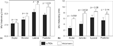
Differences in mural thickening are shown (a) between patients with congenitally corrected transposition (black bars) and healthy controls (white bars) at rest in septal, anterior, lateral and posterior segments. ccTGA: congenitally corrected transposition; ns: not significant. (b) Shows the situation during dobutamine stress testing.

Three-dimensional reconstruction of the morphologically right ventricle from a patient with congenitally corrected transposition (left and middle) and the heart of a healthy control (right). Red: volume of the systemic ventricle; green: epicardium; yellow: right ventricle of a healthy control.

Graphic showing the linear correlation between mural motion and ejection fraction at rest. The 95% confidence limits of the true regression line are indicated with dashes lines. (a) Shows the results for patients with congenitally corrected transposition, while (b) shows the situation in the normal right ventricle. RVEF: right ventricular ejection fraction; r: correlation coefficient.
 You have
Access
- 26
- Cited by