Patients with pulmonary atresia and intact ventricular septum have hypertrophied right ventricles and abnormal properties of diastolic relaxation.1 Current practice is to perform a procedure to perforate the imperforate pulmonary valve after documentation that the coronary arterial circulation is not dependent on flow from the right ventricle.2, 3 Prostaglandins given at the time of diagnosis would then be discontinued at a variable period subsequent to the procedure. Multiple attempts may be required to discontinue prostaglandins, and there is currently no way to predict if the patient will continue to require another source of flow of blood to the lungs. Various factors affect the antegrade flow of blood to the lungs, including right ventricular diastolic properties. These variables will determine need for an additional source of flow. Tissue Doppler is a new modality that is able to quantify diastolic relaxation, and is a variable that is less affected by conditions of preload. Multiple studies have validated this modality as a marker for diastolic relaxation and function.4–7 We describe a patient in whom we demonstrated improved properties of diastolic relaxation by tissue Doppler subsequent to radiofrequency perforation of the pulmonary valve.
Case report
An infant with an uncomplicated prenatal history was born at 40 weeks gestational age. He was noted to be dusky a few hours after birth, and the saturation of oxygen, measured transcutaneously, was documented to be 80 percent. He was transferred to a paediatric hospital, where an echocardiogram revealed the presence of pulmonary atresia with an intact ventricular septum. The orifice of the tricuspid valve was measured at 0.97 centimetres, equating to a Z score of −0.97, and the diameter of the imperforate pulmonary ventriculo-arterial junction was measured at 0.60 centimetres, giving a Z score of −1.99. Prostaglandins were initiated.
The infant was taken to the cardiac catheterization laboratory on the first day of life. Right ventricular pressure was measured at 100 over 4, 8 millimetres of mercury, systemic pressure was 52 over 25 millimetres of mercury, and systemic saturation was 83 percent. Angiograms showed no evidence of a right ventricular-dependent coronary arterial circulation, so radiofrequency perforation of the imperforate pulmonary valve was performed. Subsequent to the procedure, right ventricular pressure was measured at 58 over 4, 10 millimetres of mercury, systemic pressure was 58 over 27 millimetres of mercury, and pulmonary arterial pressure was 40 over 22 millimetres of mercury. Systemic saturation was now 91 percent.
He was transferred to the intensive care unit on prostaglandins, which were discontinued on the eighth day of life. Systemic saturations then decreased from the high 80s to high 70s, albeit with no evidence of inadequate cardiac output. He was subsequently discharged home with no further surgical or catheter intervention.
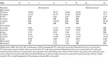
Echocardiograms were performed on the first day of life, before radiofrequency perforation, on the second, fifth and eighth days before discontinuation of prostaglandins, and on the twelfth day. We recorded the peak velocities of flow across the mitral and tricuspid valves during rapid ventricular filling, along with atrial contraction, recording analogous tissue Doppler velocities of the left and right lateral ventricular walls at the level of the atrioventricular junctions if an adequate Doppler signal was available (Table). The peak mitral ventricular and atrial blood velocities initially increased after the radiofrequency perforation, and decreased subsequent to the discontinuation of prostaglandins. The ratios of peak mitral ventricular to atrial blood velocities generally remained greater subsequent to perforation compared to the values prior to perforation. We were unable to obtain signals for the velocity of flow of blood across the tricuspid valve. Peak mitral ventricular and atrial tissue velocities increased after radiofrequency perforation, with a greater overall increase in ventricular velocity. Peak tricuspid ventricular tissue velocity also increased subsequent to perforation, with a slight decrease after discontinuing the prostaglandins. Peak tricuspid atrial tissue velocity remained relatively stable before and after the procedure. The ratios of peak mitral ventricular to atrial tissue Doppler, and peak tricuspid ventricular to atrial tissue Doppler, increased after the radiofrequency perforation.
Table. Echocardiographic data.

Discussion
The ideal therapeutic goal is to achieve biventricular physiology in patients with pulmonary atresia and an intact ventricular septum. This process is initiated by opening the right ventricular outflow tract, and then determining if an additional source of flow of blood to the lungs is needed. Parameters are available that predict if a patient will eventually be a candidate for biventricular as opposed to univentricular circulations,8, 9 but no parameters are thus far available to predict the success of discontinuing treatment with prostaglandins. In our patient, we recorded changes in tissue Doppler measurements before and after radiofrequency perforation of the imperforate pulmonary valve that we believe may help guide the decision regarding timing of discontinuation of prostaglandins.
When measuring the velocities of flow of blood across the mitral valve, a marked increase was noted after perforation. The velocities then decreased after the prostaglandins were stopped. This is because these parameters are affected by preload. When the prostaglandins were discontinued, there was decreased preload to the left atrium, with preload supplied entirely by antegrade flow across the newly opened pulmonary valve. Despite the variation in absolute velocities, there was also an increase in the ratio of peak mitral ventricular to atrial blood velocity after perforation, suggesting improved left ventricular diastolic function. This may imply some ventricular diastolic interdependence, since the only intervention was performed on the right side of the heart.
In regard to the measurement of tissue Doppler velocities, all values generally increased after perforation, with an overall greater increase in the peak velocities of rapid ventricular filling. This made the respective ratios of ventricular to atrial tissue velocity greater subsequent to perforation, suggesting an immediate improvement in diastolic relaxation properties of the respective ventricles. Left ventricular diastolic properties were also improved as indicated by these measurements, again suggesting some ventricular interdependence. There was less absolute variation in tissue Doppler velocities compared to the values for flow of blood across the atrioventricular valves, a finding that can be explained by the lack of dependence on preload of tissue Doppler measurements.
Theoretically, therefore, tissue Doppler measurements may aid in the decision when to discontinue prostaglandins. If, after perforation, the ratio of ventricular to atrial tissue velocities remains similar to the initial values, this may be a marker that diastolic properties have not improved sufficiently to allow sufficient antegrade flow through the pulmonary valve for the right ventricle to be the sole source of flow to the lungs. Conversely, if the ratio subsequent to perforation is greater, this may suggest that the right ventricle is able to relax adequately, and that the patient only needs antegrade flow to the lungs. Our report is constrained, of course, in that we studied a single patient, and made only a limited number of measurements. We are unable to comment on tissue Doppler measurements that might have predicted failure, since our patient was successfully weaned from prostaglandins at the first attempt. We submit, nonetheless, that tissue Doppler measurements may aid in the acute management of these patients. Further studies are needed to determine the usefulness of this new echocardiographic method in such patients.