Introduction
The post-auricular inlay technique known as tympanoplasty was introduced by Glasscock in 1973.Reference Rizer 1 This technique (with minor variations) is now a standard procedure for perforation closure worldwide. A success rate of more than 90 per cent has been reported for tympanoplasty using a temporalis fascia graft.Reference Jackson, Kaylie, Glasscock, Brackmann, Shelton and Arriaga 2 However, poor surgical outcomes for anterior and subtotal perforations are an inherent problem of the inlay technique.Reference Tek, Karaman, Uslu, Habeşoğlu, Kılıçarslan and Durmuş 3 – Reference Onal, Uguz, Kazikdas, Gursoy and Gokce 7
To address this problem, onlay tympanoplasty was developed to repair anterior and subtotal perforations. This technique provides excellent visualisation of the anterior tympanomeatal angle, a preserved healing plane (i.e. the residual tympanic membrane remains in situ) and no reduction in middle-ear space.Reference Hosamani, Ananth and Medikeri 4 , Reference Mokhtarinejad, Okhovat and Barzegar 8 – Reference Lee, Auo and Kang 10 However, it has several disadvantages. Firstly, because the tympanic membrane is only approximately 130 µm thick, with a squamous layer measuring about 30 µm, a lamina propria measuring 100 µm and mucosal layer of 1 µm or less,Reference Govaerts, Jacob and Marquet 11 removing the entire outer squamous layer is time-consuming and requires a high level of surgical skill.Reference Rizer 1 , Reference Lee, Auo and Kang 10 Other complications include graft lateralisation, blunting of the anterior sulcus, epithelial pearl formation, delayed healing and cholesteatoma formation.Reference Faramarzi, Hashemi and Rajaee 9 , Reference Packer, Mackendrick and Solar 12 Hence, the technique is not universally employed.
A number of surgical modifications to address the issue of poor graft uptake in anterior and subtotal perforations have been reported. Recent notable studies are over-underlay tympanoplasty (Kartush et al., 2002), modified window shade tympanoplasty (Schraff et al., 2005), mediolateral graft tympanoplasty (Jung et al., 2005), the anterior flap technique (D'Eredità et al., 2009) and loop overlay tympanoplasty (Lee et al., 2010).Reference Jung and Park 6 , Reference Lee, Auo and Kang 10 , Reference Kartush, Michaelides, Becvarovski and LaRouere 13 – Reference D'Eredità and Lens 15 . Using a modified anterior tagging technique, Hosamani et al. recently reported a success rate of 96 per cent for underlay tympanoplasty.Reference Hosamani, Ananth and Medikeri 4 Creation of a pocket between the anterior canal skin and its underlying bony canal wall for graft placement was described by Faramarzi et al. in 2012.Reference Faramarzi, Hashemi and Rajaee 9 A three-flap technique (Roychaudhuri et al., 2004) and an inferior flap technique (Gavriel et al.) have also been used for closing anterior and subtotal perforations, with impressive results.Reference Roychaudhuri 16 , Reference Gavriel and Eviatar 17
Another technique reported for closing anterior and large perforations is circumferential subannular tympanoplasty. This is an underlay technique in which the graft, instead of being tucked blindly under the tympanic membrane remnant, is placed directly over the annulus under microscopic vision and then overlapped with the tympanomeatal flap. However, there is a marked paucity of literature on this technique: an extensive search of the PubMed and Medline databases identified only four relevant studies.Reference Mokhtarinejad, Okhovat and Barzegar 8 , Reference Mishra, Sonkhya and Mathur 18 – Reference Potsic, Winawer and Marsh 20 Therefore, a prospective study of this technique in anterior and subtotal perforations was performed.
Materials and methods
Study design
A prospective observational study was conducted at the ENT Department, Lady Hardinge Medical College and Associated Hospitals, New Delhi, from April 2013 to April 2016. The study was approved by the Medical Division of the University Board of Studies, University of Delhi, New Delhi. The study sample comprised 58 adult patients of both sexes suffering from chronic suppurative otitis media mucosal disease; all provided informed consent. Inclusion criteria were the presence of an anterior or subtotal perforation in the pars tensa for a minimum period of six months, the presence of a dry ear for a period of four weeks and an air–bone gap (ABG) of less than 30 dB.Reference Alkan, Baylanĉiĉek, Sözen, Baŝak and Dadaŝ 21 Exclusion criteria were the presence of cholesteatoma in the ear, the presence of granulation tissue in the ear, previous ear surgery, having only one ear with hearing and hearing loss out of proportion to the perforation size.
Procedure
All patients were clinically evaluated and pure tone audiography was performed prior to surgery. After routine testing for pre-anaesthetic clearance, all patients underwent circumferential subannular tympanoplasty using a slightly modified version of conventional type I tympanoplasty with a post-auricular approach. A temporalis fascia graft was used and surgery was performed under local anaesthesia.
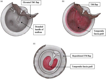
First, a post-auricular incision was made and the temporalis fascia graft was removed. The margins of the perforation were then freshened and a tympanomeatal flap was elevated from an 11 o’ clock to a 1 o’ clock position at 5 mm lateral to the fibrous annulus of the eardrum (Figure 1a). The handle of the malleus was also denuded. After inspecting the middle ear and ossicular chain, Gelfoam® was placed into the middle ear to provide graft support. The temporalis fascia graft was then directly placed onto the annulus lateral to the handle of malleus (Figure 1b). Finally, the tympanomeatal flap was repositioned to cover the graft (Figure 1c). Gelfoam® packing was added to the external auditory canal and the wound was closed in two layers.
Pictorial representation of circumferential subannular tympanoplasty (right ear). (a) Elevation of the tympanomeatal (TM) flap (from the 11 o'clock to the 1 o'clock position). (b) Placement of the temporalis fascia graft onto the annulus, all around the circumference and lateral to the handle of malleus. (c) Repositioning of the tympanomeatal flap.

All patients were given a course of antibiotics (amoxicillin) along with anti-allergy (levocetirizine) and analgesic (diclofenac plus paracetamol) drugs post-operatively. Stitch removal and pack removal were performed on post-operative day 7 and all medications were withdrawn thereafter. All patients were followed up in the ENT out-patient department at three weeks, three months, six months and one year post-operatively. All patient data were recorded on a standardised proforma for statistical analysis.
Statistical analysis
As the sample size was small, significant differences in percentage outcomes were confirmed using the Mantel–Haenszel χ2 test and Fisher's exact test, where applicable, in Epi Info version 7 (Division of Health Informatics & Surveillance, Centers for Disease Control and Prevention, Atlanta, Georgia, USA; https://www.cdc.gov/epiinfo/index.html).
Results
The study group comprised 32 male and 26 female patients. Surgical success was defined as an intact graft at the end of 1 year and failure as residual perforation. In accordance with the guidelines of the Committee on Hearing and Equilibrium of the American Academy of Otolaryngology Head and Neck Surgery, the post-operative ABG and number of decibels of ABG closure were assessed after one year. 22
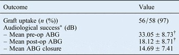
The overall success rate was 97 per cent (56 out of 58 patients had successful graft uptake; Table I). Hearing was evaluated in these 56 patients: the mean pre-operative ABG was 33.05 dB; the mean post-operative ABG was 18.12 dB; and the mean ABG closure was 14.69 dB. Thus, there was a significant improvement in hearing (p = 0.002; Table I).
Outcomes for circumferential subannular tympanoplasty

Data are means ± standard deviation unless otherwise indicated. *Evaluated in patients with successful graft uptake (n = 56). † p = 0.002. Pre-op = pre-operative; post-op = post-operative
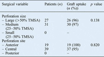
The effect of perforation size and site on tympanoplasty success were evaluated. Perforations were classified as large (involving more than 50 per cent of the tympanic membrane surface area), medium (involving 25–50 per cent of the surface area) or small (involving less than 25 per cent of the surface area).Reference Singh, Sidhu, Sharma and Singh 23 A total of 27 patients had large perforations: of these, 26 had successful graft uptake (96 per cent). A total of 31 patients had medium-sized perforations: of these, 30 had successful graft uptake (97 per cent). There was no significant difference in graft uptake rate for large and medium-sized perforations (p = 0.138; Table II). Thus, the surgical success of tympanoplasty was independent of perforation size.
Relation of perforation size and site to tympanoplasty success

*Not statistically significant. TMSA = tympanic membrane surface area
Perforations were classified as anterior, central or posterior with respect to the handle of malleus.Reference Youngs, Ludman and Wright 24 Of the 58 patients in the study group, 19 had anterior and 39 had central perforations. Of the 19 patients with anterior perforations, all had successful graft uptake; of the 39 patients with central perforations, 37 had successful graft uptake. There was no significant difference in graft uptake between groups (p = 0.820; Table II). Hence, the perforation site had no effect on tympanoplasty outcome.
No patient in this study experienced a surgical complication such as intra-operative bleeding, facial nerve palsy, chorda tympani nerve injury, wound haematoma, perichondritis, epithelial pearl formation, granulation tissue formation at the tympanomeatal flap or sensorineural hearing loss. Two graft failures were recorded; these probably resulted from technique failure because no post-operative infection was observed in either patient. One of the two patients underwent revision tympanoplasty by the same surgical technique, with a good surgical outcome.
Discussion
Poor success rates have previously been reported for anterior and subtotal perforations. The causes are reported to be poor vascularisation of the anterior half of the tympanic membrane, inadequate access to the anterior annulus for graft placement, and shrinkage of the temporalis fascia graft.Reference Hosamani, Ananth and Medikeri 4 , Reference Mokhtarinejad, Okhovat and Barzegar 8 – Reference Lee, Auo and Kang 10 , Reference Gavriel and Eviatar 17 , Reference Applebaum and Deutsch 25
For anterior perforations, it is also possible that placing a dry graft into the moist environment of middle ear causes shrinkage and therefore loss of surface contact with the anterior margin of the perforation (if not tucked in adequately), leading to tympanoplasty failure.Reference Alkan, Baylanĉiĉek, Sözen, Baŝak and Dadaŝ 21 , Reference England, Strachan and Buckley 26 – Reference Singh, Dhawan, Jha and Aggarwal 28 It is important to note that in conventional underlay tympanoplasty, anterior tucking of the graft under tympanic membrane remnant is a blind procedure. Furthermore, the margin size needed for adequate tucking has not been defined. It is also pertinent to note that subtotal perforations have an anterior element, which may explain their lower success rate compared with smaller perforations.Reference Lee, Kelly and Mills 29
There are few reports of circumferential subannular tympanoplasty: a literature search identified only four studies.Reference Mokhtarinejad, Okhovat and Barzegar 8 , Reference Mishra, Sonkhya and Mathur 18 – Reference Potsic, Winawer and Marsh 20 The salient features of these studies are compared in Table III. These data show that the graft uptake success rate of 97 per cent achieved in the present study is comparable to the success rates of 95–100 per cent reported in other studies. It is important to note that the present study had a 100 per cent success rate for anterior perforations. In terms of audiological results, Mokhtarinejad et al. reported a post-operative ABG of less than 30 for all patients, whereas Mishra et al. reported a hearing gain of 10–30 dB in 95 per cent of cases.Reference Mokhtarinejad, Okhovat and Barzegar 8 , Reference Mishra, Sonkhya and Mathur 18 Güneri et al. recorded a post-operative decrease in ABG (mean ± standard deviation 10 ± 7.9 dB) in their case series, and Potsic et al. reported that 90 per cent of their cohort (41 children) had an ABG of 20 dB or less.Reference Güneri, Ecevit and Erdağ 19 , Reference Potsic, Winawer and Marsh 20 Comparable hearing results were obtained in the present study. Although this technique has been used in paediatric patients with good results, it is prudent to note that the use of tympanoplasty in children is controversial.Reference Potsic, Winawer and Marsh 20
Published studies on circumferential subannular grafting

*Paediatric cohort.
Circumferential subannular tympanoplasty has several distinct features that make it suitable for anterior and subtotal perforations. As the graft is placed directly onto the bony annulus anteriorly under microscopic vision and not tucked blindly under the tympanic membrane remnant, there is adequate support to the graft anteriorly, which prevents it falling away, facilitates epithelial migration and thus leads to better graft uptake. To compensate for physiological shrinkage of a temporalis fascia graft, a minimum of 3 mm overlap all around the circumference of the annulus at graft placement under microscopic vision is recommended to compensate for physiological shrinkage of the temporalis fascia graft. This technique also provides an adequate graft bed all around the circumference of the annulus, with the handle of malleus in the middle for ideal anchorage. It facilitates ossicular chain reconstruction by leaving the medial surface of the malleus free for direct placement of the prosthesis.
Two important observations should be noted. Firstly, the present study used the post-auricular approach, which gives a 20 per cent larger viewing area compared with conventional endomeatal and endaural routes.Reference Robinson, Ludman and Wright 30 Further, no patient required drilling of the anterior wall for graft placement. Secondly, no graft lateralisation was observed despite the graft being placed lateral to the handle of malleus. This observation has also been reported for grafts placed lateral to handle of malleus.Reference Mokhtarinejad, Okhovat and Barzegar 8 , Reference Kartush, Michaelides, Becvarovski and LaRouere 13 , Reference Roychaudhuri 16 Mishra et al. also reported graft lateralisation in only 1 per cent of patients when the graft was placed lateral to the handle of malleus using a circumferential technique.Reference Mishra, Sonkhya and Mathur 18 In 2014, Rogha et al. reported no differences in the tympanoplasty success rate for graft placement medial or lateral to the handle of malleus.Reference Rogha, Berjis, Taherinia and Eshaghian 31 It is likely that lateralisation was not observed in this study because the graft was supported by the annulus all around the circumference and held firmly by the tympanomeatal flap. In addition, the annulus was packed against the bony sulcus tympanicus in the anterior part with Gelfoam® to recreate a sharp anterior tympanomeatal angle to prevent blunting and graft lateralisation.
-
• Graft uptake is poor for anterior and subtotal perforations with conventional underlay tympanoplasty
-
• Circumferential subannular tympanoplasty is a modified inlay technique in which the graft is placed onto the annulus under microscopic vision instead of being blindly tucked under the tympanic membrane remnant
-
• This technique had a 97 per cent surgical success rate with significant hearing improvement
-
• Circumferential subannular tympanoplasty can overcome the issue of poor graft uptake for anterior and subtotal perforations
However, the present study had some limitations: the patient cohort was small; the study was not randomised; the results were not analysed in a blinded manner; and there was no control group. Further, information from observational studies is subject to potential bias (e.g. selection bias) and confounding. Data reflects the experience of a single institution and might have been influenced by the expertise of the operating surgeon, in this case the principal investigator (a senior ENT faculty member). Nevertheless, the strength of this study lies in its prospective nature, which allowed data to be accurately assessed without depending on patient recollection. Unusually, the study also incorporated a statistical analysis, which provided outstanding data validation. Moreover, data were specific for tympanoplasty, and did not include related procedures such as ossiculoplasty. The true value of this study lies in the evaluation of outcomes for anterior and subtotal perforations using this modified technique over a long follow-up period of one year. This study thus combines the realities of clinical practice with rigorous data analysis while adding to the sparse medical literature on circumferential subannular grafting.
Conclusion
Circumferential subannular tympanoplasty provides a solution for the principal disadvantage of inlay technique: poor surgical outcomes for repairing anterior and subtotal perforations. The results provide a useful foundation for future research in this area.