Implications
Oocyte activation is accompanied by a transient increase of calcium levels. Calcium ions activate calcium-dependent proteins which are important for the process of oocyte activation. One of these is calcineurin, a calcium/calmodulin-dependent protein phosphatase which is involved in the process of oocyte activation in invertebrates and lower vertebrates. However, the exact role of calcineurin has not been investigated in mammals. This work studied the role of calcineurin in the regulation of porcine oocyte activation. The results could lead to a better understanding of oocyte activation signalling process in mammals.
Introduction
Mammalian oocyte activation involves a set of processes that includes the transformation of a mature oocyte into a zygote, which is able to initiate embryogenesis. The resumption and completion of meiosis, cortical granule exocytosis and some maternal proteins degradation occur during activation, which is under physiological conditions caused by a sperm (reviewed in Horner and Wolfner, Reference Horner and Wolfner2008). The sperm carries a specific enzyme, phospholipase C-ζ (Saunders et al., Reference Saunders, Larman, Parrington, Cox, Royse, Blayney, Swann and Lai2002), which stimulates the formation of diacylglycerol and inositol 1,4,5-trisphosphate from phosphatidyl-inositol 4,5-bisphosphate. Inositol 1,4,5-trisphosphate binding to its receptors on the endoplasmic reticulum causes the release of free calcium ions (Miyazaki et al., Reference Miyazaki, Yuzaki, Nakada, Shirakawa, Nakanishi, Nakade and Mikoshiba1992). Calcium oscillations cause cyclin B degradation, cyclin B regulates the activity of metaphase-promoting factor (MPF), and the inactivation of cytostatic factor, which allows the oocyte to enter the second meiotic anaphase stage (Lorca et al., Reference Lorca, Cruzalegui, Fesquet, Cavadore, Mery, Means and Doree1993). Mitogen-activated protein kinase (MAPK) activity decreases shortly after a fall in the level of MPF activity (Moos et al., Reference Moos, Visconti, Moore, Schultz and Kopf1995). MAPK activity is important for the regulation of the meiotic cell cycle (Lee et al., Reference Lee, Miyano and Moor2000) and its decrease is probably necessary for the formation of pronuclei (Miyano et al., Reference Miyano, Dai, Lee, Kano and Moor2000).
The increase in intracellular calcium also causes a cortical reaction, during which enzymes released from cortical granules into the perivitelline space induce zona pellucida modification and thus prevent further sperm penetration into the oocyte (Bleil et al., Reference Bleil, Beall and Wassarman1981). Shortening and depolymerization of the actin filaments are necessary for the fusion of cortical granules with the oolemma (Ducibella, Reference Ducibella1996). One of the transducers of the calcium signal system is the calcium/calmodulin-dependent serine/threonine protein phosphatase 2B, calcineurin, which may also be involved in calcium signalling in mammalian oocyte activation. Calcineurin is composed of two subunits – A subunit, which has a catalytic function, and B subunit, which has a regulatory function (Klee et al., Reference Klee, Draetta and Hubbard1988). In mammals, calcineurin A is expressed mainly in the brain but has also been identified in other tissues, such as bone, heart and kidneys. Calcineurin B has been detected in mammalian testis (reviewed in Rusnak and Mertz, Reference Rusnak and Mertz2000). In pigs, calcineurin was first described on the lymphocyte cytoplasmic membrane (Chantler, Reference Chantler1985), and subsequently also in other tissues and cells – in sperms (reviewed in Rusnak and Mertz, Reference Rusnak and Mertz2000), in muscles (Depreux et al., Reference Depreux, Scheffler, Grant, Bidwell and Gerrard2010) and in porcine oocytes (Tůmová et al., Reference Tůmová, Petr, Žalmanová, Chmelíková, Kott, Tichovská, Kučerová-Chrpová, Hošková and Jílek2013).
In somatic cells, calcineurin transforms calcium signals to intracellular processes. It mainly participates in signal transduction from the cell membrane to the nucleus, where it regulates transcription (Klee et al., Reference Klee, Draetta and Hubbard1988; Liu et al., Reference Liu, Farmer, Lane, Friedman, Weissman and Schreiber1991). In oocytes, the role of calcineurin has been demonstrated during the regulation of meiotic maturation (Drosophila – Takeo et al., Reference Takeo, Tsuda, Akahori, Matsuo and Aigaki2006 and Reference Takeo, Hawley and Aigaki2010; pig – Petr et al., Reference Petr, Chmelikova, Zalmanova, Tumova, Kheilova, Kucerova-Chrpova and Jilek2013). There is no information about calcineurin involvement in mammalian and porcine oocyte activation, even though the important role of this phosphatase has been demonstrated in Xenopus (Mochida and Hunt, Reference Mochida and Hunt2007; Nishiyama et al., Reference Nishiyama, Yoshizaki, Kishimoto and Ohsumi2007) and Drosophila (Krauchunas et al., Reference Krauchunas, Sackton and Wolfner2013) oocyte regulation, where it was confirmed that active calcineurin is necessary for oocyte exit from the second meiotic metaphase stage. Calcineurin phosphatase is responsible for the anaphase-promoting complex/cyclosome dephosphorylation and cyclin B degradation during oocyte activation (Mochida and Hunt, Reference Mochida and Hunt2007; Nishiyama et al., Reference Nishiyama, Yoshizaki, Kishimoto and Ohsumi2007; Krauchunas et al., Reference Krauchunas, Sackton and Wolfner2013).
Regarding to calcineurin is an evolutionarily very conservative protein, we can hypothesize, that it participates in the regulatory processes of oocyte activation, not only in invertebrates and lower vertebrates, but also in mammals. The aim of our study was to verify the hypothesis that calcineurin protein phosphatase plays a role in the regulation of important processes during the activation of porcine oocytes. The work aimed to map calcineurin cellular localization and to determine the effect of calcineurin inhibition on oocyte activation and cortical reaction, and the effect of calcineurin on the activation of the regulatory kinases MPF and MAPK.
Material and methods
Oocyte collection and in vitro maturation
Ovaries were collected from gilts (Large White×Landrace, slaughter weight 110 kg, 6 months old) slaughtered in a slaughterhouse in Příbram and Český Brod, Czech Republic. Within 1 h after slaughtering, the ovaries were transported to the laboratory in a saline solution (0.9% NaCl) at 38°C. Cumulus oocyte complexes were obtained by the aspiration of follicles with a diameter of 2 to 5 mm using a 20 G needle. Only oocytes with compact cumulus cells and homogeneous cytoplasm were used for further experiments. These intact oocytes were then washed three times in a M199 culture medium (catalogue number M4530; Sigma-Aldrich, Munich, Germany) which was modified with calcium lactate (0.06 mg/ml), sodium pyruvate (0.25 mg/ml), gentamicin (0.025 mg/ml), HEPES (1.5 mg/ml), 13.5 IU equine chorionic gonadotropin: 6.6 IU human chorionic gonadotropin/ml (P.G. 600; Intervet, Boxmeer, the Netherlands) and 10% foetal serum (GibcoBRL; Life Technologies, Paisley, UK). Oocytes were cultured in 1.0 ml of the maturation M199 medium for 48 h at 39°C under a 5.0% CO2 atmosphere to the second meiotic metaphase stage.
Parthenogenetic activation and in vitro fertilization of oocytes
After in vitro maturation, cumulus cells were removed from matured oocytes by pipetting through a glass pipette, and denuded oocytes were activated by exposure to 25 μM of calcium ionophore A23187 for 5 min (Jilek et al., Reference Jilek, Huttelova, Petr, Holubova and Rozinek2001).
For in vitro fertilization, the denuded MII oocytes were washed in TALP medium. The TALP medium was prepared according to the protocol published by Romar et al. (Reference Romar, Coy, Gadea and Rath2005). It was equilibrated at 39°C under a 5.0% CO2 atmosphere. Groups of 50 oocytes were transferred into each well of a four-well dish, which contained 500 µl of TALP, and were equilibrated 30 min before the introduction of spermatozoa. Sperm-rich fractions of semen were collected from fertility-tested boars (Large White×Landrace) and transported to the laboratory diluted at 1 : 1 in Beltsville Thawing Solution (Pursel and Johnson, Reference Pursel and Johnson1975). Samples of semen (0.5 ml) were placed on a Percoll (Pharmacia, New York, USA) gradient (45% and 90% v/v) and centrifuged at 600×g for 30 min. The sperm pellet was resuspended in 5 ml of TALP medium and centrifuged again to remove the Percoll at 150×g for 10 min. The final sperm concentration was adjusted to 105 cells per 1 ml. The oocytes were co-cultured with spermatozoa for 20 min. They were then washed and cultured for an additional 18 h to the zygote stage.
Treatment of oocytes with calcineurin inhibitors
Cyclosporin A (CsA) and hymenistatin I (HS-I) were used as calcineurin inhibitors. The stock solution of 8 mM was diluted in dimethyl sulfoxide. Preliminary control experiments showed that the addition of pure DMSO at the same concentrations as used during the culture of the oocytes with calcineurin inhibitors had no significant effect on oocyte maturation in vitro. Concentrations of CsA and HS-I were selected based on our previous experiments, in which the potential of CsA and HS-I to block meiotic maturation of porcine oocytes was confirmed (Petr et al., Reference Petr, Chmelikova, Zalmanova, Tumova, Kheilova, Kucerova-Chrpova and Jilek2013).
Evaluation of oocyte activation
After incubation, the oocytes were fixed in a solution of acetic acid and ethanol (1 : 3, v/v) for at least 24 h. The oocytes were then stained with 1% orcein and were evaluated by phase contrast microscope, and activation was defined when the oocytes reached the pronuclear stage.
Calcineurin antibodies specificity confirmation
The specificity of antibodies was confirmed by Western blotting. Oocytes at the stage of second meiotic metaphase were mechanically denuded of their cumulus cells. Cumulus cells were processed into separate samples. Oocytes and cumulus cells were washed three times in PBS buffer (Sigma-Aldrich) and lysed in lyses buffer (0.15 ml of 10% Triton X-100, 0.05 ml 10% SDS, 4.3 ml of PBS) enriched with 0.5 ml of the protease inhibitor cocktail Complete Mini (Roche, Sigma-Aldrich) and frozen. Before electrophoretic separation, the samples were diluted with 20 µl of SDS sample buffer and 5 µl of 0.1N NaOH. Proteins were separated by SDS-PAGE with 4% stacking gel and 12.5% separating gel. Pure calcineurin protein (Sigma-Aldrich) was used in 5 ng amounts as a positive control. After separation according to molecular weight, the proteins were transferred to a nitrocellulose membrane (Hybond; Amersham Pharmacia Biotech, GE Healthcare Life Sciences, Little Chalfont, Buckinghamshire, UK). The prestained molecular weight standard (Bio-Rad, Montreal, Canada) was used for verifying protein overprinting to a membrane. After overnight blocking in 2% milk in PBS containing 0.1% Tween 20, the membrane was incubated for 2 h with anti-calcineurin antibody (catalogue numbers C1956 and C0581; Sigma-Aldrich) at a concentration of 1 : 10 000 (anti-A), or 1 : 3000 (anti-B). The membranes were then incubated with the secondary mouse IgG antibody (Calbiochem, La Jolla, California, USA) at a concentration of 1 : 30 000. The transferred proteins to a nitrocellulose membrane were detected using the ECL Advanced Western blotting detection kit (Amersham Pharmacia Biotech). Each sample contained proteins from 250 oocytes or their cumulus cells (Supplementary Figure S1).
Immunocytochemical analysis
The immunocytochemical staining was performed according to the protocol published by Tůmová et al. (Reference Tůmová, Petr, Žalmanová, Chmelíková, Kott, Tichovská, Kučerová-Chrpová, Hošková and Jílek2013). The primary mouse anti-calcineurin antibody A or B (catalogue numbers C1956 and C0581; Sigma-Aldrich), and the secondary anti-mouse IgG antibody conjugated with fluorescein-5-isothiocyanate (FITC, catalogue number F0257; Sigma-Aldrich) were used. For chromatin visualization a dye 4’, 6-diamidino-2-phenylindole (DAPI, catalogue number D8417; Sigma-Aldrich) was used. For cortical granule co-localization with calcineurin protein, the oocytes were additionally labelled with peanut agglutinin lectin (PNA) conjugated with Alexa Fluor 568 (10 μg/ml, 30 min, catalogue number L32458; Invitrogen/Molecular Probes, Thermo Fisher Scientific, Pardubice, Czech Republic).
The oocytes, which were mounted on slides, were analysed using a laser confocal microscope (equipped with a 405, 488 and a 532 solid-state lasers, Leica SPE; Manheim, Germany). The FITC, Alexa Fluor 568 and DAPI fluorescence were scanned at an emission wavelength of 520, 603 and 461 nm, respectively. The 488 and 532 nm solid-state lasers were set up consistently for all analysed oocytes. Images from the confocal microscope were analysed using NIS Elements Image Analysis system (Laboratory Imaging, Prague, Czech Republic). A macro for mean signal intensity of FITC fluorescence in the perichromosomal/pronuclear area, cytoplasm and the cortical region of the analysed oocytes was used. The cytoplasm and cortical region were scanned in the equatorial area of the oocyte (the largest diameter section). If the chromatin was localized on the oocyte periphery, the measured fluorescence in this area was considered as a signal in the nucleus rather than a signal in the cortical area. Data from each oocyte’s measured area were compared with the mean intensity of FITC fluorescence in the whole oocyte, and thus the relative fluorescence intensity was determined in different areas of the oocyte (perichromosomal area/pronucleus, cortex area and cytoplasm).
To quantify the co-localization, the RGB images of the oocyte equatorial level were analysed using the co-localization NIS Elements Image Analysis system. The degree of overlapping between FITC (calcineurin staining) and Alexa Fluor 568 (cortical granules staining) was quantified through the use of Pearson’s correlation and Manders’ co-localization coefficients. Pearson’s correlation coefficient ranges from −1.0 to 1.0 and measures the degree of correlative variation of the two channels. Manders’ co-localization coefficients reach values of 0 to 1.0 and provide the portion of the fluorescence intensity in the red channel that coincides with some fluorescence intensity in the green channel (Manders et al., Reference Manders, Verbeek and Aten1993).
Evaluation of metaphase-promoting factor/mitogen-activated protein kinase activity
Denuded oocytes were used for kinase activity assay. The oocytes were cultured in the presence of cyclosporin A. The assay methods were performed according to the scheme published by Kubelka et al. (Reference Kubelka, Motlik, Schultz and Pavlok2000). The method was slightly modified (Kheilová et al., Reference Kheilová, Petr, Žalmanová, Kučerová-Chrpová and Řehák2015). Briefly, after treatment with CsA, the oocytes were washed four times in 0.01% polyvinyl alcohol in PBS, and were placed into 5 μl of extraction buffer. The buffer contained 40 mM 3-[n-morpholino] propanesulfonic acid (pH 7.2), 20 mM paranitrophenyl phosphate, 40 mM β-glycerolphosphate, 10 mM ethylene glycol tetraacetic acid, 0.2 mM ethylenediaminetetraacetic acid, 2 mM dithiothreitol, 0.2 mM Na3VO4, 2 mM benzamidine, 40 μg/ml leupeptin and 40 μg/ml aprotinin. The samples were stored at –80°C until further use. Cdc2 and MAPK activity were assayed using the specific substrates histone H1 and myelin binding protein, respectively. The samples were homogenized and incubated for 30 min at 30°C with a 5 μl reaction mixture (100 mM 3-[n-morpholino] propanesulfonic acid, 20 mM paranitrophenyl phosphate, 40 mM β-glycerolphosphate, 10 mM ethylene glycol tetraacetic acid, 0.2 mM ethylenediaminetetraacetic acid, 20 mM MgCl2, 5 μM protein kinase A inhibitor, 2 mM benzamidine, 40 μg/ml leupeptin, 40 μg/ml aprotinin, 600 μM ATP, 2 mg H1/ml, 3 mg myelin binding protein/ml) and 500 μCi/ml [γ-32P] ATP (GE Healthcare Life Sciences). The reaction was terminated by the addition of 10 μl Laemmli sample buffer, and samples of oocytes were boiled for 3 min. Histon H1 and myelin binding protein were separated using SDS-PAGE with 4% stacking gel and 15% separating gel. After that they were stained by Comassie Blue R250, destained overnight and dried. The signal intensity of the phosphorylated substrates histone H1 and myelin binding protein was measured and evaluated through the analysis of images MultiGauge 2.0 software. The activity level of MPF and MAPK was derived from the signal intensity. The values of MPF/MAPK activity were related to the activity values of oocytes at the second meiotic metaphase stage (48 h of in vitro culture).
Assessment of cortical granules
Cortical granules were visualized according to published protocols by Romar et al. (Reference Romar, Coy, Gadea and Rath2005). Lectin (10 μg/ml), which was obtained from Arachis hypogaea conjugated with the dye Alexa Fluor 568 (catalogue number L32458; Invitrogen/Molecular Probes), was used for cortical granule visualization. Chromatin was stained with DAPI dye. A Leica SPE laser confocal microscope (Manheim), equipped with 405 nm and 532 nm solid-state lasers, was used for oocyte analysis. Alexa Fluor 568 fluorescence was scanned at an emission wavelength of 603 nm and DAPI dye at 461 nm. The 532 nm solid-state laser was set up consistently (30%, gain 950, 1.1 μm thickness of section) for each experimental group. The equator of the oocyte (the largest diameter section) was used for further image analysis using NIS-Elements image analysis (Laboratory Imaging, Prague, Czech Republic). The quantitative measurement of the sum of the cortical granule brightness (Alexa Fluor 568 fluorescence brightness) was taken by selecting the PNA-Alexa Fluor 568 stained portion (area occupied by the sum of cortical granules) in the space immediately below the oolemma. The data of the brightness sum of cortical granules were calculated for each oocyte. The degree of cortical granule exocytosis was evaluated based on the sum of the cortical granule brightness and these values were related to the values of the control group (the sum of cortical granule brightness 100 000 was calculated as 1). The oocytes were classified into four different groups: complete exocytosis of cortical granules (oocytes with 0 to 0.25), medium cortical granule exocytosis (oocytes with 0.25 to 0.40), low cortical granule exocytosis (oocytes with 0.40 to 0.75) and no cortical reaction (oocytes over 0.75).
Statistical analysis
The data were analysed by a one-way ANOVA, post hoc Tukey’s test (Statistica program, version 12.0). The P-value <0.05 was considered to indicate statistical significance.
Experimental design
Experiment 1
The objective of this experiment was to localize the catalytic A and regulatory B subunits of calcineurin in porcine activated oocytes. After in vitro maturation oocytes were denuded from their cumulus cells and divided into three groups. The first aliquot was directly fixed to immunocytochemical analysis (MII group of oocytes). The second group of oocytes (parthenogenetically activated oocytes group) was activated by 25 μM of calcium ionophore A23187 for 5 min. Oocytes in this experimental group were after activation washed three times in the medium without any supplement and were incubated for an additional 18 h to the pronucleus stage. The last group of oocytes was fertilized in vitro and cultivated for an additional 18 h to the zygote stage (zygote group). A total of 20 oocytes were analysed in each experimental group. The experiment was four times repeated.
Experiment 2
The objective of this experiment was to study the effect of calcineurin inhibition on porcine oocyte activation. After 46 h of in vitro maturation, the oocytes were denuded from cumulus cells by pipetting and one half of the oocytes were randomly incubated for 2 h in a medium containing 8 μM of cyclosporin A (CsA group) or in the supplement-free culture medium (Control group). After CsA treatment, a portion of oocytes was activated by exposure to calcium ionophore A23187 at a concentration of 25 μM for 5 min (CsA+Ca-Io group). One half of the matured oocytes in the cyclosporine-free medium was also exposed to calcium ionophore (Ca-Io group) as a positive control of oocyte activation. After activation, the oocytes were washed and incubated with 0 or 2 μM CsA for 24 h to pronucleus stage. For verification of the calcineurin inhibitor effect, hymenistatin I (HS-I) was used as another inhibitor of calcineurin. The oocytes matured for 46 h were treated with 8 μM of HS-I, activated using calcium ionophore A23187 and incubated with 2 μM of HS-I for the next 24 h. All oocytes in all the experimental groups were incubated for the same time. After incubation, the oocytes were fixed and stained to evaluate the activation rate. The number of oocytes used in each experimental group was thirty for each repetition. This experiment was repeated four times.
Experiment 3
The aim of this experiment was to study the effect of calcineurin inhibition on MPF and MAPK levels during porcine oocyte activation. The oocytes matured for 46 h were denuded and incubated for 2 h with 8 μM CsA, and subsequently were activated by calcium ionophore A23187, then cultivated for another 12 or 24 h. After that, the kinase activity assays were performed. The number of oocytes used in each experimental group was 20. This experiment was replicated four times.
Experiment 4
The objective of this experiment was to co-localize the cortical granules with calcineurin subunits A and B in matured porcine oocytes. After 48-h of in vitro maturation, the denuded oocytes without zona pellucida, were stained for calcineurin A or calcineurin B and subsequently for cortical granules, and were analysed using a laser confocal microscope. The number of oocytes used in each experimental group was 20. There were four repetitions of the experiment.
Experiment 5
The aim of this experiment was to study the effect of calcineurin inhibition on cortical granule exocytosis. The experimental groups (CsA+Ca-Io, CsA, Ca-Io, Control) were obtained according to the same scheme as in experiment 2. After in vitro maturation or after different treatments, the oocytes were incubated for cortical reaction for 1.5 h. Then they were stained on cortical granule exocytosis and analysed using a laser confocal microscope. The brightness of the cortical granules was measured in each oocyte. The number of oocytes used in each experimental group was 30. This experiment was replicated four times.
Results
Experiment 1: calcineurin localization in in vitro matured and activated porcine oocytes
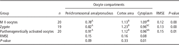
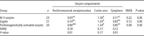
The aim of this experiment was to localize calcineurin protein in the two subunits calcineurin A and calcineurin B in mature porcine oocytes and in oocytes that were parthenogenetically activated or were activated by in vitro fertilization, and to determine whether these groups of oocytes have a different cellular localization of the calcineurin enzyme subunits. Immunocytochemical evaluation revealed the presence of both calcineurin subunits in the oocytes. In oocytes at the second meiotic metaphase stage calcineurin A and B were localized mainly in the cortical area (Supplementary Figure S2 a). After in vitro fertilization, in zygotes and in the parthenogenetically activated oocytes, the fluorescence intensity of the protein in the cortical area remained unchanged (Supplementary Figure S2 b and c; Tables 1 and 2). Calcineurin was also observed in the area of cytoplasm, particularly in the neighbourhood of pronuclei (Supplementary Figure S2 b and c). After activation, calcineurin A fluorescence intensity was in the cytoplasm of zygotes and parthenogenetically activated oocytes decreased and calcineurin B fluorescence intensity increased (Tables 1 and 2). Since the calcineurin fluorescence of parthenogenetically activated and in vitro fertilized oocytes did not differ we therefore, by reason of elimination of the effect of sperm calcineurin in in vitro fertilized oocytes, continued in the other experiments with only parthenogenetically activated oocytes.
Calcineurin A relative fluorescence intensity in in vitro matured and activated porcine oocytes

M II=oocytes at second metaphase stage; n=number of oocytes assessed.
The data are presented as mean and RMSE.
Meiotic spindle area was observed in oocytes at the second meiotic metaphase stage; pronuclei were observed in IVF or parthenogenetic embryos.
Significant differences (P<0.05) in observed oocyte categories (within columns) are marked with different superscripts a, b, c and between oocyte compartments (perichromosomal area or pronucleus, cortex area, cytoplasm) at given development stages of the oocytes (within rows) are marked with different superscripts A,B,C.
Calcineurin B relative fluorescence intensity in in vitro matured and activated porcine oocytes

M II=oocytes at second metaphase stage; n=number of oocytes assessed.
The data are presented as mean and RMSE.
Meiotic spindle area was observed in oocytes at the second meiotic metaphase stage; pronuclei were observed in IVF or parthenogenetic embryos.
Significant differences (P<0.05) in observed oocyte categories (within columns) are marked with different superscripts a, b, c and between oocyte compartments (perichromosomal area or pronucleus, cortex area, cytoplasm) at given development stages of the oocytes (within rows) are marked with different superscripts A, B, C.
Experiment 2: the effect of calcineurin inhibition on porcine oocyte activation
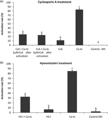
In this experiment we tested whether calcineurin inhibition prevents mature oocytes from exiting from the second meiotic metaphase, to complete meiosis and form pronuclei after calcineurin inhibitor treatment by CsA and HS-I. In mature oocytes that were parthenogenetically activated by Ca-Io and cultured in vitro other 24 h, pronuclei were observed in more than 80% (Figure 1). After oocyte treatment with calcineurin inhibitors during oocyte activation, the percentage of activated oocytes significantly decreased.
Oocyte activation results in in vitro matured porcine oocyte after treatment with calcineurin inhibitors and calcium ionophore. The graphs show the oocyte activation rate after treatment with cyclosporin A and/or calcium ionophore A23187 (a) or with hymenistatin I and/or calcium ionophore A23187 (b). Activation was considered to have occurred when oocytes reached the pronuclear stage. Different letters a,b indicate significant differences among groups (P<0.05). CsA=cyclosporin A; Ca-Io=calcium ionophore A23187; MII=oocyte at second metaphase stage; HS-I=hymenistatin I.

Experiment 3: the effect of calcineurin inhibition on metaphase-promoting factor and mitogen-activated protein kinase levels during oocyte activation
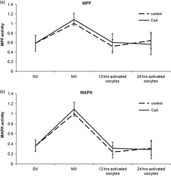
The aim of this experiment was to determine the effect of calcineurin inhibition on the level of MPF and MAPK during porcine oocyte activation. Oocytes 46 h in vitro cultured were treated with CsA and then parthenogenetically activated and cultured for an additional 12 or 24 h. During oocyte activation, MPF and MAPK activity decreased; this decrease, however, was not statistically significant compared to those oocytes not treated with CsA (Figure 2).
MPF and MAPK activity in porcine oocytes after treatment with cyclosporin A and calcium ionophore. The graphs show the activity of MPF (a) and MAPK (b) in porcine oocytes cultivated with or without cyclosporin A during oocyte activation. The kinase activity was related to oocytes at the second meiotic metaphase stage cultivated for 48 h. No significant differences were found between control and CsA groups (P<0.05). MPF=metaphase-promoting factor; MAPK=mitogen-activated protein kinase; GV=oocytes at germinal vesicle stage; MII=oocyte at second metaphase stage; CsA=cyclosporin A.

Experiment 4: the co-localization of cortical granules with calcineurin
The aim of this experiment was to co-localize calcineurin subunits A and B (staining with FITC) with cortical granules (staining with Alexa Fluor 568) in matured oocytes (Supplementary Figure S3). The Manders’ co-localization coefficient showed values of 0.455 in the calcineurin A group and 0.569 in the calcineurin B group. Manders’ coefficients indicate that 45.5% and 56.9%, respectively, of the FITC-stained fraction in the image overlapped Alexa Fluor 568. Pearson’s correlation coefficient was 0.32 in the calcineurin A group and 0.46 in the calcineurin B group. Values of Pearson’s coefficient of below 0.5 indicate that the co-localization is not significant.
Experiment 5: the effect of calcineurin inhibition on cortical granule exocytosis in the porcine oocyte
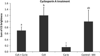
The aim of this experiment was to determine whether calcineurin protein is involved in cortical granule exocytosis. Cortical granules were observed in oocytes in control groups (CsA and Control-MII, Supplementary Figure S4 b and d). When the oocytes were treated with Ca-Io, parthenogenetic activation and cortical reaction subsequently occurred, so it was not possible to localize cortical granules that were below the oolemma (Supplementary Figure S4 c). However, when the oocytes were exposed to CsA before parthenogenetic activation cortical granule exocytosis did not occur (Supplementary Figure S4 a). Oocytes treated with CsA, and subsequently activated with Ca-Io showed similar values of cortical granule brightness as the oocytes at the second meiotic metaphase stage (Figure 3). In comparison in oocytes treated by CsA before parthenogenetic activation, the sum of cortical granule brightness was significantly lower in oocytes that were activated by Ca-Io only, which confirmed higher level of cortical granule exocytosis. Oocyte treatment with CsA before parthenogenetic activation led to reduced level of cortical reaction compared to oocytes in which calcineurin protein was not inhibited by cyclosporine A (Supplementary table S1).
Cortical granule brightness in in vitro matured porcine oocyte after treatment with cyclosporin A and calcium ionophore. The data are presented as mean±SEM. RMSE is 0.37. Different letters a,b,c indicate significant differences among groups (P<0.05). CG=cortical granules; CsA=cyclosporin A; Ca-Io=calcium ionophore A23187; MII=oocytes at second metaphase stage.

Discussion
We have demonstrated in this study the involvement of calcium-dependent protein phosphatase 2B, calcineurin, in the process of porcine oocyte activation. To the best of our knowledge, this is the first evidence of the role of calcineurin in the activation of mammalian oocytes. Both the calcineurin subunits catalytic A and regulatory B were localized in oocytes using immunocytochemical methods. Both subunits were detected in oocytes at the second meiotic metaphase stage and also after their spontaneous activation by the spermatozoa during in vitro fertilization and after parthenogenetic activation by Ca-Io. The presence of calcineurin was previously reported in pigs only in growing and maturing oocytes (Tůmová et al., Reference Tůmová, Petr, Žalmanová, Chmelíková, Kott, Tichovská, Kučerová-Chrpová, Hošková and Jílek2013). The involvement of calcineurin into the regulation of meiotic maturation was also confirmed (Petr et al., Reference Petr, Chmelikova, Zalmanova, Tumova, Kheilova, Kucerova-Chrpova and Jilek2013). In this study, we localized both subunits of calcineurin, subunit A and B, in the pronuclei of the zygotes after in vitro fertilization and parthenogenetic activation. Stronger fluorescence intensity was also recorded in the surroundings of the pronuclei, especially in the case of calcineurin A. Calcineurin A in the pronuclei area could affect microtubule network which is essential for the pronuclei migration to the centre of the egg (Schatten, Reference Schatten1994). It is known that calcineurin can affect phosphorylation status of the cytoskeleton components (Silverman-Gavrila and Charlton, Reference Silverman-Gavrila and Charlton2009).
The role of calcineurin in the activation process had previously been demonstrated only in the oocytes of the fly Drosophila (Krauchunas et al., Reference Krauchunas, Sackton and Wolfner2013) and the frog Xenopus (Mochida and Hunt, Reference Mochida and Hunt2007; Nishiyama et al., Reference Nishiyama, Yoshizaki, Kishimoto and Ohsumi2007), the involvement of calcineurin in the regulation of meiosis of these oocytes has also been confirmed. Our results indicate that calcineurin can regulate mammalian oocytes in the same way as it does in Drosophila and Xenopus oocytes.
If the calcineurin was inhibited by CsA or HS-I before porcine oocytes activation, we would notice a reduced activation rate of oocytes. The same effect of calcineurin inhibition was also observed during the activation of Xenopus oocytes (Nishiyama et al., Reference Nishiyama, Yoshizaki, Kishimoto and Ohsumi2007), which demonstrates that calcineurin is necessary for oocytes to exit from the second meiotic block.
For the oocytes exit from the metaphase II is also necessary Cyclin B degradation related to active anaphase-promoting complex (cdc2), which causes the decrease of MPF activity and subsequent MAPK activity (Moos et al., Reference Moos, Visconti, Moore, Schultz and Kopf1995; Yamamuro et al., Reference Yamamuro, Kano and Naito2008). In our study, we have not observed the effect of calcineurin inhibition on the level of MPF and MAPK activity in oocytes after parthenogenetic activation. These results would suggest that calcineurin participates in pronuclei formation after oocyte activation through other signalling mechanisms rather than through the direct regulation of MPF and MAPK activity. The influence of alternative calcineurin target systems in oocyte activation also indicates the fact that in Xenopus oocytes calcineurin inhibition suppresses the majority of cdc20 dephosphorylation, while MPF inactivation by cyclin B degradation is delayed but not completely inhibited (Mochida and Hunt, Reference Mochida and Hunt2007). A recent study by Feine et al. (Reference Feine, Hukasova, Bruinsma, Freire, Fainsod, Gannon, Mahbubani, Lindqvist and Brandeis2014) has shown that calcineurin in Xenopus oocytes activates anaphase-promoting complex/cyclosome and causes Aurora Borealis kinase (Bora) degradation during the process of fertilization, because it dephosphorylates threonine 52, which subsequently leads to cyclin-dependent kinase and polo-like kinase-1 inactivation. In pig oocyte, calcineurin is involved in signalling in a noticeably different way.
We observed in our study that calcineurin inhibition before porcine oocyte activation prevented cortical reaction. This would suggest that calcineurin is involved in the cortical granule exocytosis process. We did not observe any changes in the fluorescence of calcineurin in the cortical region of porcine oocytes after their activation by a sperm or parthenogenetic activation. Moreover, simultaneous staining of mature oocytes with anti-calcineurin antibodies and PNA lectin did not confirm co-localization of calcineurin A or B with cortical granules. Therefore, these results suggest that calcineurin is probably not included in the cortical granules content, but occurs in their region, below the oolemma. This observation is in agreement with previous results showing that subcortical secretory vesicles distinct from cortical granules contain calcium-related proteins released during pig oocyte activation (Saavedra et al., Reference Saavedra, Mondéjar, Coy, Betancourt, González-Márquez, Jiménez-Movilla, Avilés and Romar2014). The influence of calcineurin on cortical granule exocytosis may be related to the activity of actin filaments, which are attached to cortical granules below the oolemma (Longo and Chen, Reference Longo and Chen1985). The fusion of cortical granules with the oolemma is a calcium-dependent process in which calmodulin as a regulatory molecule may be involved (Ducibella, Reference Ducibella1996). Intracellular calcium oscillations temporarily activate calcium/calmodulin-dependent kinase II (Dupont, Reference Dupont1998) and calcineurin (Nishiyama et al., Reference Nishiyama, Yoshizaki, Kishimoto and Ohsumi2007). Both of these proteins are probably involved in cortical granule exocytosis (Abbott and Ducibella, Reference Abbott and Ducibella2001; Nishiyama et al., Reference Nishiyama, Yoshizaki, Kishimoto and Ohsumi2007) but are activated independently (Nishiyama et al., Reference Nishiyama, Yoshizaki, Kishimoto and Ohsumi2007). It is known that cortical granule exocytosis leads to their movement toward the oolemma thanks to the shortening and depolymerization of the actin filaments (Ducibella, Reference Ducibella1996), when calcineurin probably plays an important role during actin dephosphorylation (Silverman-Gavrila and Charlton, Reference Silverman-Gavrila and Charlton2009).
Based on our results, it is clear that the protein phosphatase 2B is present in porcine oocytes and is involved in the process of pronuclei formation after fertilization and parthenogenetic activation, and that calcineurin also participates in regulation of cortical granule exocytosis. The regulation process is apparently MPF- and MAPK-activity independent. Further explanatory experiments are necessary for better understanding of calcineurin role in different activation processes.
Acknowledgements
The authors thank Dr Manuel Avilés from the Department of Cell Biology and Histology, Faculty of Medicine, University of Murcia for oocyte analysis using an electron microscope. We thank Proof-Reading Service for their editorial assistance with this manuscript. This work was supported by the National Agency of Agriculture Sciences (NAZV QJ1510138), the Ministry of Agriculture (MZeRO0714), the Czech University of Life Sciences in Prague (CIGA 20132035), and the MINECO and FEDER (grant AGL2012-40180-C03-01).
Supplementary material
To view supplementary material for this article, please visit http://dx.doi.org/10.1017/S1751731116000884