


INTRODUCTION
One of the main challenges faced by most of the production systems using different genotypes is to precisely meet their energy and protein requirements, which has been shown to be different between Bos taurus and Bos indicus breeds (NASEM 2016; Gomes et al. Reference Gomes, Busato, Ladeira, Johnson, Galvão, Rodrigues and Chizzotti2017). Since skeletal muscle growth is potentially different between these breeds, it is reasonable to believe that protein turnover rate may contribute to discrepancies in energy expenditure and protein accretion. Protein turnover is an energetically expensive cellular process due to the high amount of adenosine triphosphate (ATP) required to operate the proteasome system, as well as the amount of energy spent during protein synthesis (Cruzen Reference Cruzen2013).
The Calpain proteolytic system has been identified as a major contributor to the regulation of skeletal muscle growth as well as for the post-mortem meat tenderization process (Koohmaraie et al. Reference Koohmaraie, Kent, Shackelford, Veiseth and Wheeler2002). The calpain/calpastatin system consists of two calcium-requiring proteases known as μ-calpain (CAPN1) and m-calpain (CAPN2) and its inhibitor, calpastatin (CAST), with both playing an essential role in skeletal muscle apoptosis, protein turnover, myogenesis and metabolism (Campbell & Davies Reference Campbell and Davies2012). Recently, the calpain/calpastatin proteolytic systems have been investigated at mRNA level (Rubio Lozano et al. Reference Rubio Lozano, Alfaro-Zavala, Sifuentes-Rincón, Parra-Bracamonte, Braña Varela, Medina, Pérez Linares, Ríos Rincón, Sánchez Escalante, Torrescano Urrutia and Figueroa Saavedra2016). However, the mRNA expression of genes does not always explain the differences in myofibrillar proteolysis in skeletal muscle of Bos taurus and Bos indicus cattle (Giusti et al. Reference Giusti, Castan, Dal Pai, Arrigoni, Rodrigues Baldin and De Oliveira2013). Thus, due to inconsistencies regarding mRNA levels of CAPN1 and myofibrillar proteolysis of skeletal muscle in Bos taurus and Bos indicus, a post-transcriptional regulatory mechanism such as a change in the abundance of an alternative polyadenylated variant of the CAST transcript has been proposed (Nattrass et al. Reference Nattrass, Cafe, McIntyre, Gardner, McGilchrist, Robinson, Wang, Pethick and Greenwood2014). However, the molecular factors underlying the differences in proteolysis between Bos taurus and Bos indicus animals are still not fully understood.
Therefore, the objective of the present study was to investigate the influence of transcription and abundance of calpain and calpastatin as well as calpastatin activity in myofibrillar proteolysis of the skeletal muscle of Nellore and Angus cattle.
MATERIAL AND METHODS
Ethical approval
All animal procedures were approved by the Animal Care and Use Committee of the Department of Animal Science at Universidade Federal de Viçosa, Brazil (19/2013-CEUAP).
Animals and experimental diet
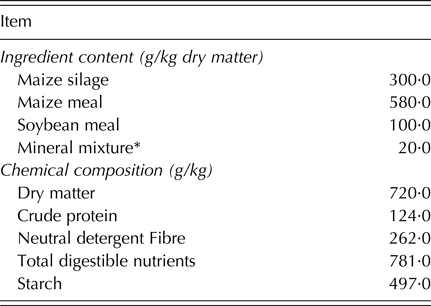
A contemporary group of 20-month-old Nellore (n = 6; initial body weight (iBW) = 373 ± 37·3 kg) and Angus (n = 6; iBW = 383 ± 23·9 kg) bulls raised under the same grazing conditions in a high quality Brachiaria decumbens pasture were confined in individual pens and fed ad libitum for a total of 84 days during the finishing phase. The feeding management used was chosen to be as representative as possible to the feeding conditions commonly observed in Brazilian beef systems. Chemical composition and ingredient proportion of the experimental diets are presented in Table 1.
Ingredient content and chemical composition of experimental diet

* Mineral mixture: 45·0 g/kg; magnesium = 7·5 g/kg; phosphorus = 11·0 g/kg; copper = 104 mg/kg; zinc = 344 mg/kg; selenium = 0·83 mg/kg; Virginiamycin = 140·0 mg/kg; Monensin = 120·0 mg/kg.
Slaughter, carcass pH measurement and sample collection
At the end of the feeding phase, all animals were harvested. Pre-harvest handling was in accordance with good animal welfare practices, and harvesting procedures followed the Brazilian Sanitary and Industrial Inspection Regulation for Animal Origin Products. After exsanguination, carcass pH (initial) was measured in the longissimus muscle (LM) between the 11th and 12th ribs using a portable pH meter for meat (Hanna Instruments, Woonsocket, RI, USA) at the left-hand side of the carcass. Following the pH measurement, a sample of LM was quickly collected from each animal and snap frozen in liquid nitrogen. Samples were then powdered in liquid nitrogen, placed in cryovials, and kept at −80 °C until total RNA isolation and protein extraction. Another LM sample was collected from each animal directly after slaughter and used immediately for calpastatin isolation and analysis of myofibrillar fragmentation index (MFI) as described later in this section. Carcass ultimate pH was measured after 24 h post-mortem chill using the same pH meter used for initial pH measurements. Carcass ultimate pH was measured on the left-hand side of the carcass at the LM, between the 11th and 12th ribs, after 24 h post-mortem chill (Hanna Instruments, Woonsocket, RI, USA).
Gene expression analysis
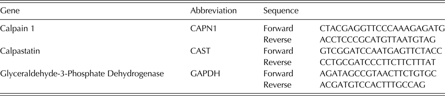
Total RNA (1 µg) was extracted from 0·5 g of powdered tissue samples using Trizol® reagent (Invitrogen, Carlsbad, CA, USA). The RNA integrity (RIN) was evaluated by capillary electrophoresis using a RNA 6000 Nano kit and a 2100 Bioanalyser System (Agilent Techonologies, Santa Clara, CA, USA). Samples with RIN >7·0 were treated with DNAse I, Amplification Grade (Invitrogen, Carlsbad, CA, USA) and reverse transcribed into cDNA using the GoScript Reverse Transcription System (Promega, Madison, WI, USA). The primer sets used are shown in Table 2. Quantitative polymerase-chain reaction (qPCR) was performed on a 7300 Real-Time PCR System (Applied Biosystems, Carlsbad, CA, USA) using GoTaq kit (Promega, Madison, WI, USA) and the following cycle parameters: 95 °C for 3 min and 40 cycles at 95 °C for 10 s and 60 °C for 30 s. The amplification efficiency ranged from 0·90 to 0·99. After amplification, a melting curve (0·01 °C/s) was used to confirm product purity. Gene expression values were calculated and expressed relative to glyceraldehyde-3-phosphate dehydrogenase (GAPDH), as described by Livak & Schmittgen (Reference Livak and Schmittgen2001).
List of primers

Calpain and Calpastatin abundance
Whole muscle protein was extracted in Lysis buffer (10 mm Tris pH 7·2; 0·5% Triton X-100; 10% Glycerol; 0·5% Dithiothreitol; 0·5 mm Phenylmethanesulfonyl fluoride and 0·5 mm Benzamidine). Protein content was measured by Bradford Protein Assay (Bio-Rad, Hercules, CA, USA), and an equal amount of protein was separated by 10% dodecyl sulphate-polyacrylamide gel electrophoresis. Proteins were transferred to nitrocellulose membranes and blocked with blocking solution (3% bovine serum albumin w/v in tris-buffered saline with triton-X100 solution – TBSt) for 1 h with gentle agitation at room temperature. Membranes were then incubated with the following primary antibodies against to μ-Calpain (no. MA3-940), Calpastatin (no. MA3945) (Thermo Scientific, Rockford, IL, USA), and β-tubulin (no. 2128) (Cell Signaling Technology Inc, Danvers, MA, USA). Primary antibodies were incubated at 1 : 1000 diluted in the blocking solution for 16 h at 4 °C with gentle agitation. After incubation with primary antibodies, membranes were washed three times at room temperature with TBSt and then incubated with the appropriate horseradish peroxidase secondary antibody (goat anti-mouse) at 1 : 5000 dilution, for 1 h at room temperature with gentle agitation. Then, membranes were washed three times (5 min each) with TBSt, developed with Clarity™ ECL substrate (Bio-Rad, Hercules, CA, USA), scanned with c-Digit Blot scanner, and analysed with Image Studio (LI-COR Inc., Lincoln, NE, USA). Band density of target proteins was normalized according to β-tubulin content.
Calpastatin activity
Calpastatin activity was measured as described by Cruzen et al. (Reference Cruzen, Paulino, Lonergan and Huff-Lonergan2014). Fresh finely minced 5-g samples were taken directly after slaughter and extracted immediately using 3 vol (w/v) of ice-cold extraction buffer containing 100 mm Tris–HCl, 10 mm ethylenediaminetetraacetic acid (EDTA), pH 8·3. Immediately before use, 0·1% 2-mercaptoethanol (MCE), 2 µ m E-64, and 500 mg/l trypsin inhibitor were added to the buffer. Samples were homogenized using a tissue homogenizer in three 30-s bursts. The resulting homogenate was centrifuged at 25 000 g for 20 min at 4 °C, and the supernatant was filtered through cheesecloth and dialysed in 40 volumes of tris-EDTA 2-mercaptoethanol (TEM: 40 mm Tris–HCl, 1 mm EDTA, pH 7·4, with 0·1% 2-mercaptoethanol). Once dialysis was complete, samples were again centrifuged at 25 000 g for 20 min at 4 °C and the supernatant filtered through cheesecloth.
Dialysed and filtered samples were then loaded onto a 20 ml Q-Sepharose Fast Flow (GE Healthcare Biosciences, Pittsburgh, PA, USA) anion exchange column previously equilibrated with TEM. After washing the loaded column with 10 volumes (200 ml) TEM, calpastatin, was eluted using a linear gradient of 60 to 400 mm potassium chloride (KCl) in TEM using an ÄKTA prime automated liquid chromatography system (GE Healthcare Bio-Science Corp., Piscataway, NJ, USA) with a flow rate of 2·0 ml/min and fraction volume of 2·5 ml.
The activity of calpastatin-containing fractions was determined using casein as substrate (Koohmaraie Reference Koohmaraie1990). A sample from each eluted fraction was brought to 1 ml with Tris-EDTA (TE, 40 mm Tris–HCl, 1 mm EDTA, pH 7·4). For calpastatin activity, approximately 0·4 units of previously purified porcine lung m-calpain in TE were included. One millilitre of casein buffer (100 mm Tris-acetate 7 mg/ml casein, and 1 mm sodium azide, pH 7·5, with 0·2% MCE added just before use) was added, followed by 100 µl of 200 mm CaCl2. Samples were briefly vortexed and incubated in a water bath at 25 °C for 1 h. Following incubation, the reaction was stopped with 2 ml of 5% trichloroacetic acid, vortexed, and centrifuged at 1500 g for 20 min at 25 °C. The absorbance at 278 nm of the supernatant was measured and compared to the blank (no eluant, no porcine lung m-calpain) and to a positive control (no eluant, with porcine lung m-calpain) (Koohmaraie et al. Reference Koohmaraie, Shackelford, Wheeler, Lonergan and Doumit1995). One unit of calpastatin activity was defined as the amount required to inhibit 1 unit of porcine lung m-calpain (Koohmaraie Reference Koohmaraie1990).
Skeletal muscle proteolysis
MFI was determined on LM samples collected at the moment of slaughter according to Olson et al. (Reference Olson, Parrish and Stromer1976) and later modified by Culler et al. (Reference Culler, Parrish, Smith and Cross1978). Four grams of minced muscle were homogenized for 30 s in 10 vol (v/w) of a 2 °C isolating medium consisting of 100 mm KCl, 20 mm potassium phosphate, 1 mm EDTA, 1 mm magnesium chloride (MgCl), and 1 mm sodium azide (NaN3). The homogenate was centrifuged at 1000 g for 15 min and then the supernatant was decanted. The sediment was then resuspended in 10 vol (v/w) of isolating medium using a stirring rod, centrifuged again at 1000 g for 15 min and the supernatant decanted. The sediment was resuspended in 2·5 vol (v/w) of isolating medium and passed through a polyethylene strainer (18 mesh) to remove connective tissue and debris. An additional 2·5 vol (v/w) was used to facilitate passage of myofibrils through the strainer. The protein concentration of the myofibril suspension was determined by the biuret method (Gornall et al. Reference Gornall, Bardawill and David1949). An aliquot of the myofibril suspension was diluted with an isolating medium to reach a protein concentration of 0·5 ± 0·05 mg/ml. Protein concentration was again determined by the biuret method. The diluted myofibril suspension was stirred and poured into a cuvette; absorbance of this suspension was measured immediately at 540 nm then absorbance was multiplied by 200 to give a MFI for each sample.
Statistical analysis
Data were analysed using the fixed-effect model as follows:
 $$Y_{ij} = \mu + B_i + IW_{(i)j} + e_{ij}$$
where Y
ij
is the phenotype of the individual j from Breed i; μ is the overall mean; B
i
is the ith level of the fixed effect of Breed; IW
(i)j
is the fixed-effect of the covariate initial body weight (iBW) within Breed; and e
ij
is the random error associated with Y
ij
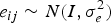
, distributed as
 $e_{ij} \sim N(I,\sigma_e^2)$
.
Prior to the final analyses, the residuals from the analysis of each trait were assessed for normality using Shapiro–Wilk's test. Once normality was met (P > 0·05), the effect of the covariate iBW within breed was tested and removed from the final analyses when P > 0·10. Least-squares means were estimated for the effect of Breed.
Additionally, a general linear model was used to evaluate the principal effects on MFI, considering fixed effects of breed and the interaction of breed with Calpastatin and Calpain protein expressions and Calpastatin activity. Further analysis was conducted to test which covariates had a significant impact on the variation in MFI.
All analyses were performed using SAS 9·4 (Statistical Analysis System Institute, Inc., Cary, NC, USA).
RESULTS
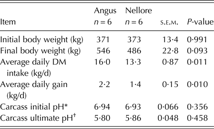
No difference was observed for iBW (P = 0·991) between breeds (Table 3). Although the number of animals used in the present study limit a deep investigation on performance variables, a greater average daily intake (P = 0·012) and average daily gain (P = 0·013) was observed in Angus compared with Nellore cattle (Table 3). As a consequence, the final BW tended to be greater in Angus compared with Nellore cattle (P = 0·095; Table 3). No difference was observed for carcass initial (P = 0·362) and ultimate pH (P = 0·467; Table 3).
Performance and carcass pH of Angus and Nellore cattle

s.e.m., standard error of the means.
* Carcass pH measured at the moment of slaughter.
† Carcass pH measured after 24 h post-mortem chill.
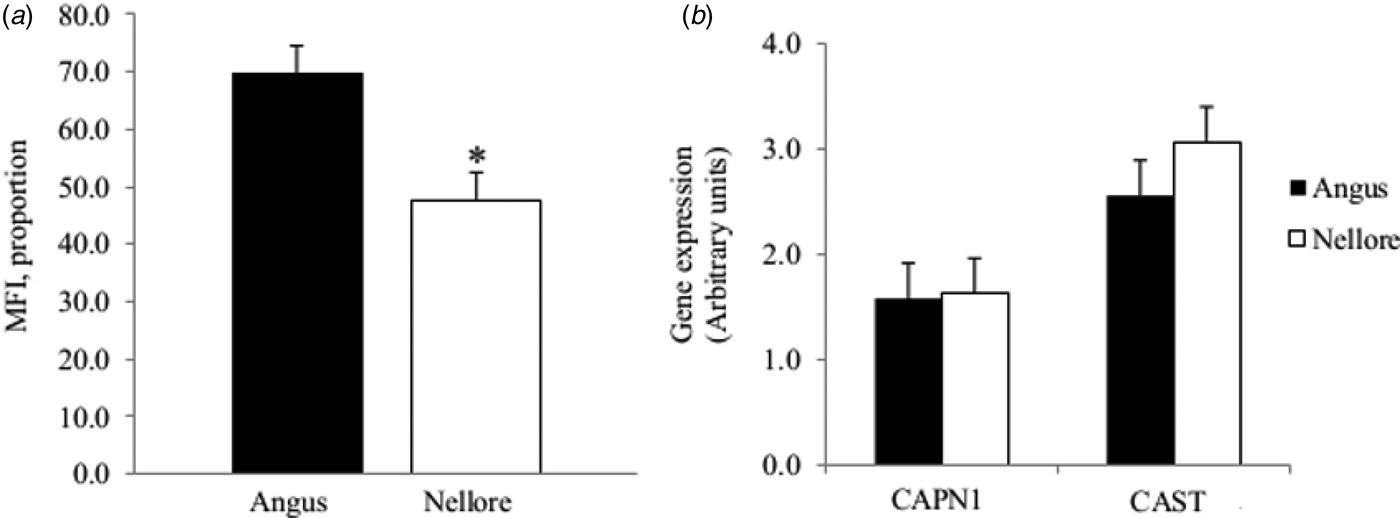
A greater MFI was observed in beef from Angus compared with Nellore cattle (P = 0·031; Fig. 1(a)). On the other hand, similar values were observed between treatments for mRNA expression of “CAPN1”(P = 0·817) and “CAST” (P = 0·413, Fig. 1(b)).
Extension of proteolysis post-mortem and mRNA expression of genes that encode the Calpain and Calpastatin enzymes in skeletal muscle of Nellore and Angus cattle. (a) Extension of proteolysis post-mortem measured by the quantification of myofibrillar fragmentation index (MFI; Angus = 69; Nellore = 48; s.e.m. = 4·8) in longissimus muscle; (b) mRNA expression of Calpain 1 (CAPN1; Angus = 27·3; Nellore = 27·1; s.e.m. = 0·33) and Calpastatin (CAST; Angus = 25·4; Nellore = 24·1; s.e.m. = 0·69). Differences were considered at P < 0·05 (*).

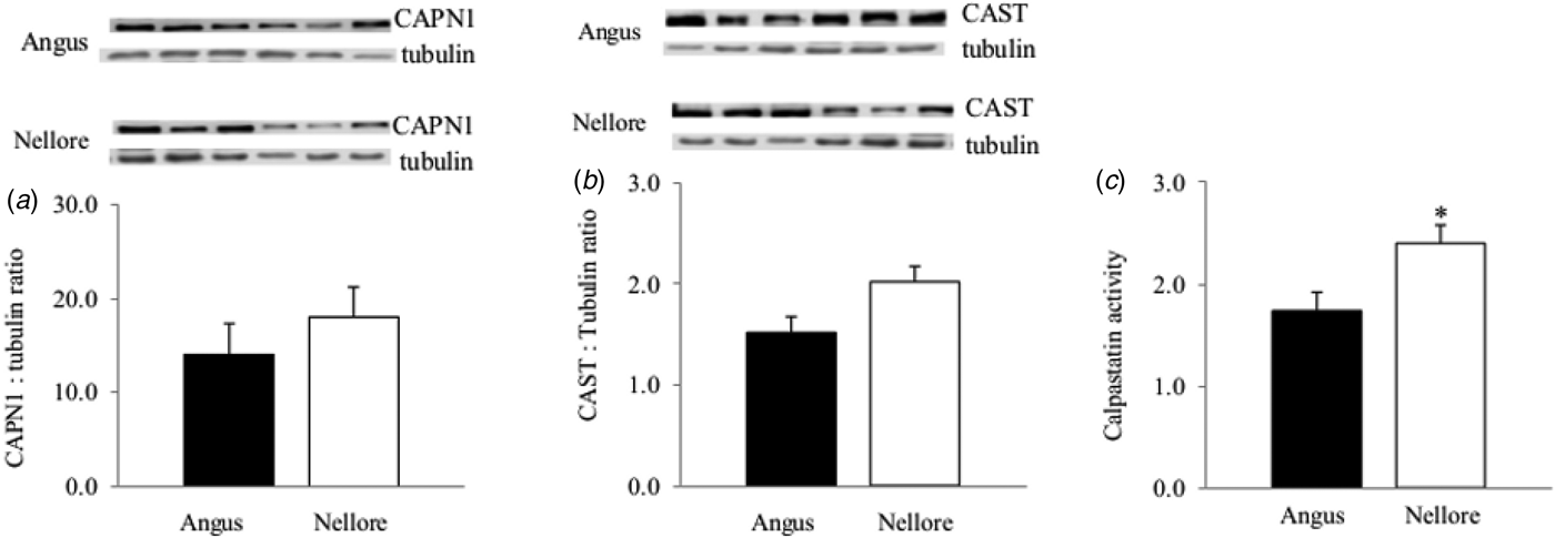
As similar values for mRNA expression of CAPN1 and CAST were observed between treatments, it was hypothesized that discrepancies in proteolysis post-mortem (measured by MFI) may have occurred due to differences in the CAPN1 and/or CAST abundance in skeletal muscle of both breeds. However, no distinctions were observed either in the abundance of CAPN1 (P = 0·639) and CAST (P = 0·141) in skeletal muscle of Nellore and Angus cattle (Figs. 2(a ) and ( b ), respectively).
Abundance of Calpain and Calpastatin and activity of Calpastatin in skeletal muscle of Nellore and Angus cattle. Abundance of Calpain (a); Angus = 14; Nellore = 17; s.e.m. = 3·2) and Calpastatin (b); Angus = 1·5; Nellore = 2·0; s.e.m. = 0·15) in longissimus muscle evaluated by Western-blot using Tubulin as a loading control; (c) Calpastatin activity measure in longissimus muscle (Angus = 1·8; Nellore = 2·4; s.e.m. = 0·18). Differences were considered at P < 0·05 (*).

Based on the previous results, calpastatin activity seemed to provide the best explanation of differences in post-mortem proteolysis of both breeds. As a greater activity of calpastatin would decrease the action of calpain, a lower MFI would then be observed. Indeed, the skeletal muscle of Nellore cattle had a greater calpastatin activity (P = 0·035) compared with skeletal muscle of Angus cattle (Fig. 2(c)). The analysis of covariates for MFI indicated that breed is responsible for 70·3 of the variation observed in calpastatin activity (P = 0·010), with no significant effect being observed neither for Calpain nor Calpastatin abundance (P = 0·329 and P = 0·131, respectively).
DISCUSSION
Skeletal muscle mass accumulation, the muscle biology translational goal of beef cattle production systems, may be controlled by several molecular events. Among them, increased rate of proteolysis may represent a key step that could potentially impair skeletal muscle growth (Krawiec et al. Reference Krawiec, Frost, Vary, Jefferson and Lang2005) and consequently compromise the efficiency of muscle deposition in beef cattle. The findings of the present study have demonstrated that post-mortem skeletal muscle myofibrillar fragmentation is higher for Angus cattle compared with Nellore. It has been suggested in a previous study using the same animals that differences in protein turnover rates between these breeds may have contributed to differences in energy expenditure (Gomes et al. Reference Gomes, Busato, Ladeira, Johnson, Galvão, Rodrigues and Chizzotti2017). Thus, the first result obtained in the present work endorses a possible reason for the discrepancy in nutrient requirements for maintenance between Angus and Nellore cattle.
The next question to be answered is what causes the differences in myofibrillar proteolysis in skeletal muscle of Bos taurus and Bos indicus. To answer that question a great body of evidence has been amassed to evaluate the mRNA abundance and/or the allelic as well as genotype frequencies of CAPN1 and CAST and their polymorphism to assess the possible mechanisms related to myofibrillar proteolysis in the skeletal muscle (Curi et al. Reference Curi, Chardulo, Mason, Arrigoni, Silveira and De Oliveira2009, Reference Curi, Chardulo, Giusti, Silveira, Martins and De Oliveira2010; Giusti et al. Reference Giusti, Castan, Dal Pai, Arrigoni, Rodrigues Baldin and De Oliveira2013; Nattrass et al. Reference Nattrass, Cafe, McIntyre, Gardner, McGilchrist, Robinson, Wang, Pethick and Greenwood2014). Although most of these studies were developed to investigate beef tenderness, their results may reflect the effects of these genes on proteolysis in a live skeletal muscle tissue. Nattrass et al. (Reference Nattrass, Cafe, McIntyre, Gardner, McGilchrist, Robinson, Wang, Pethick and Greenwood2014) speculated that the level of gene expression of CAPN1 and CAST would ultimately influence the activity of the proteins encoded by these genes (Calpain and Calpastatin), thus leading to the difference in the extent of skeletal muscle proteolysis. However, a genome scan performed by Tizioto et al. (Reference Tizioto, Decker, Taylor, Schnabel, Mudadu, Silva, Mourao, Coutinho, Tholon, Sonstegard, Rosa, Alencar, Tullio, Medeiros, Nassu, Feijo, Silva, Torres, Siqueira, Higa and Regitano2013) found a small effect of single nucleotide polymorphisms (SNPs) in CAPN1 and CAST in proteolysis and consequently Warner–Bratzler shear force on beef from Nellore cattle. On the other hand, it has been suggested that increased CAST activity in Bos indicus skeletal muscle is one of the main factors that impairs proteolysis of the skeletal muscle (Pringle et al. Reference Pringle, Williams, Lamb, Johnson and West1997). The main limitation of these studies is that none of them was able to evaluate CAST and CAPN1 at transcriptional, post-transcriptional and post-translational levels at the same time, which may be the main cause of inconsistencies from one study to another.
In the present study, no differences in mRNA expression for CAPN1 and CAST in skeletal muscle of Nellore and Angus cattle were found. Such an observation suggests that the main effect of this proteolytic system in post-mortem proteolysis of skeletal muscle from Nellore and Angus, at slaughter, may not occur due to their transcriptional activity. As post-transcriptional modifications may occur, differences (or a lack of them) in gene expression may not lead to differences in protein abundance. Indeed, in the observations for mRNA abundance, CAST and CAPN1 protein abundance did not differ among treatments, which strongly suggests that the changes in myofibrillar proteolysis between Nellore and Angus are not controlled by a post-transcription event. Conversely, a greater activity of Calpastatin was observed in skeletal muscle of Nellore compared with Angus cattle, indicating that post-translation modifications of the Calpain/Calpastatin system, not the quantity of these enzymes, might be the main cause of differences in proteolysis post-mortem of skeletal muscle of Nellore and Angus cattle. Although Calpain I activity was not measured in the present study, Ferguson et al. (Reference Ferguson, Jiang, Hearnshaw, Rymill and Thompson2000) reported a decrease in beef tenderness in Bos indicus genotype at a similar Calpain I activity, and supporting the present results, a higher (P < 0·001) Calpastatin activity in Bos indicus compared with Bos taurus cattle.
It should be noted that the results of calpastatin activity as well as the skeletal muscle proteolysis, might be affected by pre-slaughter handling (Wulf et al. Reference Wulf, Emnett, Leheska and Moeller2002). However, the results observed for carcass pH were within the normal pH range at the moment of slaughter, being close to the physiological pH (7·15; Harmer et al. Reference Harmer, McKenna, Sutton, Snow, Ruell, Booth, Thompson, Mackay, Stathis, Crameri, Carey and Eager2000), indicating that animals did not experience pre-slaughter stress that would affect the results of calpastatin activity.
A possible post-translational modification that regulates calpastatin activity is phosphorylation (Averna et al. Reference Averna, De Tullio, Passalacqua, Salamino, Pontremoli and Melloni2001). As reviewed by Cruzen (Reference Cruzen2013), bovine calpastatin has seven serine residues that may be phosphorylated, which may be the mechanism by which calpastatin activity is controlled, since it has also been shown that phosphorylation of calpastatin is a reversible process (Averna et al. Reference Averna, De Tullio, Passalacqua, Salamino, Pontremoli and Melloni2001). Since Nellore and Angus cattle have different origins and consequently seem to have different adaptabilities in a tropical environment, further investigations may consider the change in chromatin structure that would cause alternative splicing leading to changes in the number of phosphorylation sites of calpastatin.
In summary, these data strongly suggest that the greater post-mortem proteolysis in skeletal muscle of Angus compared with Nellore cattle is not caused by differences in expression of genes encoding Calpain and Calpastatin, or by the abundance of these enzymes, but is due to the greater Calpastatin activity in skeletal muscle of Nellore cattle. These findings warrant further investigation of the factors underlying the post-translational changes in the Calpain/Calpastatin system that may lead to the differences in protein turnover and ultimately nutrient requirements between Angus and Nellore cattle.
The present study was funded by Fundação de Amparo a Pesquisa do Estado de Minas Gerais (FAPEMIG, grant PPM-0036615) and Conselho Nacional de Desenvolvimento Científico e Tecnológico (CNPq), and Instituto Nacional de Ciência e Tecnologia – Ciência Animal (INCT-CA).