Global prevalence of obesity in school-age children and adolescents has significantly increased in recent decades.Reference Kimm and Obarzanek 1 – Reference Ogden, Flegal, Carroll and Johnson 3 Along with obesity, there are disorders with cardiovascular repercussions, such as abnormalities in lipoprotein metabolism and lipid profile, insulin–glucose imbalance, sodium and water retention,Reference Schaub and von Mutius 4 – Reference Freedman, Dietz, Srinivasan and Berenson 8 disorders of the aldosterone–angiotensin–renin system, and increase in the systemic blood pressure, mainly in patients with central obesity.Reference Poirier, Giles and Bray 9 , Reference Goodfriend and Calhoun 10 Heart disease due to obesity has been described since 1933.Reference Edvardsen, Helle-Valle and Smiseth 11 Obesity modifies aspects of the cardiovascular system so as to preserve body homoeostasis.Reference Troyo and Rosas-Peralta 12 Increase in body mass at the expense of adipose tissues requires a greater cardiac output and intra-vascular volume expansion to meet the metabolic demands.Reference Poirier, Giles and Bray 9 , Reference Goodfriend and Calhoun 10 The end-diastolic pressure and volume of the left ventricle increase, altering the pressure/volume curve that finally causes ventricular dilatation. The volume of the dilated ventricular chamber increases inappropriately with respect to the stress exercised in the left ventricular wall, favouring diastolic dysfunction. The myocardium adapts itself by increasing the contractile elements and subsequently the myocardial mass, resulting in left ventricular hypertrophy, which is frequently eccentric. If this haemodynamic disorder continues, it could also cause left ventricle systolic dysfunction with reduction in left ventricle ejection fraction,Reference Edvardsen, Helle-Valle and Smiseth 11 distortion of the cavities, and disruption of the regulation of the mitral sub-valvular apparatus with mitral deficiency and secondary growth of the left atrium.Reference Poirier, Giles and Bray 9 , Reference Goodfriend and Calhoun 10
As the initial changes secondary to obesity may go unnoticed, the aim of the present study was to evaluate the left ventricular function and structure with conventional echocardiography and to analyse the longitudinal deformity of the left ventricle using myocardial-tracking signals in a group of severely obese adolescents.
Materials and methods
We carried out a cross-sectional descriptive study at the Children Hospital of Mexico Federico Gómez. Patients with severe obesity from 11 to 17 years of age were included in this study. Severe obesity was defined as a body mass index>40 kg/m2 or>35 kg/m2 with co-morbidities such as insulin resistance by homoeostatic index>5 IU, sleep apnoea, type 2 diabetes mellitus, hypertriglyceridaemia>150 mg/dl, hypercholesterolaemia>200, and high-density lipoprotein<40 mg/dl. Patients with genetic syndromes or dysmorphias and those who had some systemic disease such as global developmental delay that could make difficult or modify the echocardiographic evaluation, central nervous system disorders, epilepsy, hypothalamic and thyroid diseases, nephritic syndrome, hepatic or renal insufficiency, asthma, ingestion of alcohol, drug abuse, or use of illegal substances, as well as those with history of high blood pressure or those with a blood pressure>95th percentile for height and gender at the first visit were excluded from this study.
Convenience sampling was carried out. All the patients who met the inclusion criteria were invited to participate. Clinical records were reviewed, and demographic, clinical treatment, and follow-up information was gathered. Echocardiographic studies were carried out with transthoracic echocardiograms using a Vivid 7 (GE Medical Systems, Horten, Norway) echocardiograph and a 4 MHz transducer following the recommendations from the American Echocardiography Society. Standard evaluation of the left ventricle was carried out, which included measures of diameters, volumes, valvular function, fractional shortening, and left ventricle ejection fraction using the Simpson method.
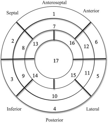
For left ventricle longitudinal deformation analysis, the transducer was placed in the apical position and the patient in the left lateral decubitus position. Images were obtained on four-chamber, three-chamber – long apical axis – and two-chamber echocardiographic views. Images were obtained at end expiration. Echocardiograph parameters were programmed until a high-quality image was obtained. Grey scales were obtained between 60 and 90 photograms/s. Image analysis was carried out using the commands and screen of the echocardiograph itself. Longitudinal deformation analysis of the 17 ventricular segments was analysed. For this, the software used myocardial signal tracking and delineated the myocardial thickness from the endocardium to the epicardium. Segments were manually checked in real-time, and the endocardial outline was adjusted until good coverage of the movement of the myocardial signals was obtained. Tracking of the myocardial signals describes the deformation of the myocardium generated by the tension; the result provided is expressed as a negative value and describes the degree of shortening – in percentage – that the myocardial fibre experiences during contraction – strain. The curves of the longitudinal deformation reflect the average value of the deformation of each segment.
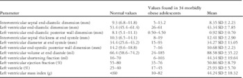
Abnormal values in the standard echocardiographic evaluation of the left ventricle were defined as values lower than the 5th percentile of the normal paediatric population that are shown in Table 1.Reference Kampmann, Wiethoff and Wenzel 13 , Reference Vogel, Staller and Bühlmeyer 14 Abnormal values in the global longitudinal deformation of the left ventricle were defined as values lower than the 5th percentile of the normal paediatric populationReference Marcus, Mavinkurve-Groothuis and Barends 15 and abnormal values in the longitudinal deformation by segments as the values lower than the 5th percentile found in a population of 817 healthy individuals with 194 individuals<18 years of age.Reference Takigiku, Takeuchi and Izumi 16
Left ventricular evaluation by normal conventional bi-dimensional echocardiogram and values found in 34 morbidly obese adolescents.

Results
A total of 53 adolescents met the inclusion criteria, but only 40 agreed to participate in this study; six of them were excluded due to technical difficulties in obtaining quality echocardiographic images for the analysis. This report includes 34 patients – 21 males and 13 females – from 11 to 17 years of age – 13±3 years – with a body mass index ranging from 35.22 to 53.5 kg/m2 body surface (SC) – 41.2±5.3 m2 body surface. Values obtained by the conventional echocardiographic evaluation of the left ventricle of these adolescents are shown in Table 1. Comparing these with the normal values, 52% had a left ventricle ejection fraction<55% and, of these, half were<45%, shortening fraction (FS) was reduced in 38%, the left ventricular end-diastolic diameter was increased in 70.5% of patients, 22.9% showed an increase in the thickness of the interventricular septum at end diastole, 35% had left ventricle posterior wall thickness increased at end diastole, and 32.3% had an increase in the left ventricular mass.
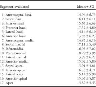
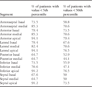
The analysis of the left ventricle longitudinal deformation using myocardial-tracking signals is shown in Table 2 and Figure 1. Among all, 74% of the patients had a value of the global left ventricle longitudinal deformation<5th percentile of the healthy population,Reference Takigiku, Takeuchi and Izumi 16 and in 78.9% of the cases the value was<50th percentile (Table 3). Apical segments were the most affected, specifically the anterior apical segment, which had a reduced longitudinal deformation in 79.4% of the cases, compared with the 5th percentile. Although the basal region was the least affected, 59.7% of the cases had reduced deformity values in the segments of this region.
Segments for analysis of the longitudinal deformation of the left ventricle using myocardial-tracking signals.

Analysis of left ventricular longitudinal deformation using myocardial signal tracking according to segments in 34 morbidly obese adolescents.

Analysis of left ventricular longitudinal deformation using myocardial signal tracking by segments in 34 morbidly obese adolescents.

Discussion
Obesity is a condition that is increasing both in industrialised countries as well as in developing countries, affecting children and adults alike. Without intervention, 80% of obese adolescents will continue to be obese in their adult years.Reference Mahoney, Burns and Stanford 17 Obesity is recognised as a direct cause of major cardiovascular diseases that cause increased morbidity and mortality in this group of patients.
A variety of changes and adaptations in cardiac structure and function occur in patients with obesity due to the excessive accumulation of adipose tissue. Since 1933, Smith and WilliusReference Rocha, Victor, Braga, Barbosa e Silva and Becker Mde 18 have described cardiomyopathy of obesity in autopsies of obese patients, and the first studies using echocardiography were performed beginning in 1978.
Severe obesity causes an increase in the total blood volume, which in turn causes dilation of the left ventricular chamber, left ventricle hypertrophy, and an increase of the myocardial mass.Reference Poirier, Giles and Bray 19 Haemodynamic overload is seen in these patients, with increase in the left ventricle afterload. Right ventricle afterload can also increase due to mechanical disorders caused by interventricular asynchrony such as the association of respiratory disorders during sleep, which is common in these patients.
In a previous study in asymptomatic adults with severe obesity, it was reported that left ventricle hypertrophy showed a positive correlation with body mass index and with the duration of obesity and diastolic dysfunction was found in 54% of the cases.Reference Rocha, Victor, Braga, Barbosa e Silva and Becker Mde 18 The high prevalence of metabolic syndrome associated with disorders diagnosed with echocardiography such as ventricular hypertrophy and diastolic dysfunction justifies the evaluation of these patients at earlier ages.
Evaluation of ventricular function by echocardiography plays an important role in the study of pathological re-modelling associated with left ventricle dysfunction.Reference Garcia, Perez and Garcia 20 Using conventional echocardiogram, in this study, we found that a group of asymptomatic adolescents with severe obesity presented abnormalities in the structure and left ventricle systolic function, similar to studies performed in adults. Despite such early ages, a significant number of these cases already had increased thickness of the interventricular septum and increase in the left ventricle lateral wall, with high ventricular mass, dilation of the left ventricle, and reduced systolic function.
In two-dimensional echocardiography, myocardial signals correspond to the bright spots resulting from the reflection and interference of the ultrasound in the myocardial tissue. By analysing the movement of these signals frame by frame during the cardiac cycle, speckle tracking, it is possible to evaluate the degree of left ventricle longitudinal deformation.Reference Rocha, Victor, Braga, Barbosa e Silva and Becker Mde 18 , Reference Phan, Shivu, Abozguia, Gnanadevan, Ahmed and Frenneaux 21 – Reference Dandel, Lehmkuhl, Knosalla, Suramelashvili and Hetzer 23 This method represents a simplified, objective, and angle-independent modality for fine quantification of myocardial deformation,Reference Sun, Lee and Wu 24 , Reference Hurlburt, Aurigemma and Hill 25 which is important because sole measurement of the left ventricular ejection fraction and the systolic volume could remain unchanged in patients with incipient heart failure.Reference Rocha, Victor, Braga, Barbosa e Silva and Becker Mde 18 , Reference Phan, Shivu, Abozguia, Gnanadevan, Ahmed and Frenneaux 21 To the best of our knowledge, this is the first report of the analysis of the left ventricle longitudinal deformation by means of myocardial signal tracking in severely obese adolescents. This analysis has allowed the evaluation of the left ventricle by regions and segments. We were able to observe that>75% of the cases had a reduced longitudinal deformation in the apical region segments. The average segments affected per individual was 5.8, but it is notable that 100% of the adolescents had a reduced longitudinal deformation in at least some of the 17 segments. Data were compared with the values reported in a study performed in a healthy Japanese population, taking into account that it is one of the reports with the largest number of young patients in the world. Under this precept, we must take into consideration the inherent inter-racial differences, which constitute a limitation of our study because we do not have reference values for healthy Mexican adolescents. Another limitation of this study is the lack of an inter- and intra-observer variability analysis.
As some of these patients showed no abnormalities during the evaluation of the systolic left ventricle function using the usual echocardiographic parameters, such as left ventricle ejection fraction and fractional shortening, segment analysis of the longitudinal deformation could be a valuable tool for the early detection of ventricular abnormalities in asymptomatic patients, whereas the number of patients was small, and therefore generalisation of the study results is difficult. The population included was a little-studied group. Published reports both in children as well as in adults are generally centred on obese individuals, whereas in this report we include only severely obese adolescents with a body mass index>35 kg/m2 body surface. This probably explains the high degree of ventricular mechanism disorders compared with other previously reported studies.
Obesity represents a multi-organ disease state that gives rise to the deregulation of a large number of functions. Early recognition of organ involvement, mainly the heart, and its appropriate follow-up will allow us to directly influence the search for clinical improvement that also improves the cardiovascular prognosis for these adolescents.
More than half of the 34 severely obese adolescents showed abnormalities in the systolic structure and function of the left ventricle, evaluated using the usual two-dimensional echocardiography techniques, but all of them showed reduction in the longitudinal deformation in at least some of the 17 left ventricle segments. Apical regions of the left ventricle segments were the most frequently affected. Analysis of the longitudinal deformation of the left ventricle using myocardial-tracking signals can be a useful tool in the evaluation and monitoring of ventricular function in obese adolescents.
Acknowledgements
None.
Financial Support
This research received no specific grant from any funding agency, commercial, or not-for-profit sectors.
Conflicts of Interest
None.
Ethical Standards
The authors assert that all procedures contributing to this work comply with the ethical standards of the Helsinki Declaration of 1975, as revised in 2008, and has been approved by the Hospital Infantil de México Federico Gómez.