

Case Report
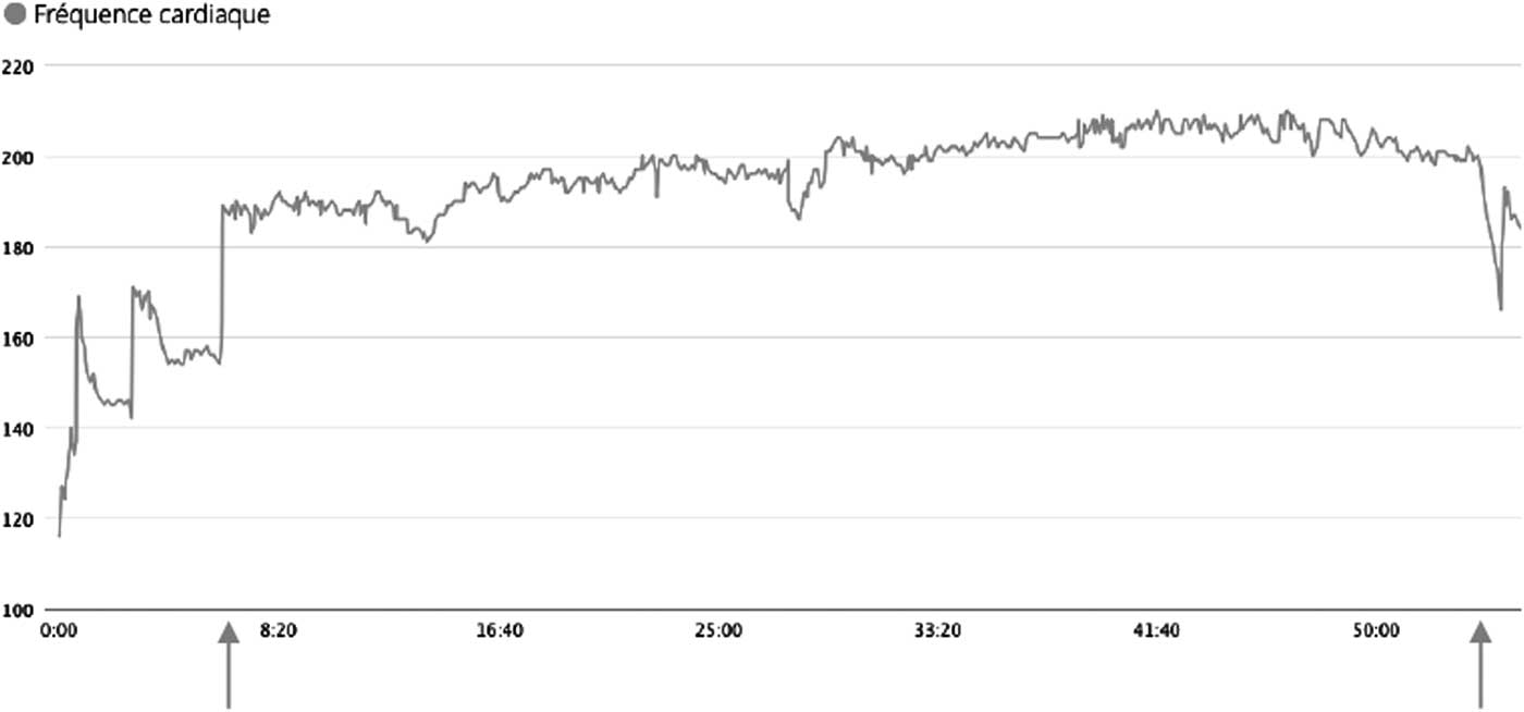
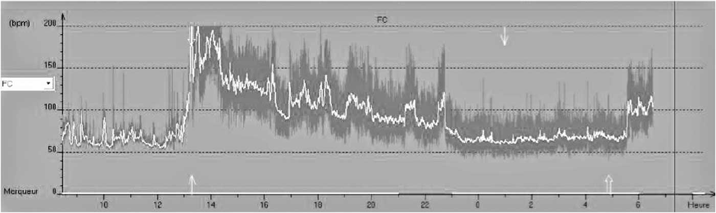
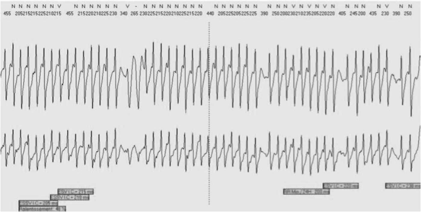
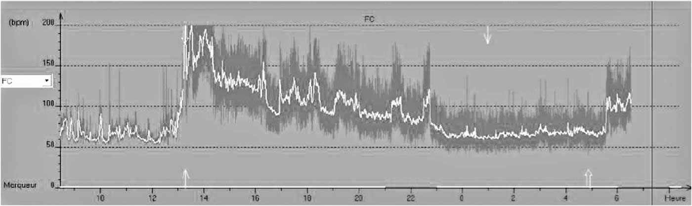
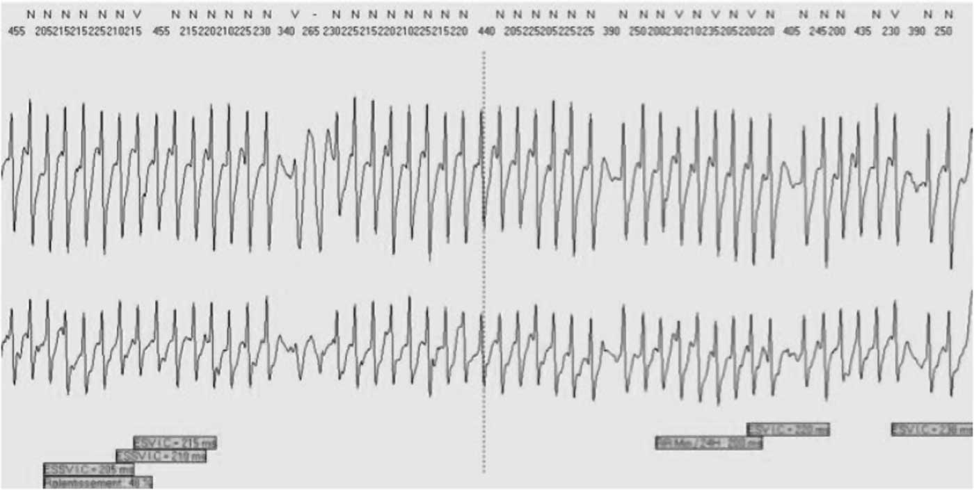
The authors report the clinical case of a frequent runner, 45-year-old male, with neither personal nor family-linked medical history of heart or neurological disorders. He measured 182 cm and weighed 89 kg. During a 10-kilometer-long event, the patient had difficulties maintaining his usual race pace in the very first kilometer. During the last kilometer, he reported complete amnesia with troubles walking, and he did not remember crossing the finish line. While being initially taken care of by the Red Cross (American Red Cross; Washington, DC USA), the patient passed out. His hemodynamic parameters were normal and no hyperthermia was found. The patient was thereafter transferred to the Bégin Military Teaching Hospital’s (Saint-Mandé, France) emergency department. During the clinical examination, the hemodynamic parameters were stable with apyrexia. The patient did not show any neurological focal points. The cardiovascular examination presented no notable features. The electrocardiogram (ECG) showed a sinus rhythm with benign early repolarization in inferior and lateral leads. There was no ST segment depression or elevation, and the QT interval was normal. A transthoracic echocardiogram and cardiac magnetic resonance imaging did not show structural heart disease. The left ventricular ejection fraction was normal without wall motion abnormalities. On the biological level, a high sensitive cardiac troponin was highlighted with a peak at 220ng/L. Concurrent creatinine phosphokinase (CPK) was 378 ui without signs of renal failure. The interrogation of his heart rate monitor (HRM) watch showed 200 bpm tachycardia beginning in the first kilometer and increasing up to 220 bpm during the last kilometer (Figure 1). The heart rate turned to normal at the end of the race. To be sure of the reliability of the values indicated by the watch, a stress test was led with the patient wearing his HRM. No significant differences on heart rate recording were shown during that test. The patient was asked to wear a Holter-ECG (Holter Research Laboratory; Helena, Montana USA) while practicing a training session (Figure 2 and Figure 3). This examination allowed for the diagnosis of an adrenergic paroxysmal atrial fibrillation (AF) with an impressive auriculo-ventricular conduction over 260 bpm. The patient was first treated with beta-blockers, followed a few weeks later by an AF ablation.
Heart Rate Recorded by the Cardio Frequency Meter. Note: Start of tachycardia at the 7th minute of exercise. Stop at the 54th minute.

Holter-ECG Record. Note: Start of the training at 1:00pm. Start of a tachycardia over 200 bpm at 1:15pm. Stop of the training at 2:45pm, with slowdown of heart rate and back in sinusal rhythm at 4:30pm. Abbreviation: ECG, electrocardiogram.
Extract of Holter-ECG Record at the Peak of Tachycardia. Note: Atrial fibrillation at reliable ventricular fibrillation more than 260 bpm (cycle at 225ms). Abbreviation: ECG, electrocardiogram.
Discussion
This case highlights that non-medical devices, such as connected watches, can be helpful to diagnose arrhythmias. Nowadays, HRM watches or other connected watches are more and more worn by sportsmen. Physicians in emergency departments should be aware of the interest to interrogate these devices, as is the case in this case report. There are two types of heart rate monitoring devices: those using electrodes on the skin surface towards the cardiac apex, and wristbands using a transcutaneous technique on the wrist thanks to the photo plethysmograph principle. The precision of these devices remains open to debate due to the fact that they have never been independently tested, and also because there exists a reference technique such as an electrocardiographic record during rest, and during a physical strain. To the best of the authors’ knowledge, only Vasconcellos et alReference Vasconcellos, Seabra, Cunha, Montenegro, Bouskela and Farinatti 1 tested the precision of a HRM device and a photo plethysmograph technique compared to a medical grade electrocardiographic record, and the test was conducted on 14 obese teenagers. The results did not show any substantial differences between all these techniques. A larger-scale study seems necessary before being able to validate these devices.
This patient presented two AF risks:Reference Miller, Aronis and Chrispin 2 excessive weight and inadequate physical training. However, the link between physical activity and lowered AF risk is not linear. Hence, even though the conclusions of the Qureshi et al’s FIT projectReference Qureshi, Alirhayim and Blaha 3 seem to be without appeal, they assert that regular physical training reduces the risk of AF, even more so in the case of obese patients; other studies nuance that statement. For instance, whereas Tromso’s prospective study conducted by Morseth et alReference Morseth, Graff-Iversen and Jacobsen 4 on nearly 20,000 patients showed a lowered risk of AF with patients practicing a moderate physical activity compared to those practicing none, this risk increased significantly with patients practicing a high-intensity physical activity. Those results are correlated with Kim et al’s results:Reference Kim, Kim and Park 5 they observed that sportsmen having an AF during a physical strain have a maximal maximum volume of oxygen higher than the group with no arrhythmia. Independently, an AF happening during a physical activity, and stopping spontaneously when resting, is a well-known pathologyReference Coelho, Palileo, Ashley, Swiryn, Petropoulos and Welch 6 , Reference Furlanello, Bertoldi, Dallago, Galassi, Fernando and Biffi 7 whose physiopathological roots have yet to be fully explained. O’Keefe et alReference O’Keefe, Patil, Lavie, Magalski, Vogel and McCullough 8 suggest that extreme practices such as marathons, ultra-trails, and long biking races cause higher heart pressures and result in micro-lesions. Those micro-lesions can reverse themselves after a week or so, but their multiplication can cause a myocardial fibrosis responsible for disorders such as AF. Those paroxysmal AF episodes can evolve into constant AF in some people.Reference Hoogsteen, Schep, Van Hemel and Van Der Wall 9