Among adults with CHD, those with residual cyanosis are among the most complex patients with a very high risk of cardiovascular complications and premature death. Reference Diller, Kempny and Alonso-Gonzalez1,Reference Greutmann, Tobler and Kovacs2 These patient cohorts comprise patients with shunt lesions and subsequent irreversible pulmonary hypertension (Eisenmenger syndrome) and patients with unrepaired cyanotic heart defects. The latter group includes patients with cyanotic defects who were left untreated and those who had undergone palliative procedures (e.g. aorto-pulmonary shunts) but without later intracardiac repair. Among patients with Eisenmenger syndrome, there is a high incidence of patients with Down syndrome (trisomy 21). Reference Kempny, Hjortshoj and Gu3
Cyanotic heart disease is a multi-systemic disorder. Reference Chaix, Gatzoulis, Diller, Khairy and Oechslin4 Haematologic abnormalities and disorders of the coagulation system are common and contribute substantially to long-term morbidity and mortality, either by bleeding or thrombo-embolic complications. Reference Hjortshoj, Kempny and Jensen5,Reference Daliento, Somerville and Presbitero6 Secondary erythrocytosis (polyglobulia) is a normal adaptation of the human body to maintain oxygen transportation capacity in situations with low systemic arterial oxygen partial pressure. In contrast, the pathophysiology of thrombocytopaenia in cyanotic heart disease, albeit frequently observed, is less well understood. Reference Lill, Perloff and Child7,Reference Martin-Garcia, Arachchillage and Kempny8 Recently, an elegant mouse model has improved our understanding for the important role of the lung in platelet biogenesis. Reference Lefrancais, Ortiz-Munoz and Caudrillier9 In cyanotic heart disease, impaired fragmentation of megakaryocytes within the lungs due to increased bypassing of the pulmonary circulation in case of worsening right-to-left shunting has been raised as a hypothesis for thrombocytopaenia but remains unproven. As thrombocytopaenia has been identified as a potent risk marker in patients with cyanotic heart disease, a better understanding of its pathophysiology may be important. Reference Martin-Garcia, Arachchillage and Kempny8
The aims of this study were therefore to evaluate the association of thrombocytopaenia in adults with cyanotic CHDs with the underlying cardiac physiology and baseline characteristics and to determine predictors of changes in platelet counts over time in order to assess potential mechanisms for progressive thrombocytopaenia.
Methods
Patient population
All patients with cyanotic heart disease were identified from our clinical database. This included patients with Eisenmenger syndrome and patients with unrepaired or palliated cyanotic heart defects, with or without concomitant pulmonary hypertension. We also included patients with intracardiac shunts and severe, irreversible pulmonary arterial hypertension with desaturation below 90% during exercise. All patients, who had at least one clinic visit at our centre and in whom at least one full haematogram was available for review, were included in this analysis. The study was approved by the local ethics committee.
Study measures
Baseline information was derived from chart review and included type of the underlying defect/physiology (Eisenmenger syndrome versus unrepaired or palliated cyanotic defects), presence of trisomy 21, previous reparative or palliative procedures, previous complications, and resting oxygen saturations. Recorded cardiovascular complications included arrhythmias, heart failure, haemoptysis, thrombo-embolic complications, brain abscesses, and infective endocarditis.
Determinants of blood work included haemoglobin level, haematocrit, mean corpuscular erythrocyte volume, leucocyte count, and platelet count. Normal ranges of haemoglobin levels were defined according to our institutional laboratory as 134–170 g/L for males and 117–153 g/L for females. Thrombocytopaenia was defined as a platelet count below 143 G/L. When available, ferritin levels were recorded and levels below 30 µg/L were determined abnormal. As ferritin levels were not available in all patients, iron deficiency was defined as mean corpuscular erythrocyte volume below 80 fl and/or ferritin levels below 30 µg/L in the absence of known microcytic haemoglobin disorders such as thalassemia.
Associations of platelet counts and presence of thrombocytopaenia with various patient characteristics and haematologic characteristics were evaluated at baseline and during follow-up. To determine the impact of progression of the underlying cardiac disease on changes in platelet counts, we analysed the correlation between changes in haemoglobin levels and oxygen saturations with changes in platelet counts during follow-up. This analysis was conducted for the entire cohort and in addition in eight selected patients to determine the impact of disease progression on platelet counts on the individual patients’ level. All eight patients had a follow-up duration of at least 5 years and serial blood work available.
The association of thrombocytopaenia on all-cause mortality was analysed during follow-up.
Statistics
Categorical variables are reported as frequencies and percentages and continuous variables as mean and standard deviation or median and interquartile range as appropriate. For comparisons between groups, we used Chi-square or Fisher’s exact tests and for categorical variables Student’s t-test or Mann–Whitney tests as appropriate. Factors associated with thrombocytopaenia were assessed by linear or logistic regression. Survival probability was calculated using the Kaplan–Meier method. To test the impact of thrombocytopaenia on survival, the log-rank test was used. A two-sided p-value of <0.05 was considered statistically significant. All statistical analyses were performed using a commercially available software package (IBM SPSS Statistics, Version 22).
Results
Patient population
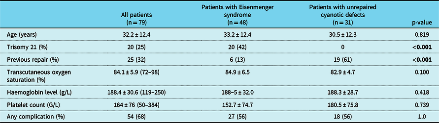
A total of 79 cyanotic adults with at least one clinic visit and blood work performed at our institution were identified from our clinical database and comprised the study group. The majority of patients had Eisenmenger syndrome (61%) and 25% of all patients had Down syndrome. Baseline characteristics, specified for patients with Eisenmenger syndrome or unrepaired cyanotic defects, are outlined in Table 1. Compared to patients with Eisenmenger syndrome, more patients with unrepaired cyanotic defects had undergone previous surgical palliation (61% versus 6%, p < 0.0001) and none of the patients with unrepaired cyanotic defects had trisomy 21.
Baseline characteristics, specified for patients with Eisenmenger syndrome and unrepaired cyanotic defects

The bold values highlight the statistically significant differences between groups (p <0.05, as outlined in the methods section).
Of patients with unrepaired or palliated cyanotic defects without Eisenmenger physiology, 18 (58%) had underlying pulmonary atresia, 11 (36%) had single ventricle physiology with pulmonary valve stenosis, 1 (3%) had a common arterial trunk after pulmonary artery banding, and 1 (3%) had undergone staged Norwood palliation for hypoplastic left heart syndrome but was left with massive veno-venous collaterals. The type of palliative surgical procedures within the group of cyanotic patients without Eisenmenger syndrome were shunt operations in 14 patients (74%), pulmonary artery banding in 3 patients (16%), partial repair in 1 patient (5%), and Norwood palliation for hypoplastic left heart syndrome in 1 patient (5%).
There were no other differences in baseline characteristics between patients with Eisenmenger syndrome and those with unrepaired cyanotic defects. Ferritin levels were available in 59 patients (75%) at baseline and in 57 patients (81%) during follow-up. Cardiovascular complications were extremely common, affecting more than two-thirds of all patients at baseline and/or during follow-up. Most prevalent were arrhythmias (24 patients, 30%), ischaemic strokes/paradoxical systemic embolism (17 patients, 22%), haemoptysis (15 patients, 19%), brain abscesses, and heart failure (each 8 patients, 10%).
Haematologic abnormalities
Baseline
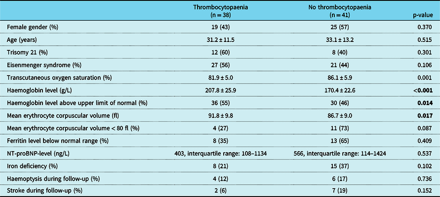
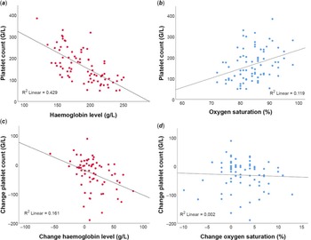
As expected, the majority of patients had marked secondary erythrocytosis with a mean haemoglobin level of 188.4 ± 30.6 g/L. Female patients had lower haemoglobin levels compared to males (182.2 ± 26.6 g/L versus 196.2 ± 33.8 g/L, p = 0.043) without significant differences in baseline resting oxygen saturations (84.4 ± 6.8% versus 83.9 ± 5.1%, p = 0.74). At baseline, almost half of all patients (38/79, 48%) had thrombocytopaenia. A comparison of patients with and without thrombocytopaenia is illustrated in Table 2. Patients with thrombocytopaenia had lower transcutaneous oxygen saturations and higher haemoglobin levels compared to those without. There was a strong inverse correlation between platelet count and haemoglobin concentration (R = −0.655, R2 = 0.429, p < 0.0001) and a weaker correlation between platelet count and transcutaneous oxygen saturation (R = 0.345, R2 = 0.119, p = 0.002) illustrated in Figure 1 (panels a and b). This effect was stronger for patients with iron deficiency, compared to those without (R2 = 0.536 for patients with evidence of iron deficiency versus R2 = 0.383 for patients without iron deficiency, p = <0.0001 for both). Patients with thrombocytopaenia had higher mean corpuscular erythrocyte volume compared to patients without (91.8 ± 9.8 fl versus 86.7 ± 9.0 fl, p = 0.017) suggesting that iron depletion may be associated with higher platelet counts; however, this did not reach statistical significance.
Baseline characteristics specified for patients with and without thrombocytopaenia

The bold values highlight the statistically significant differences between groups (p <0.05, as outlined in the methods section).
Correlation between platelet count, haemoglobin level, and oxygen saturation.
Panel a : Scatter plot illustrating correlation between platelet count and haemoglobin level at baseline. Panel b : Scatter plot illustrating correlation between platelet count and oxygen saturation at baseline. Panel c : Scatter plot illustrating correlation between change in platelet count and change in haemoglobin during follow-up. Panel d : Scatter plot illustrating correlation between change in platelet count and change in oxygen saturation during follow-up.

As outlined in Table 2, there were no significant differences in platelet counts between patients with and without trisomy 21. All patients with trisomy 21 had Eisenmenger syndrome. When associations with thrombocytopaenia were analysed within the subgroup of patients with trisomy 21, results mirrored findings of the entire cohort. Those with thrombocytopaenia had a trend towards lower resting oxygen saturation (84.6 ± 3.7% versus 89.1 ± 6.8%, p = 0.081), significantly higher haemoglobin levels (210.9 ± 17.9 g/L versus 164.8 ± 23.1 g/L, p < 0.0001) and a higher mean corpuscular volumes (100.0 ± 5.2fl versus 91.5 ± 10.8fl, p = 0.028). No significant differences were found regarding the presence of iron deficiency at baseline and regarding frequencies of haemoptysis or haemorrhagic events during follow-up.
Follow-up
In 70 patients (89%), at least one clinical follow-up visit with follow-up blood work was available with a median follow-up duration of 9.4 years (interquartile range: 3.4–15.2 years). During follow-up, mean resting transcutaneous oxygen saturation decreased from 84.1 ± 5.9% to 82.3 ± 5.8%, haemoglobin levels increased from 188.4 ± 30.6 to 201.2 ± 30.9 g/L, and platelet counts decreased from 164 ± 76 to 136 ± 60 G/L (p < 0.0001 for all comparisons). As illustrated in Figure 1 (panels c and d), there was a significant inverse correlation between increase in haemoglobin levels and decrease in platelet counts during follow-up (R = −0.401, R Reference Greutmann, Tobler and Kovacs2 = 0.161, p = 0.001) but no correlation between changes in transcutaneous oxygen saturations and platelet counts (R = 0.043, R Reference Greutmann, Tobler and Kovacs2 = 0.002, p = 0.726).
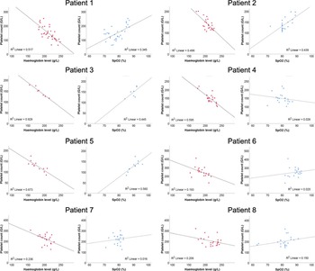
Eight patients, currently under follow-up, with a follow-up duration of at least 5 years (median 19.0 years, interquartile range: 12.3–21.1 years) and multiple serial measurements of haemoglobin levels and platelet counts were selected for correlation of platelet counts with haemoglobin levels and oxygen saturations over time. Per patient, a median of 21 pairs of measurements (interquartile range: 14–24 pairs) were available for analysis. As shown in Figure 2, in all eight individual patients there was a significant inverse correlation between platelet counts and haemoglobin concentration over time (R value: 0.44–0.96, R Reference Greutmann, Tobler and Kovacs2 value: 0.19–0.93, p < 0.05 for all patients) but only in three patients a significant correlation between platelet counts and resting oxygen saturations was found (patients 1, 3 and 5).
Scatter plots illustrating correlation of platelet counts and haemoglobin levels and platelet counts and oxygen saturation during follow-up in eight individual patients.

Of patients with Eisenmenger syndrome, at baseline 6/48 (13%) and at last follow-up 20/44 patients (45%) were on specific medical therapy for pulmonary hypertension. Most common drugs were endothelin receptor antagonists (33%), phosphodiesterase 5-inhibitors (19%), and prostacyclin (6%). Only four patients (8%) were on combination therapy. There were no differences between patients with and without specific medical treatment for pulmonary arterial hypertension regarding the presence of thrombocytopaenia at baseline (67% versus 55%, p = 0.683) and during follow-up (63% versus 71%, p = 0.745).
Survival estimates
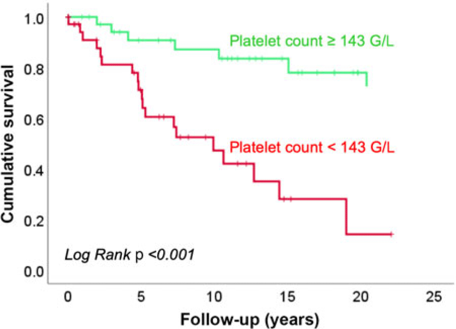
A total of 26 patients (33%) died during follow-up at a mean age of 41.0 ± 11.7 years. As depicted in Figure 3, there was a significant difference in survival estimates for patients with and without thrombocytopaenia at baseline (log-rank test p-value < 0.0001).
Kaplan–Meier survival estimates for patients with thrombocytopaenia (red line) and without thrombocytopaenia (green line).

Discussion
In this study in adults with cyanotic CHD, we confirm findings from previous studies, demonstrating a strong inverse correlation between haemoglobin levels and platelet counts. Reference Lill, Perloff and Child7,Reference Martin-Garcia, Arachchillage and Kempny8 During follow-up, a change in platelet counts was accompanied by a significant inverse change in haemoglobin levels, but there was no significant correlation between changes in transcutaneous oxygen saturations and changes in platelet counts. Thrombocytopaenia was strongly associated with all-cause mortality. By analysing long-term data on changes of haematologic parameters, our study adds to the understanding of thrombocytopaenia in cyanotic CHD.
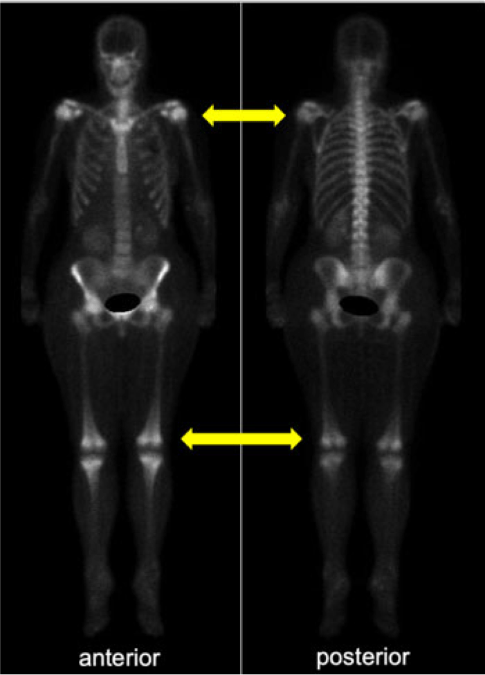
In healthy adults, haematopoiesis is typically confined to bone marrow in ribs, sternum, vertebrae, and bones of the pelvis. Figure 4 represents a bone scan of a 58-year-old patient with Eisenmenger syndrome due to a large, non-restrictive ventricular septal defect. Resting oxygen saturations were around 70% and the patient had marked secondary erythrocytosis (haemoglobin level 194 g/L) with moderate thrombocytopaenia (91 G/L). The bone scan shows increased activity at the ends of the long bones (arrows), illustrating the expansion of haematopoetic bone marrow. This phenomenon is however also seen in patients with hereditary haemoglobinopathies that are not accompanied by thrombocytopaenia. Thus, a mere imbalance within the haematopoetic bone marrow towards increased erythropoiesis and a corresponding decrease in thrombopoiesis cannot be the explanation for thrombocytopaenia in patients with cyanotic CHDs.
Whole body bone scan of a 58-year-old patient with Eisenmenger syndrome: Increased activity is noted at the ends of the long bones (yellow arrows), consistent with expansion of haematopoetic bone marrow as a consequence of marked secondary erythrocytosis.

Previous studies postulated that altered megakaryocyte metabolism was the major contributor to low platelet counts in cyanotic patients. Reference Lill, Perloff and Child7 Indeed, a recently published mouse model underscores the importance of the lung in platelet biogenesis. Reference Lefrancais, Ortiz-Munoz and Caudrillier9 This model demonstrates that megakaryocytes migrating out of the bone marrow into the lungs release platelets and contribute substantially to the overall platelet production. Reference Lefrancais, Ortiz-Munoz and Caudrillier9 In cyanotic patients, intracardiac right-to left shunting leads to a substantial amount of systemic venous blood bypassing the pulmonary circulation. This led to the hypothesis that the severity of the underlying pathophysiology and, hence, the degree of right-to-left shunting, characterised by hypoxaemia and cyanosis, are the main determinants for the severity of thrombocytopaenia and thus may explain the strong prognostic capability of thrombocytopaenia. By adding a longitudinal perspective, both, on the level of the entire cohort and on the level of individual patients, we were able to demonstrate that changes in platelet counts over time were significantly associated with inverse changes of haemoglobin levels but not associated with changes in transcutaneous oxygen saturations. This observation suggests that the severity of right-to-left shunting alone may not be the sole determinant and driver of thrombocytopaenia in adults with cyanotic CHD. Among patients with Eisenmenger syndrome, specific medical therapy for pulmonary arterial hypertension was not associated with the presence of thrombocytopaenia at baseline and during follow-up.
Chromosome 21 is thought to play an important role in haematopoiesis. It is well known that patients with Down syndrome are at an increased risk for developing haematologic malignancies, such as acute leukaemia. Reference Saida10 A recent study suggested that the mechanism leading to lower platelet counts in patients with Down syndrome was not related to abnormalities in the platelet maturation, as seen in non-Down-syndrome patients with Eisenmenger syndrome, but related to bone marrow deficiency. Reference Martin-Garcia, Arachchillage and Kempny8 In our analysis, the pattern of thrombocytopaenia and its associations, however, followed the pattern of the entire cohort.
As seen in other studies, thrombocytopaenia at baseline was strongly associated with all-cause mortality in our cohort. Reference Martin-Garcia, Arachchillage and Kempny8 Although thrombocytopaenia is likely not the driver of mortality, its potential as an important, potentially independent marker of risk needs further consideration and study.
Limitations
This study has inherent limitations, arising from its retrospective nature. Given the nature of our data, it is not possible to derive a direct cause–effect relationship explaining the inverse correlation between haemoglobin levels and thrombocyte counts. For a better understanding of thrombocytopaenia in cyanotic heart disease, further mechanistic studies including more sophisticated haematologic tests were required. To increase the validity of thrombocytopaenia as an independent prognostic marker, larger patient cohorts, which allow more detailed statistical analysis, are required to draw firm conclusions.
Conclusions
Our findings suggest a direct inverse correlation between platelet counts and haemoglobin levels in adults with cyanotic heart disease – without clear association to the severity of intracardiac right-to-left shunting. Further studies are required to explore the mechanisms of thrombocytopaenia in cyanotic heart disease and its potential as an independent marker of risk.
Acknowledgement
None.
Financial support
This research received no specific grant from any funding agency, commercial, or not-for-profit sectors.
Conflicts for interest
None.