INTRODUCTION
Stylasterid corals are colonial hydroids characterized by a rigid skeleton composed of calcium carbonate (coenosteum). Their colonies are generally erect and branched and only the Pacific genus Stylantheca shows an encrusting habitus. The colony is polymorphic, with gastrozooids and dactylozooids retractable into particular skeleton depressions named respectively gastropores and dactylopores (Moseley, Reference Moseley1881; Broch, Reference Broch1942; Cairns, Reference Cairns1983; Bouillon et al., Reference Bouillon, Gravili, Pagés, Gili and Boero2006). The distribution of gastrozooids and dactylozooids over the colony may be irregular or limited to certain regions. In several genera such as Stylaster, they are arranged in circles (cyclosystems) where one gastrozooid is surrounded by several dactylozooids and a central style (gastrostyle) is present on the bottom of the gastropore cavity (Moseley, Reference Moseley1881; Broch, Reference Broch1942; Cairns, Reference Cairns1983; Bouillon et al., Reference Bouillon, Gravili, Pagés, Gili and Boero2006).
The gonophores develop inside internal or superficial globose skeletal structures named ampullae. The sperm are released from the male ampullae and enter into the female one; when mature the planula arises from the ampulla and settles on a suitable substrate leading to the formation of a new colony (Moseley, Reference Moseley1878; Hickson, Reference Hickson1890; England, Reference England1926; Broch, Reference Broch1942; Ostarello, Reference Ostarello1973, Reference Ostarello and Mackie1976; Fritchman, Reference Fritchman1974).
The early development stages of the new coenosteum after the planulae settlement are known only in few species. Ostarello (Reference Ostarello1973) studied the natural history of Stylaster californicus (cited as Allopora californica) observing that, after its release, the planula generally crawls for a short time around the parental colony settling close to it. Nevertheless, some new colonies are born well away from the female, demonstrating that a certain degree of dispersal before settling is possible (Ostarello, Reference Ostarello and Mackie1976). A newly settled colony is flat. However, traces of the depression where the first cyclosystem will form are visible even at this early stage (Ostarello, Reference Ostarello1973).
Recruits of Stylaster roseus, showing gastropores and dactylopores, were recorded for the first time on experimental carbonate tiles in the Mexican Caribbean (Ruiz-Zárate & Arias-González, Reference Ruiz-Zárate and Arias-González2005). The most exhaustive study, concerning the planula structure, its metamorphosis and the development of the early stages of the colony, was carried out by Fritchman (Reference Fritchman1974). He observed that the larva of the encrusting species Stylantheca petrograpta (cited as Allopora petrograpta) attaches through the anterior side and rapidly flattens into a disc 1.5–2 mm in diameter. A series of roughly concentric elevations appear on the upper surface two hours after the attachment. Once they disappear, a large number of internal vesicles arise within the ectoderm that is meanwhile emerging through the involution of the lower surface. The vesicles gradually disappear leaving the endoderm organized into a reticulum of cords sheathed with ectoderm. Skeletal material appears between the ectodermal layers within twenty-four hours, and during the following two weeks, a miniature crater is formed developing in the centre a gastrozooid. Along the rim of the crater, the dactylozooids appear each one in its own dactylopore.
The aim of this work is to describe the skeletal morphology of the early stages of the colonies of Stylaster sp. settled on artificial panels placed along a coral reef in the Bunaken Marine Park (North Sulawesi, Indonesia). Moreover the different phases of development of new cyclosystems in young colonies are shown.
MATERIALS AND METHODS
The study area is located on the reef in front of the Siladen Island (North Sulawesi, Indonesia). Stylaster sp., characterized by slender colonies, is particularly abundant on the vertical portions of the reef exposed to strong currents.
During July 2008, 36 experimental plastic tiles (10×3 cm) were fixed by SCUBA diving on the reef. They were positioned at 5–20 m depth close to adult colonies of this species in order to enhance the probability of planulae settling. The tiles were collected by diving during October 2008 and immediately fixed in neutralized 4% formaldehyde. In the laboratory, each tile was examined under a stereomicroscope and the early growth stages of Stylaster sp. were photographed.
For scanning electron microscopy (SEM) analysis, small portions of tiles with settled colonies were rinsed, treated with sodium hypochlorite for 10 minutes, rinsed again and dried. The samples were then coated with gold-palladium in a Balzer Union evaporator, and examined with a Philips XL20 scanning electron microscope.
RESULTS
The Indonesian stylasterids were the object of intensive study during the Siboga expedition (Hickson & England, Reference Hickson and England1905). Hickson & England collected 7 species belonging to the genus Stylaster: S. eximius, S. bellus, S. densicaulis, S. gemmascens, S. amphiheloides, S. bilobatus and S. multiplex (Boschma, Reference Boschma1957; Cairns, Reference Cairns1983). In particular specimens of S. eximius were recorded from the shallow waters of North Sulawesi. Due to the geographical congruence and the sharing of several morphological features such as the shape of the colony, the colour, the surface longitudinally striated, the cyclosystem diameter and the presence of blunt spines especially over the ampullae it is very likely that our material is conspecific with S. eximius of Hickson & England. According with Boschma (Reference Boschma1957), Cairns (Reference Cairns1986) stated that the taxon S. eximius, described by Kent (Reference Kent1871) is a junior synonymous with the Atlantic S. duchassaingi Pourtalés Reference Pourtalés1867, but he excluded from this species the specimens of S. eximius identified by Hickson & England. In this way probably our specimens belong to a species until now not formally described.
Description of the adult colonies of Stylaster sp.
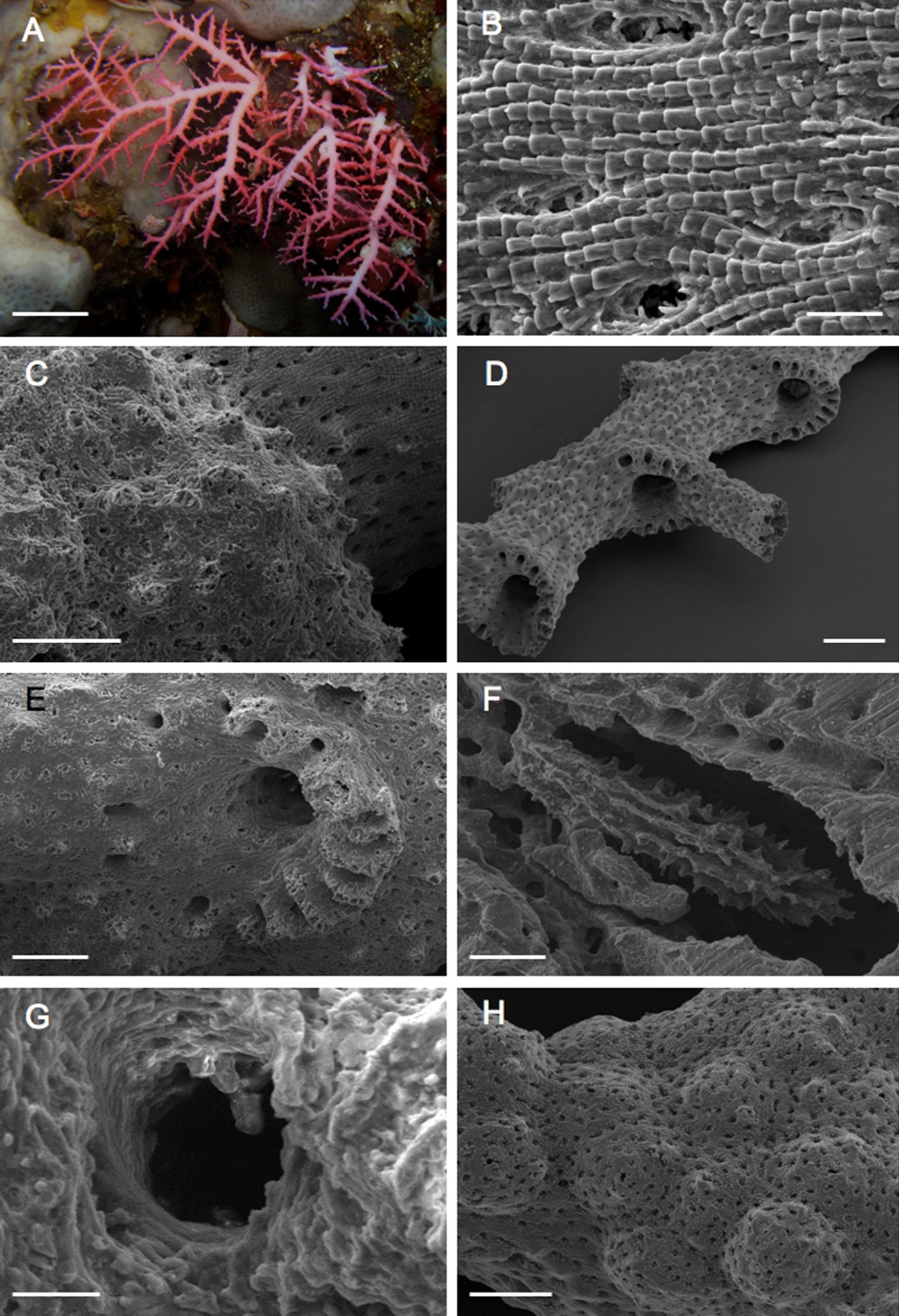
Adult colonies of this species are generally uniplanar and delicate, up to 7.5 cm tall and 8 cm wide. Branching is frequent, producing a lacy pattern (Figure 1A). Branch tips are slender and each tip supports a terminal cyclosystem. Coenosteum is orange or deep rose, linear–imbricate in texture and periodically penetrated by deep, elliptical pores (up to 40×25 µm) usually with small teeth, about 5–10 µm long, projecting inward toward the centre of pore (Figure 1B). Apically perforate, conical to cylindrical papillae (up to 70 µm tall and up to 70 µm in diameter) usually scattered over coenosteum and ampullae (Figure 1C).
Adult colonies of Stylaster sp. (A) Group of colonies in situ; scanning electron microscope photographs of: (B) coenosteum showing a linear–imbicate texture; (C) apically perforate papillae scattered over an ampulla; (D) branch with sympodially arranged cyclosystems; (E) magnification of a cyclosystem; (F) gastrostyle; (G) dactylostyles; (H) group of ampullae. Scale: A, 2 cm; B, G, 25 µm; C, E, 200 µm; D, 500 µm; F, 100 µm; H, 250 µm.
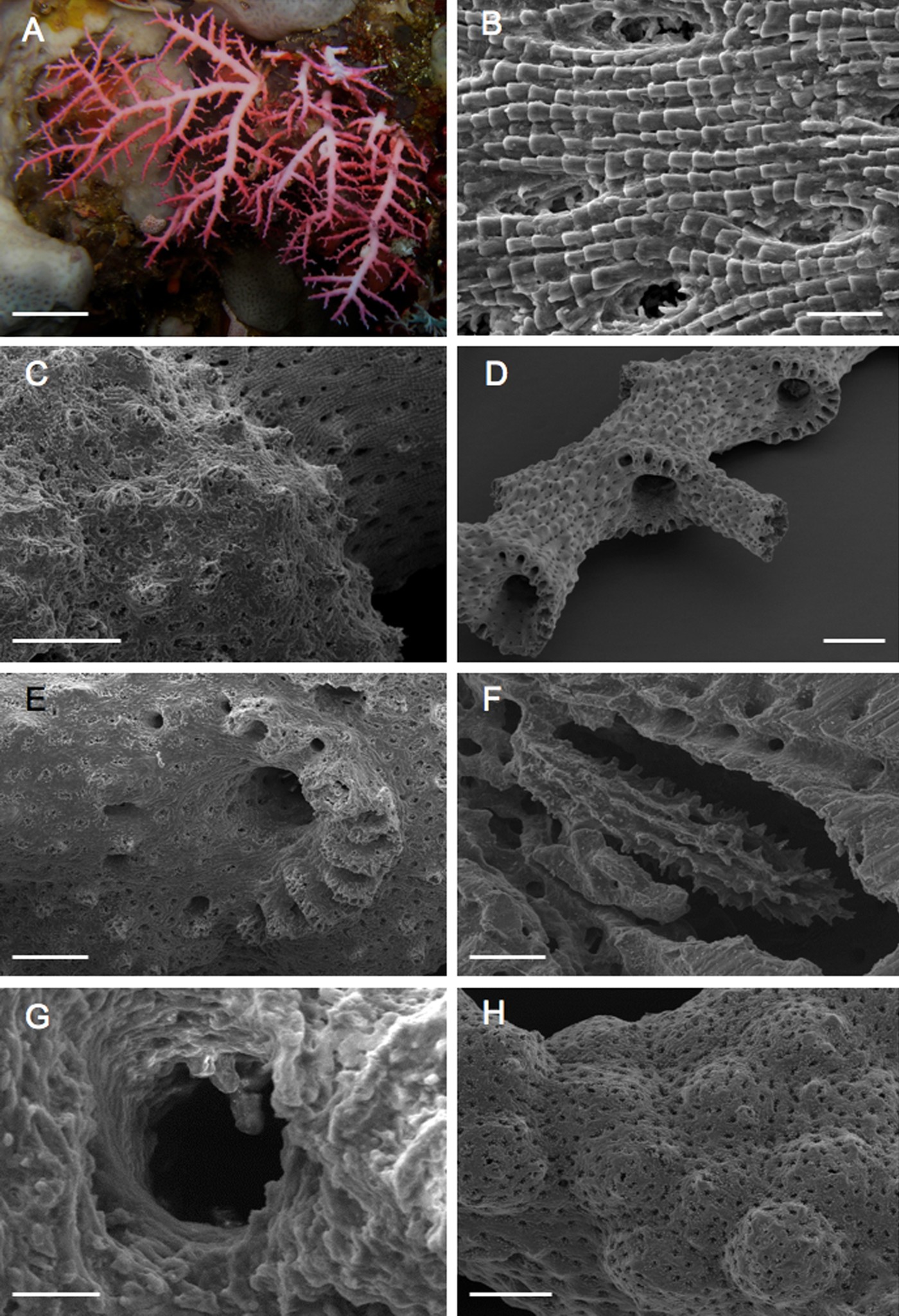
Cyclosystems (about 800 µm in diameter) are closely spaced and exclusively sympodially arranged (type C) (Figure 1D); they are restricted to the lateral branch edges, except on the branching axils where they sometimes occur on the anterior face. Cyclosystems on distal branches are round to elliptical, usually without a diastema. As a branch increases in diameter, cyclosystems become irregular in shape and develop broad abcauline diastemas. Based on 160 cyclosystems, the number of dactylopores per cyclosystem ranges from 7 to 21, the average being 14 (Figure 1E). The gastropore tube is cylindrical, up to 540 µm in diameter. A ring palisade is present in the gastropore tube at the level of the gastrostyle tip, composed of irregularly shaped elements up to 50 µm long. Gastrostyle is up to 600 µm tall, about 120 µm in diameter at the central level and about 80 µm in diameter at the apex. Styles are ornamented with linearly arranged spines up to 25 µm long, occasionally elevated in low ridges (Figure 1F). Spines are multi-tipped and often bifurcate. Dactylopore slits are about 50–60 µm wide. Dactylostyles are rudimentary, being composed of a single row of widely spaced blunt cylinders up to 20 µm tall and up to 10 µm in diameter (Figure 1G). Ampullae are hemispherical, about 500 µm in diameter and usually grouped (Figure 1H).
Description of the early growth stages
After three months, on the plastic tiles several young colonies in different phases of development were recorded allowing a detailed reconstruction of the early stages of growth of the species. The largest observed colonies were about 4 mm high and composed of 10 cyclosystems.
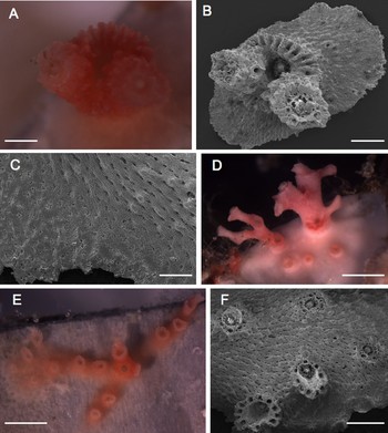
Unfortunately no newly settled planulae were observed and the youngest stage was represented by a well-developed primary cyclosystem symmetrically budding two secondary cyclosystems in a very early phase of growth (Figure 2A, B). Comparing the gastropore of the central cyclosystem with that of an adult colony, it appears less deep with the gastrostyle clearly visible from the surface. On the contrary, the dactylopores were similar to the adult both in shape and number.
Early growth stages of Stylaster sp. (A) Stereomicroscope and (B) scanning electron microscope photographs of primary cyclosystem budding two secondary cyclosystems; (C) basal disc showing the linear–imbricate texture in the older portion, a smooth surface in the growing distal zone and an irregular transitional zone between them; stereomicroscope photographs of the enlarged basal disc producing (D) round and (E) branching flattened crusts from which new cyclosystems arise; (F) scanning electron microscope photograph of new cyclosystems arising from the basal disc. Scale: A, B, F, 500 µm; C, 200 µm; D, E, 2 mm.

The primary cyclosystem arose from a basal disc, oval in shape and measuring 2.8×1.8 mm in diameter (Figure 2B). It was circular, 1.3 mm in diameter and about 520 µm high. The gastropore, about 400 µm in diameter, was surrounded by about 20 dactylopores and the gastrostyle apex was visible from the outside (Figure 2B). The basal disc and cyclosystem surfaces were covered with typical deep elliptical pores (Figure 2C). The linear–imbricate coenosteal texture characterizing the adult colony was already present in the oldest portions while it was still absent in the growing distal zones of the basal disc, showing instead a smooth surface with randomly distributed pores. A transitional zone, characterized by an irregular imbricate texture and a still irregular pore distribution, linked the younger portions to the older, completely organized portions (Figure 2C).
The secondary cyclosystems probably originated contemporaneously each one from a dactylopore of the central cyclosystem or from the enlargement of coenosteal pores at the two opposite sides of the primary cyclosystem.
The secondary cyclosystems were 800 µm and 900 µm in diameter, 470 µm and 700 µm high respectively. The gastropore appeared partially filled by calcareous material and it was surrounded by about 10 small circular dactylopores lacking the characteristic slit shape (Figure 2B).
During the following development the basal disc of the primary cyclosystems enlarged producing round or branching flattened crusts from which new cyclosystems arose (Figure 2D–F). In one case 3 single cyclosystems together with 2 colonies in a more advanced stage of growth were observed arising from a common basal disc (Figure 2D).
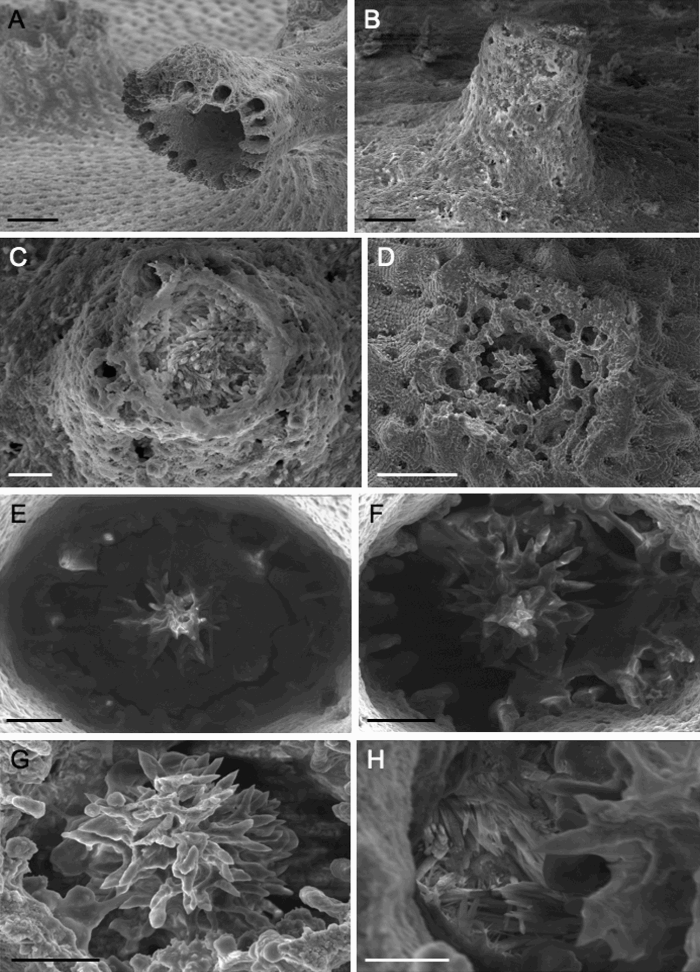
Each cyclosystem started its vertical growth producing new cyclosystems tending to be sympodially arranged. The largest observed colony was characterized by 10 cyclosystems and from the distal portion of one of them the production of the first ampulla (360 µm in diameter) was recorded (Figure 3A).
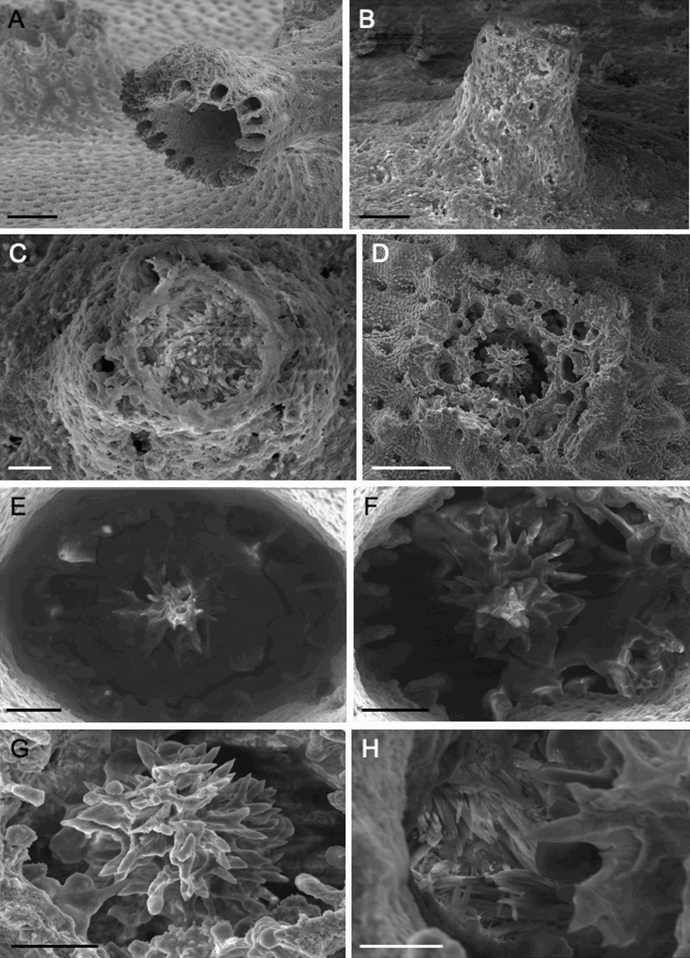
Each new cyclosystem originated as a small conical elevation 180 µm high and 120 µm in diameter (Figure 3B) with an apical circular zone characterized by a prominent margin and a calcium carbonate filled centre (Figure 3C). The surface of this plug shows the same irregularly linear–imbricate texture as the coenosteal surface surrounding the elevation. Several pores, very similar in size and shape to the coenosteal pores, were irregularly arranged around the central zone (Figure 3C). In the following growth stages the cyclosystem enlarged its diameter until 400–500 µm (Figure 3D). The gastropore was about 190–300 µm in diameter, surrounded by 9–20 dactylopores sometimes irregular in shape and size (Figure 3D).
The stopper progressively disappeared leading to the formation of the gastrostyle and the ring palisade. At the beginning only the apical gastrostyle portion was visible arising from the calcareous matrix, while many elements of the ring palisade were still fused one to each other or with the gastrostyle (Figure 3E).
Gradually the remaining matrix dissolved leaving free, in its definitive appearance, the gastrostyle and the ring palisade (Figure 3E, F). Only few elements of the ring palisade remained connected to the gastrostyle spines (Figure 3G) and the basal portion of the gastropore tube was characterized by the presence of numerous calcium carbonate crystals (Figure 3H).
Scanning electron microscope photographs of (A) first ampulla arising from a branch of a colony bearing 10 cyclosystems; (B) a new cyclosystem originating as a small volcano-like elevation; (C) volcano-like elevation apical zone with the centre filled with calcium carbonate; (D) following growth stage of the new cyclosystem; (E) apical gastrostyle portion arising from the calcareous matrix and many elements of the ring palisade still fused one to each other or with the gastrostyle; (F) calcareous matrix gradually dissolving and leaving free the gastrostyle and the ring palisade; (G) some elements of the ring palisade still connected to gastrostyle spines; (H) basal portion of the gastropore tube showing numerous calcium carbonate crystals. Scale: A, D, 200 µm; B, E, F, G, 50 µm; C, 25 µm; H, 20 µm.
DISCUSSION
Our data allowed the description of the early phases of development of Stylaster sp. Although it was impossible to describe the development of the planular polyp, the observed primary cyclosystems were about of the same size and shape of the adult ones. Starting from the first cyclosystem, colonies asexually grew according to two different ways.
The vertical growth is initially due to a dichotomous budding followed by sympodial budding giving rise to a branched colony. On the other hand the basal disc creeping growth produces a round or branching sheet running on the substrate. From this structure new cyclosystems arise. The result of this process is the production of several sister colonies arising from a common plate. Creeping colonies are very unusual within Stylasteridae, in fact only Stylantheca species are known to grow according to this strategy.
Ostarello (Reference Ostarello and Mackie1976) attributed the distribution of new colonies of Stylaster californicus to the strong tendency of the planulae to settle near the parental colony. In situ observations of Stylaster sp. indicated also for this species the formation of large and crowded groups of colonies: generally their bases appear completely separated (Figure 1A) but we may hypothesize that this situation could be at least partially produced by the separation of the colonies asexually generated from the basal disc of the primary cyclosystem.
According to Jackson (Reference Jackson, Larwoodand and Rosen1979) colonial animals are subdivided into 6 groups based on the growth forms. Vertical (trees) and encrusting (runners and sheets) forms are considered alternative modalities to face the problem of space competition in marine benthic organisms. In the case of Stylaster sp. the colonies growth using both strategies results in an unusual process of asexual reproduction that it is able to produce several colonies from a single settled planula.
A second goal of this study is the description of the production of new cyclosystems. The coenosteal pores and the coenosarc contained in them seem to play a key role in the plasticity of the colony. In fact, in the asexually generated cyclosystems, the dactylopores seem to be derived from the reorganization of the coenosteal pores present on the surface of the small cone arisen from the basal disc. Moreover, also the gastropores of the secondary cyclosystems may originate from the dactylopores of the primary one or from the coenosteal pore present on it.
The tube of newly produced gastropores is filled with calcium carbonate material that in this early stage shows the coenosteal texture typical of this species. In the subsequent stages this plug is progressively dissolved starting from its outer zone probably by the coenosarc creeping in the internal canals of the tube. The perimetric dissolution leads to the formation of both the gastropore and the gastrostyle while the ring palisade must be considered as the remains of this process. Also in mature gastropores gastrostyle spines still linked to the ring palisade elements were often observed. The numerous calcium carbonate crystals present around the base of the gastrostyle are probably an indication of this rapid remodelling activity.
Endogenous processes of skeletal dissolution are very poorly studied in cnidarians. Physiological dissolution of the calcareous stalk regularly occurs in scleractinians belonging to the family Fungiidae (Yamashiro & Yamazato, Reference Yamashiro and Yamazato1996) while it was shown that Diaseris distorta reproduces asexually by fragmentation along radially oriented slits (Yamashiro & Nishihira, Reference Yamashiro and Nishihira1994; Reference Yamashiro and Nishihira1998). The gorgonian Corallium rubrum is able to autotomize portions of branches by dissolving the calcareous inner skeleton (Russo et al., Reference Russo, Ulianich, Cicogna, Cicogna, Bavestrello and Cattaneo-Vietti1997). In scleractinians the calicoblastic cells are actively involved in the dissolution process. In fact, while the tissue layer adjacent to the skeleton is composed of flattened cells, in the region of skeletal dissolution these cells appear tall and columnar. They show many microvilli, abundant mithocondria and several different types of vesicles suggesting an active involvement in the calcium mobilization (Yamashiro & Yamazato, Reference Yamashiro and Yamazato1996). In Stylaster sp. calcium dissolution appears to be the normal process in cyclosystem formation. Broch (Reference Broch1942) suggested that the canals characterizing the stylasterid coenosteum are formed by a secondary dissolution of the calcareous substance. It is plausible that the cells composing the stolon nets deeply pervading the calcareous structure are responsible for this skeletal plasticity.
Although it is impossible to make a correct estimation, the growth rate of the young colonies may be comprised between 1.3 and 4 mm per month. The first event of sexual reproduction occurs in very small colonies that in the same period of time are also engaged in the asexual reproduction. In fact, in a colony composed of 10 cyclosystems in different growth stages and still sharing the basal disc with the sister colonies, the ampullae production was observed.
ACKNOWLEDGEMENTS
This work was financially supported by MIUR (Italian Ministry of Education, University and Research). The comments provided by Dr Helmut Zibrowius and an anonymous referee greatly improved the manuscript.