INTRODUCTION
Barnacles are distributed in all oceans, from the intertidal zone to deep sea, and attach to various substrata including rocks, sponges, corals, molluscs and floating objects. Some species are known to occur on marine vertebrates including sea turtles, dolphins and whales, and are often termed turtle barnacles and classified into the superfamily Coronuloidea (Newman & Ross, Reference Newman and Ross1976).
Collected records of these barnacles have been described in Queensland waters, Australia (Monroe & Limpus, Reference Monroe and Limpus1979), south-western Atlantic, Brazil (Young, Reference Young1991), north-western Atlantic, USA (Frick et al., Reference Frick, Williams and Robinson1998) and the Medditerrenean (Kitsos et al., Reference Kitsos, Christodoulou, Kalpakis and Noidou2003). The records regarding the hosts, collected localities, and barnacle morphology, however, are limited and scattered in Japanese waters (e.g. Hiro, Reference Hiro1936; Matsuura & Nakamura, Reference Matsuura and Nakamura1993; Hayashi & Tsuji, 2008; Hayashi, Reference Hayashi2009; Hayashi et al., in press).
The aims of this study were to review previous records to form a new collection of coronulid barnacles on marine vertebrates in Japanese waters and compensate for the lack of the descriptions regarding soft parts morphology. The morphology of the hard and soft parts areas is presented. The systematic arrangement used here follows Newman (Reference Newman and Forest1996).
MATERIALS AND METHODS
I investigated many marine vertebrates for epibionts captured on some occasions including by-catches, strandings, and landings for their breeding on the Japanese coast between 2002 and 2011. The following species of marine vertebrates were investigated: loggerhead sea turtle (Caretta caretta), green sea turtle (Chelonia mydas), hawksbill turtle (Eretmochelys imbricata), humpback whale (Megaptera novaeangliae), gray whale (Eschrichtius robustus), finless porpoise (Neophocaena phocaenoides), melon-headed whale (Peponocephala electra) and Indian Ocean bottlenose dolphin (Tursiops aduncus).
Numerous specimens were removed from various parts of host body and fixed in 99% ethanol. Collected materials were identified in the laboratory and examined specimens were deposited in Fujukan, the Museum of the University of Ryukyus (RUMF-ZC), National Museum of Nature and Science Tokyo (NMNS) and Scripps Institution of Oceanography, California (SIO).
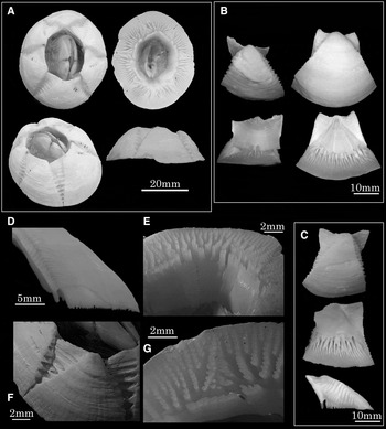
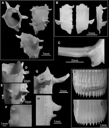
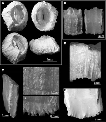
Chelonibia testudinaria (Linnaeus, 1758): (A) upper, basal, upper oblique, and lateral views; (B) rostrum with rostrolateral plates and carina (external and internal views); (C) lateral compartment (external, internal and lateral views); (D) longitudinal section of rostrum; (E) detail of rostrum (internal view); (F) crenated radii (external view): (G) detail of coupling adjacent plates (basal view). (A) RUMF-ZC-02040; (B & C) RUMF-ZC-02041; (D–G) RUMF-ZC-02021.

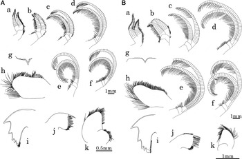
Soft parts of the bamacles (A) Chelonibia testudinaria (Linnaeus, 1758) and (B) Chelonibia caretta (Spengler, 1790): a, cirrus I; b, cirrus II; c, cirrus III; d, cirrus IV; e, cirrus V; f, cirrus VI; g, labrum; h, palp; i, mandible; j, maxilla I; k, maxilla II.

MATERIALS EXAMINED
Hahajima Island, Ogasawara, Tokyo, from green sea turtle (RUMF-ZC-02021); Yakushima Island, Kagoshima, 26 May 2006, from loggerhead turtle (RUMF-ZC-02040); Ie, Kunigani, Okinawa, 2 May 2010, from loggerhead turtle (RUMF-ZC-02041).
DESCRIPTION
Shell conical, depressed, heavy, massive and smooth; parietes thick and strong; basal septa very numerous, not ribbed or longitudinally folded, the peripheral edge not lobed or incised; base oval to round; radii rather narrow and depressed, mostly notched on the sides, rarely smooth, with characteristic toothed pattern; adherent to surface of scutes of host; tergal spur shallow, basal margin of terga very long, scutal margin of terga short, articular ridge, articular furrow and crest for depressor muscles rudimentary; tergal spur adhere to basal margin.
Cirri having many segments in each ramus. Cirrus III resembling cirrus IV. Labrum with a notch in the middle and multi-denticulated on both sides. Palp rectangular; lateral edge covered with long and fine setae; upper edge with short and fine setae. Mandible usually with five teeth, 1st to 4th large and sharp, 2nd and 3rd bifid, 5th blunt and covered a tuft of setae. Maxilla I with two large and thick spines and numerous fine setae; without a notch and cutting edge straight. Maxilla II globular; apex covered with fine setae.
NEW OCCURRENCES ON JAPANESE COAST
Ginoza, Okinawa (on Caretta caretta); Kunigami, Okinawa (on C. caretta); Yomitan, Okinawa (on C. mydas); Yakushima Island, Kagoshima (on C. caretta); Tateyama, Chiba (on C. caretta); Minabe, Wakayama (on C. caretta); Otsuchi, Iwate (on C. caretta); Hahajima Island, Ogasawara, Tokyo (on C. mydas).
PREVIOUS RECORDS ON JAPANESE COAST
Ginoza, Okinawa (on Chelonia mydas, Hayashi and Tsuji, Reference Hayashi and Tsuji2008); Yomitan, Okinawa (on C. caretta, Hayashi, Reference Hayashi2009); Chichijima Island, Ogasawara, Tokyo (on C. mydas, Tachikawa, Reference Tachikawa1995); Fukiage beach, Kagoshima (on C. caretta, Matsuutra and Nakamura, 1993); Hakata, Fukuoka (on C. caretta, Utinomi, Reference Utinomi1949).
HABITAT
Found on carapace, plastron, head and posterior legs of sea turtles.
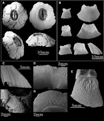
Chelonibia caretta (Spengler, 1790): (A) upper, basal, upper oblique, and lateral views; (B) rostrum with rostrolateral plates, carina and lateral plate (external and internal views); (C) longitudinal section of rostrum; (D) detail of rostrum (internal view); (E) lateral compartment (basal view); (F) narrow radii (external view); (G) detail of coupling adjacent plates (basal view). (A, D, F & G) RUMF-ZC-02019; (B, C & E) SIO-C5824.

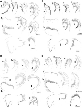
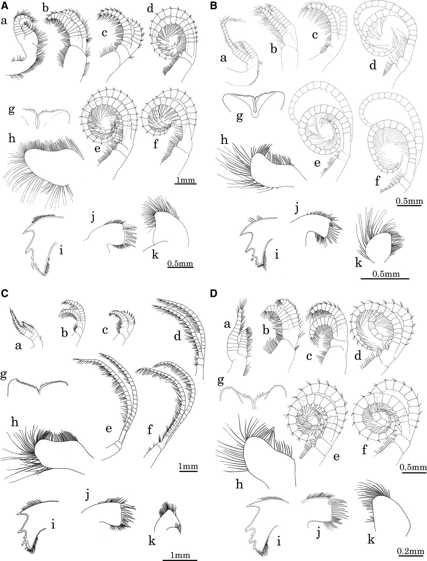
Soft parts of the bamacles (A) Coronula diadema (Linnaeus, 1758), (B) Cryptolepas rhachianecti Dall, 1872, (C) Tubicinella cheloniae Monroe & Limpus, Reference Monroe and Limpus1979, and (D) Xenobalanus globicipitis Steenstrup, 1852: a, cirrus I; b, cirrus II; c, cirrus III; d, cirrus IV; e, cirrus V; f, cirrus VI; g, labrum; h, palp; i, mandible; j, maxilla I; k, maxilla II.

MATERIALS EXAMINED
Toya, Yomitan, Okinawa, 2 February 2010, from loggerhead turtle (SCL 89.5 cm, SCW 67.4 cm, Tag No. 46604) (RUMF-ZC-02019); Tortugero, Costa Rica, July 1965 (SIO-C5824).
DESCRIPTION
Shell rounded, conical, rough, globule-convex, extremely massive and heavy, the compartments thick and strong; radii narrow, rather not developed; basal septa very numerous; parietes without cavities extending up between the interrupted, parietes solidly filled up nearly to the base; orifice eroded; scuta and terga as for C. testudinaria.
Cirri having many segments in each ramus. Cirrus III resembling cirrus IV. Labrum with a notch in the middle and multi-denticulated on both sides. Palp rectangular; lateral edge covered with long and fine setae; upper edge with short and fine setae. Mandible usually with five teeth, 1st to 4th large and sharp, 2nd and 3rd bifid, 5th blunt and covered a tuft of setae. Maxilla I with two large and thick spines and numerous fine setae; with a notch. Maxilla II globular; apex covered with fine setae.
NEW OCCURRENCES ON JAPANESE COAST
Yomitan, Okinawa (on Eretmochelys imbricata and Caretta caretta).
HABITAT
Found on turtle carapace.
REMARKS
New record for Japan.
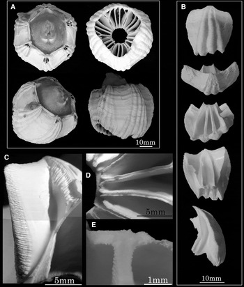
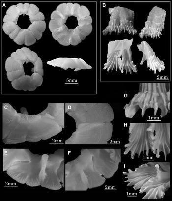
Coronula diadema (Linnaeus, 1767): (A) upper, basal, upper oblique, and lateral views; (B) rostrum (external, upper, basal, internal and lateral views); (C) detail of alae (external view); (D) detail of compartments (basal view); (E) cross-section of ‘T’ shape flange. (A) RUMF-ZC-02021; (B–E) SIO-C10012.

Soft parts of the bamacles, (A) Platylepas hexastylos (Fabricius, 1798), (B) Platylepas decorata Darwin, 1854, (C) Cylindrolepas darwiniana Pilsbury, Reference Pilsbury1916, and (D) Cylindrolepas sinica Ren, 1980: a, cirrus I; b, cirrus II; c, cirrus III; d, cirrus IV; e, cirrus V; f, cirrus VI; g, labrum; h, palp; i, mandible; j, maxilla I; k, maxilla II.

MATERIALS EXAMINED
Tosashimizu, Kochi, 4 November 2004, from humpback whale (RUMF-ZC-02021); Perth, Australia, from humpback whale (SIO-C10012).
DESCRIPTION
Shell crown-shaped, with longitudinal convex ribs, having their edges crenated; orifice hexagonal, larger than the base; radii moderately thick, very broad, having numerous transverse septa; terga absent or rudimentary; basal margin of scuta very long, occrudent margin of scuta very short, articular furrow broad, adductor ridge small.
Cirri short, strong and thick; segments wider than long; cirrus I with unequal rami, cirrus III resembling cirrus II; having some short and strong teeth on front margin of cirrus II to VI. Labrum with a notch in the middle, and swelled up on each corner; multi-denticulated on each side. Palp covered with fine setae. Mandible with many teeth; secondary teeth between main teeth; inferior corner carrying the irregular number of minute denticles and hair. Maxilla I with two strong spines followed by a distinct notch and shorter spines below; tapered to be the cutting edge. Maxilla II covered with numerous fine setae.
NEW OCCURRENCES ON JAPANESE COAST
Rumoi, Hokkaido (on Megaptera novaeangliae); Tateyama, Chiba (on Megaptera novaeangliae); Kihoku, Mie (on Megaptera novaengliae); Tosa-Shimizu, Kochi (on Megaptera novaeangliae); Shimajiri series, Katsuren peninsula, Okinawa (Pleistocene–Pliocene).
PREVIOUS RECORDS ON JAPANESE COAST
Chikura, Chiba (on Megaptera novaeangliae, Nogata & Matsumura, Reference Nogata and Matsumura2006); Kakegawa series, Shizuoka (Lower Pliocene, Hatai, Reference Hatai1939); the Masuda beds, Sakata, Akumi-gun, Yamagata (Lower Pliocene, Hatai, Reference Hatai1938).
HABITAT
Found on head, flippers, tail and ridged throat of humpback whales.
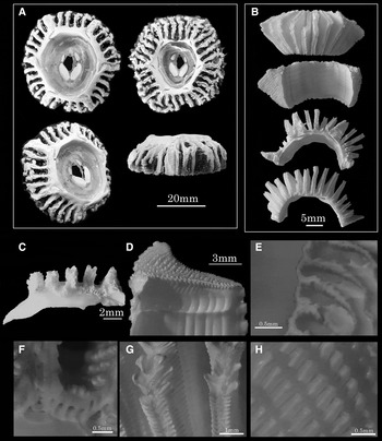
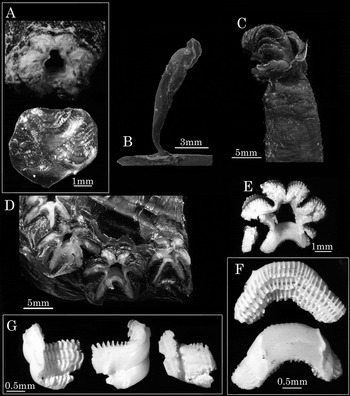
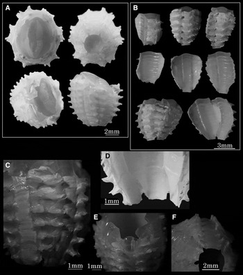
Cryptolepas rhachianecti Dall, 1872: (A) upper, basal, upper oblique, and lateral views; (B) compartments (external, internal, upper and basal views); (C) transversal section of lateral compartment; (D) radii (internal view); (E) detail of radii (lateral view); (F) detail of Figure 4C; (G) tips of radiate branches; (H) detail of imbricating shell wall. (A) RUMF-ZC-02022; (B–H) RUMF-ZC-02023.

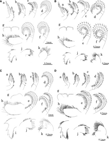
Soft parts of the bamacles, (A) Stomatolepas praegustator Pilsbry, 1910, (B) Stomatolepas transversa Nillson-Cantell, 1930, (C) Stephanolepas muricata Fischer, 1886 (type-A), and (D) Stephanolepas muricata Fischer, 1886 (type-B): a, cirrus I; b, cirrus II; c, cirrus III; d, cirrus IV; e, cirrus V; f, cirrus VI; g, labrum; h, palp; i, mandible; j, maxilla I; k, maxilla II.

MATERIALS EXAMINED
Tomiyama, Chiba, 11 May 2005, from gray whale (RUMF-ZC-02022, 02023).
DESCRIPTION
Almost wholly embedded in the host's skin, mostly on the head and back, only the opercular membrane and parts of radii or sometimes the more prominent ribs being exposed; compartments normally having thirty ribs and thirty lobes; having four ribs on each of the parietes; radial ornamentation denticulated with alaeal ornamentation of adjacent compartment; ribs on compartments finely striae vertically and crenulated at the edge; sheath very long, not quite reaching to the base, transversely grooved, and not overhanging or prominent on its basal margin; radii are as thick as the compartments and their edges are intricately sculptured with crimped sutural laminae; terga absent or rudimentary; scuta piled up by caustic layers.
Cirri short and thick; segments wider than long. Cirrus I to III unequal rami; cirrus III resembling cirrus II. Labrum with a notch in the middle; having two to three teeth on each side. Palp club-shaped and covered by fine setae. Mandible with many teeth; 1st to 3rd strong; secondary teeth between main tooth; inferior teeth covered with fine setae and minute denticles. Maxilla I with one strong spine and two notches on cutting edge; covered with shorter spines; upper and lower edge covered with fine setae. Maxilla II covered with fine setae.
PREVIOUS RECORDS ON JAPANESE COAST
Tomiyama, Chiba (Takeda & Ogino (Reference Takeda and Ogino2005) on Eschrichtius robustus).
HABITAT
Found on head and dorsal side of gray whales.
Tubicinella cheloniae Monroe & Limpus, Reference Monroe and Limpus1979: (A) upper, upper oblique, and lateral views; (B) lateral (external, internal and lateral views); (C) transverse section of compartment; (D) aperture (oblique view); (E) parietal props; (F) external surface of parietes; (G) side view of parietal prop; (H) detail of radii (lateral view); (I) basal portion of parietal plate (external and internal views). (A–H) SIO-C5813; (I) RUMF-ZC-02024.

MATERIALS EXAMINED
Toya, Yomitan, Okinawa, 2 February 2010, from loggerhead turtle (SCL 89.5 cm, SCW 67.4 cm, Tag No. 46604) (RUMF-ZC-02024); Talong Talong Basar Island, Sarawak, South China Sea, 1952, from green sea turtle (SIO-C5813).
DESCRIPTION
Having areas of horizontally flattened upward curving spines projecting from the lateral edges of the compartments; burrowing in chelonians, the orifice below the level of host scutes; shell cylindrical and porose; orifice slightly larger than basis; parietes smooth on the outer surface, having longitudinal ridges on median area and projections on sutures; sheath very long, not quite reaching to the base; radii and alae almost confused; having terga and scuta, but rudimentary.
Cirrus I and II with unequal rami; cirrus III resembling cirrus II. Labrum with a notch in the middle; having a few teeth on each side. Palp club-shaped, covered with fine setae. Mandible 4 or 5 main teeth; a secondary tooth between 2nd and 3rd, 3rd and 4th; inferior tooth covered with minute denticles and fine setae. Maxilla I having a notch on cutting edge; two strong spines above a notch and shorter spines below; upper and inferior edge covered with fine setae. Maxilla II globular and covered with fine setae.
NEW OCCURRENCES ON JAPANESE COAST
Yakushima Island, Kagoshima (on C. caretta); Yomitan, Okinawa (on C. caretta).
HABITAT
Embedded in carapace of loggerhead sea turtle.
REMARKS
New record for Japan. Ross & Frick (Reference Ross and Frick2007) established the genus Chelolepas as new for Tubicinella cheloniae, however, there is no description and comparison on soft parts between Tubicinella major and T. cheloniae. A rigid comparison of morphology and phylogenetic work including both species are wanting.
Xenobalanus globicipitis Steenstrup, 1852: (A) upper view of shell walls and attachment trace; (B) body attached to host skin; (C) detail of opercular and cirri; (D) line of shell walls without bodies (upper view); (E) shell walls without skin; (F) detail of rostrum (external and internal views); (G) lateral compartment (external, internal and lateral view). (A) NSMT-M35037; (B) NSMT-M32811; (C–D) SIO-C5867; (E–G) NSMT-M34068.

MATERIALS EXAMINED
WFP579, 15 May 1978, from Delphinus delphinus (SIO-C5867), Higashimatsushima, Miyagi, 15 November 2007, from finless purpoise (NSMT-M35037), Shirako, Chiba, 5 January 2006, from finlas porpoise (NSMT-M34068), Hasaki, Ibaraki, 25 February 2002, from melon-headed beaked whale, 200+ (NSMT-M32811).
DESCRIPTION
Shell almost rudimentary, star-formed, composed of six compartments; out of this thin, star-shaped shell a cylindrical flexible, peduncle-formed body springs, which forms the main part of the animal; with a long peduncle-formed body rising from the middle; opercular valves absent; it is narrow where coming out of the central cavity of the star, but soon acquires its full diameter; at the upper end it has a reflexed hood, and hence is broader, and this has the appearance of forming a capitulum, like that of a pedunculated Cirripedia.
Cirri short, strong and thick. Cirrus I and II with unequal rami; cirrus III resembling cirrus II; cirrus I to III having blunt apex on rami. Labrum with a notch in the middle; having no teeth and not covered with short and fine hairs on each side. Palp club-shaped and covered with fine setae. Mandible slightly rounded, with 3 main teeth and inferior teeth composed of irregular denticulated and fine setae. Maxilla I having a notch on cutting edge; two strong spines above a notch and shorter spines below; upper and inferior edge covered with fine setae. Maxilla II globular and covered with fine setae.
NEW OCCURRENCES ON JAPANESE COAST
Okumatsushima, Higashimatsushima, Miyagi (on Neophocaena phocaenoides); Shirako, Chiba (on Neophocaena phocaenoides); Kamisu, Ibaraki (on Peponocephala electra); Mikurajima Island, Tokyo (on Tursiops aduncus).
PREVIOUS RECORDS ON JAPANESE COAST
Kii peninsula (on Orcinus orca, Sakai et al., Reference Sakai, Hayashi, Murata, Yamada and Asakawa2009); Taiji, Wakayama (on Stenella attenuata, Greenwood et al., Reference Greenwood, Taylor and Gauckler1979).
HABITAT
Found on flippers, dorsal fin and tail of cetacean.
Platylepas hexastylos (Fabricius, 1798): (A) upper, basal, upper oblique, and lateral views; (B) lateral compartment (external, internal and lateral views); (C) detail of lateral plates (upper view); (D) detail of radii (upper view); (E) detail of midrib (basal view); (F) detail of juncture plates (basal view); (G–I) detail of basal edge of parietes (external, internal and oblique views). (A & C–F) RUMF-ZC-02025; (B & G–I)RUMF-ZC-02026; F, RUMF-ZC-02046.

MATERIALS EXAMINED
Otsuchi, Iwate, 24 August 2006, from loggerhead turtle (SCL 66.3 cm, SCW 52.6 cm, Tag No. 47127 and 47128) (RUMF-ZC-02025); (RUMF-ZC-02026).
DESCRIPTION
Shell low, conical, margin multilobed, and orifice small; each compartment fimbriated at the edge, having a midrib; radii narrow; growth lines the predominant sculpturing, septae visible through the wall, becoming more numerous peripherally; line of ‘midrib fold’ obvious, dividing each compartment into two main lobes, accessory ‘folds’, and inter-compartmental sutures, appear to enclose host tissue; midribs extend only slightly below level of periphery giving slightly convex base; septae visible on inner surface of outer wall towards periphery; opercular valves subequal, extend full length of orifice.
Cirrus I with unequal ramus; cirrus III resembling cirrus II. Labrum with a notch in the middle with two to three teeth on each side. Palp rectangular; lateral edge covered with long and fine setae; upper edge with short and fine setae. Mandible with 4 or 5 main teeth; a secondary tooth between 2nd and 3rd; 2nd and 3rd bifid; inferior tooth pectinated and covered with fine setae. Maxilla I having a notch on cutting edge; two strong spines above a notch and shorter spines below; upper and inferior edge covered with fine setae. Maxilla II globular and covered with fine setae.
NEW OCCURRENCES ON JAPANESE COAST
Ginoza, Okinawa (on Chelonia mydas and Caretta caretta); Ohgimi, Okinawa (C. mydas); Yomitan, Okinawa (on C. mydas and Eretmochelys imbricata); Uruma, Okinawa (E. imbricata); Ishigakijima Island, Okinawa (C. mydas and E. imbricata); Yakushima Island, Kagoshima (on C. caretta); Tateyama, Chiba (on C. caretta); Otsuchi, Iwate (on C. caretta); Hachijojima Island, Tokyo (on C. caretta); Hahajima Island, Ogasawara, Tokyo (on C. mydas); Minabe, Wakayama (on E. imbricata); Motojima beach, Tanabe, Wakayama (on C. mydas); Manazuru, Kanagawa (on C. mydas); Fukui, Fukui (on C. caretta and E. imbricata); Sakai, Fukui (on C. caretta and E. imbricata); Awara Fukui (C. caretta).
PREVIOUS RECORDS ON JAPANESE COAST
Yomitan, Okinawa (on C. caretta, Hayashi, Reference Hayashi2009); Otsuchi, Iwate (on Chelonia mydas agassizii, Hayashi et al., in press).
HABITAT
Found on carapace, plastron, head, flipper, legs and soft skin of sea turtles.
Platylepas decorata Darwin, 1854: (A) upper, basal, upper oblique, and lateral views; (B) compartments (external and internal views); (C) upper margin of parietes; (D) radii and external ornamentations (oblique view); (E) juncture of compartments (upper view); (F) rostrum (internal view); (G) detail of secondary ribs; (H) compartments (basal view). (A, D, G &H) RUMF-ZC-02027; (B, C & E) RUMF-ZC-02042.

MATERIALS EXAMINED
Ginoza, Okinawa, 20 September 2002, from green sea turtle (SCW 62.1 cm, Tag No. 29245, 28141) (RUMF-ZC-02027, 02042); D7017 (RUMF-ZC-02046).
DESCRIPTION
Shell subcircular, ring-like, shallow, steep sided, and non-porous; compartments with fine longitudinal ridges, lower edge dentate; each compartment with a midrib; radii narrow with simple septate margins; basal membrane equalling in convexity the shell; opercular valves oblong and narrow, scuta longer than terga.
Cirrus I with unequal ramus; cirrus III resembling cirrus II. Labrum with a notch in the middle and multi-denticulated on each side. Palp rectangular; lateral edge covered with long and fine setae; upper edge with short and fine setae. Mandible with 4 or 5 main teeth; a secondary tooth between 2nd and 3rd; 2nd and 3rd bifid; inferior tooth pectinated and covered with fine setae. Maxilla I having a notch on cutting edge; two strong spines above a notch and shorter spines below; upper and inferior edge covered with fine setae. Maxilla II globular and covered with fine setae.
NEW OCCURRENCES ON JAPANESE COAST
Ginoza, Okinawa (on Chelonia mydas and Eretmochelys imbricata); Ishigakijima Island, Okinawa (C. mydas and E. imbricata); Ohgimi, Okinawa (C. mydas); Yomitan, Okinawa (on C. mydas); Yakushima Island, Kagoshima (on C. mydas); Hahajima Island, Ogasawara, Tokyo (on C. mydas).
HABITAT
Found on plastron, legs and soft skin of sea turtles.
REMARKS
New record for Japan.
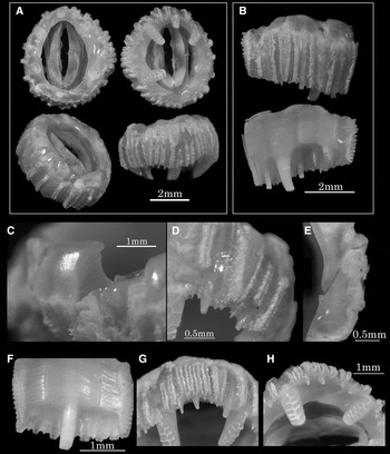
Cylindrolepas darwiniana Pilsbry, 1916: (A) upper, basal, upper oblique, and lateral views; (B–C) rostrum (external, internal and lateral views); (D) external view of compartments; (E) detail of external sculpture; (F) detail of basal edge; (G) internal view of basal edge. (A & D) RUMF-ZC-02028; (B,C & E–G) RUMF-ZC-02029.

MATERIALS EXAMINED
Toya, Yomitan, Okinawa, 2 February 2010, from loggerhead turtle (SCL 89.5 cm, SCW 67.4 cm, Tag No. 46604) (RUMF-ZC-02028, 02029).
DESCRIPTION
Form cylindrical, the orifice and base of equal size; bases of the compartments obtusely dentate, the median tooth of each compartment larger, slightly inflected; outer surface of parietes sulcated longitudinally; sheath very long, not quite reaching to the base; opercular valves as in Platylepas.
Cirrus I with unequal ramus; cirrus III resembling cirrus II. Labrum (Plate 1g) with a notch in the middle and multi-denticulated on each side. Palp rectangular; lateral edge covered with long and fine setae; upper edge with short and fine setae. Mandible with 4 or 5 main teeth; a secondary tooth between 2nd and 3rd; 2nd and 3rd bifid; inferior tooth pectinated and covered with fine setae. Maxilla I having a notch on cutting edge; two strong spines above a notch and shorter spines below; upper and inferior edge covered with fine setae. Maxilla II globular and covered with fine setae.
NEW OCCURRENCES ON JAPANESE COAST
Yomitan, Okinawa (on Caretta caretta).
HABITAT
Found on turtle carapace.
REMARKS
New record for Japan.
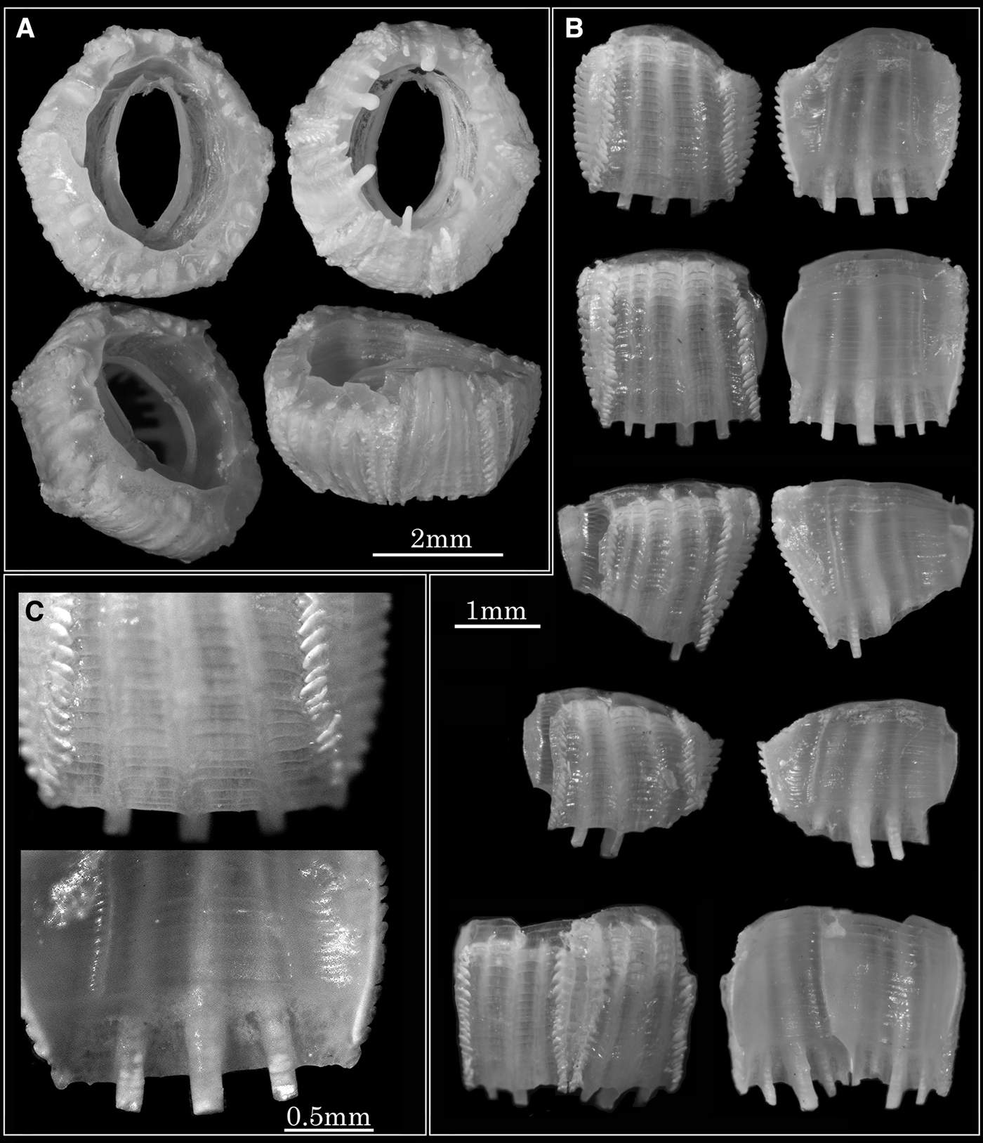
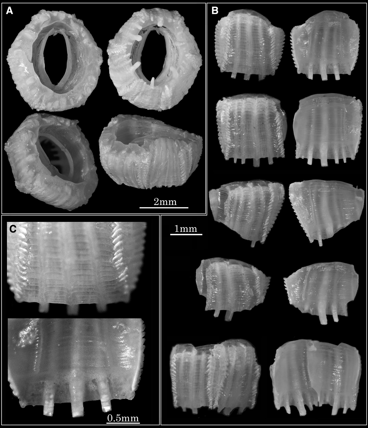
Cylindrolepas sinica Ren, 1980: (A) upper, basal, upper oblique, and lateral views; (B) compartments (external and internal views); (C) detail of rostrum (external and internal views). (A) RUMF-ZC-02001; (B)RUMF-ZC-02030; (C) RUMF-ZC-02031.
MATERIALS EXAMINED
Yomitan, Okinawa, 18 August 2008, from loggerhead turtle (RUMF-ZC-02001); Ginoza, Okinawa, 20 September 2002, from green turtle (SCW 62.1 cm, Tag No. 29245, 28141) (RUMF-ZC-02030); Hahajima Island, Ogasawara, Tokyo, 20 April 2004, from green turtle (SCL 90.5 cm, SCW 70.2 cm) (RUMF-ZC-02031).
DESCRIPTION
Shell cup-shaped, hexagonal, slightly elongated; basis almost equal to orifice in diameter; outer surface of median portion smooth, permeated with three white longitudinal ribs inside of the wall, the middle rib much thicker and projected out of basal edge of parietal plate, forming an inwardly curved blunt tooth; sheath very long, not quite reaching to the base; opercular valves as in Platylepas.
Cirrus I with unequal ramus; cirrus III resembling cirrus II. Labrum with a notch in the middle with two to three teeth on each side. Palp rectangular; lateral edge covered with long and fine setae; upper edge with short and fine setae. Mandible with 4 or 5 main teeth; a secondary tooth between 2nd and 3rd; 2nd and 3rd bifid; inferior tooth pectinated and covered with fine setae. Maxilla I having a notch on cutting edge; two strong spines above a notch and shorter spines below; upper and inferior edge covered with fine setae. Maxilla II globular and covered with fine setae.
NEW OCCURRENCES ON JAPANESE COAST
Ginoza, Okinawa (on Chelonia mydas); Ohgimi, Okinawa (C. mydas); Ishigakijima Island, Okinawa (C. mydas and Eretmochelys imbricata); Yomitan, Okinawa (on C. mydas and E. imbricata); Hahajima Island, Ogasawara, Tokyo (on C. mydas).
PREVIOUS RECORDS ON JAPANESE COAST
Yomitan, Okinawa (on C. caretta, Hayashi, Reference Hayashi2009).
HABITAT
Found on soft skin around neck and tail, and embedded in sulcus of plastrons and scales of sea turtles, not on flippers and legs.
Stomatolepas praegustator Pilsbry, 1910: (A) upper, basal, upper oblique, and lateral views; (B) lateral compartment (external, internal and upper views); (C) incomplete lateral plate (imbricating sculptures removed); (D) juncture of plates (upper view); (E) basal edge of parietes (external view); (F) detail of imbricating sculpture (external view); (G) inner view of parietes. (A–C) RUMF-ZC-02033; (D–G) RUMF-ZC-02032.

MATERIALS EXAMINED
Otsuchi, Iwate, 24 August 2006, from loggerhead turtle (SCL 83.6 cm, SCW 57.8 cm, Tag No. 47121, 47124, RUMF-ZC-02032); Yakushima Island, Kagoshima, May 2006, from loggerhead turtle (RUMF-ZC-02033).
DESCRIPTION
Shell approximately circular in dorsal aspects; wall broad and shallow bowl shaped; the diameter of the basis about half that of the orifice; parietes with series of imbricating scales; basis of compartments smooth, not denticulated; opercular plates small, smooth, not articulated.
Cirrus I with unequal ramus; cirrus III resembling cirrus II. Labrum with a notch in the middle with two to three teeth on each side. Palp rectangular; lateral edge covered with long and fine setae; upper edge with short and fine setae. Mandible with 4 or 5 main teeth; no secondary tooth between 2nd and 3rd; 2nd and 3rd bifid; inferior tooth pectinated and covered with fine setae. Maxilla I having a notch on cutting edge; two strong spines above a notch and rather short spines below; upper and inferior edge covered with fine setae. Maxilla II globular and covered with fine setae.
NEW OCCURRENCES ON JAPANESE COAST
Otsuchi, Iwate (on C. caretta); Ooamishirasato-machi, Chiba (on C. caretta); Yakushima Island, Kagoshima (on C. caretta); Yomitan, Okinawa (on C. mydas).
PREVIOUS RECORDS ON JAPANESE COAST
Seto, Wakayama (on C. caretta, Hiro Reference Hiro1936).
HABITAT
Found on throat and soft skin around neck, tail and flippers of sea turtles.
REMARKS
Hiro (Reference Hiro1936) recorded S. elegans on a female loggerhead sea turtle. Monroe (Reference Monroe1981) regarded Stomatolepas elegans as a junior synonym of S. praegustator. Frick et al. (2010) described the neotype specimens of S. praegustator and S. elegans; however, there is no description and comparison on soft parts. A rigid comparison of morphology and phylogenetic work including both species are wanting.
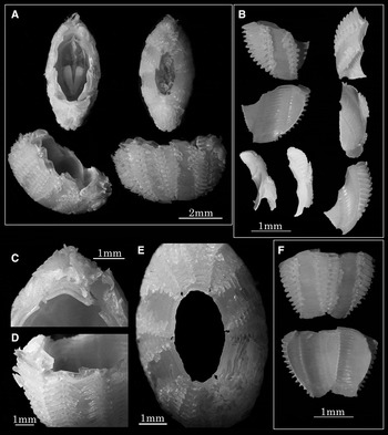
Stomatolepas transversa Nillson-Cantell, 1930: (A) upper, basal, upper oblique, and lateral views; (B) lateral (external, internal and lateral views); (C) upper edge of carina; (D) oblique view of carina; (E) basal view of whole shell; (F) compartment plates (external and inner views). (A) RUMF-ZC-02031; (B & F) RUMF-ZC-02048; (C–E) RUMF-ZC-02047.

MATERIALS EXAMINED
Hahajima Island, Ogasawara, Tokyo, 20 April 2004, from green sea turtle (SCL 90.5 cm, SCW 70.2 cm) (RUMF-ZC-02034).
DESCRIPTION
Shell boat-shaped; opercular wider than the base; plates with series of imbricating scales; rostrum and carina both very narrow but are still similar; radii well developed, externally with a series of scales that imbricate with those from the neighbouring plate; opercular plates small, smooth, not articulated.
Cirrus I with unequal ramus; cirrus III resembling cirrus II. Labrum with a notch in the middle with two to three teeth on each side. Palp rectangular; lateral edge covered with long and fine setae; upper edge with short and fine setae. Mandible with 4 or 5 main teeth; no secondary tooth between 2nd and 3rd; 2nd and 3rd bifid; inferior tooth pectinated and covered with fine setae. Maxilla I having a notch on cutting edge; two strong spines above a notch and shorter spines below; upper and inferior edge covered with fine setae. Maxilla II globular and covered with fine setae.
NEW OCCURRENCES ON JAPANESE COAST
Ishigakijima Island, Okinawa (C. mydas); Yomitan, Okinawa (on C. mydas); Hahajima Island, Ogasawara, Tokyo (on C. mydas).
PREVIOUS RECORDS ON JAPANESE COAST
Ginoza, Okinawa (on Chelonia mydas, Hayashi & Tsuji, 2008).
HABITAT
Embedded in groove of plastrons and scales of green sea turtle (Chelonia mydas).
Stephanolepas muricata Fischer, 1886. In the case of penetrated into a scale (type-A): (A) upper, basal, upper oblique, and lateral views; (B) carina, rostrum, lateral and compartments (external and internal views); (C) detail of radii (external view); (D) basal edge of compartment (internal view); (E) upper margin of parietes; (F) basal view of compartments. (A)RUMF-ZC-02035; (B) RUMF-ZC-02044; (C–F) RUMF-ZC-02043.

Stephanolepas muricata Fischer, 1886. In the case of embedded between scales (type-B): (A) upper, basal, upper oblique, and lateral views; (B) detail of radii (external view); (C) carina, rostrum and lateral (external and internal views); (D) comparison of lateral compartments of Stephanolepas muricata Fischer, 1886 between type-B (left) and type-A (right). View from above, view of ala, view of radius from outside, basal view. (A) RUMF-ZC-02036; BRUMF-ZC-02037; (C) RUMF-ZC-02038; (D) RUMF-ZC-02040 (left) and RUMF-ZC-02039 (right).

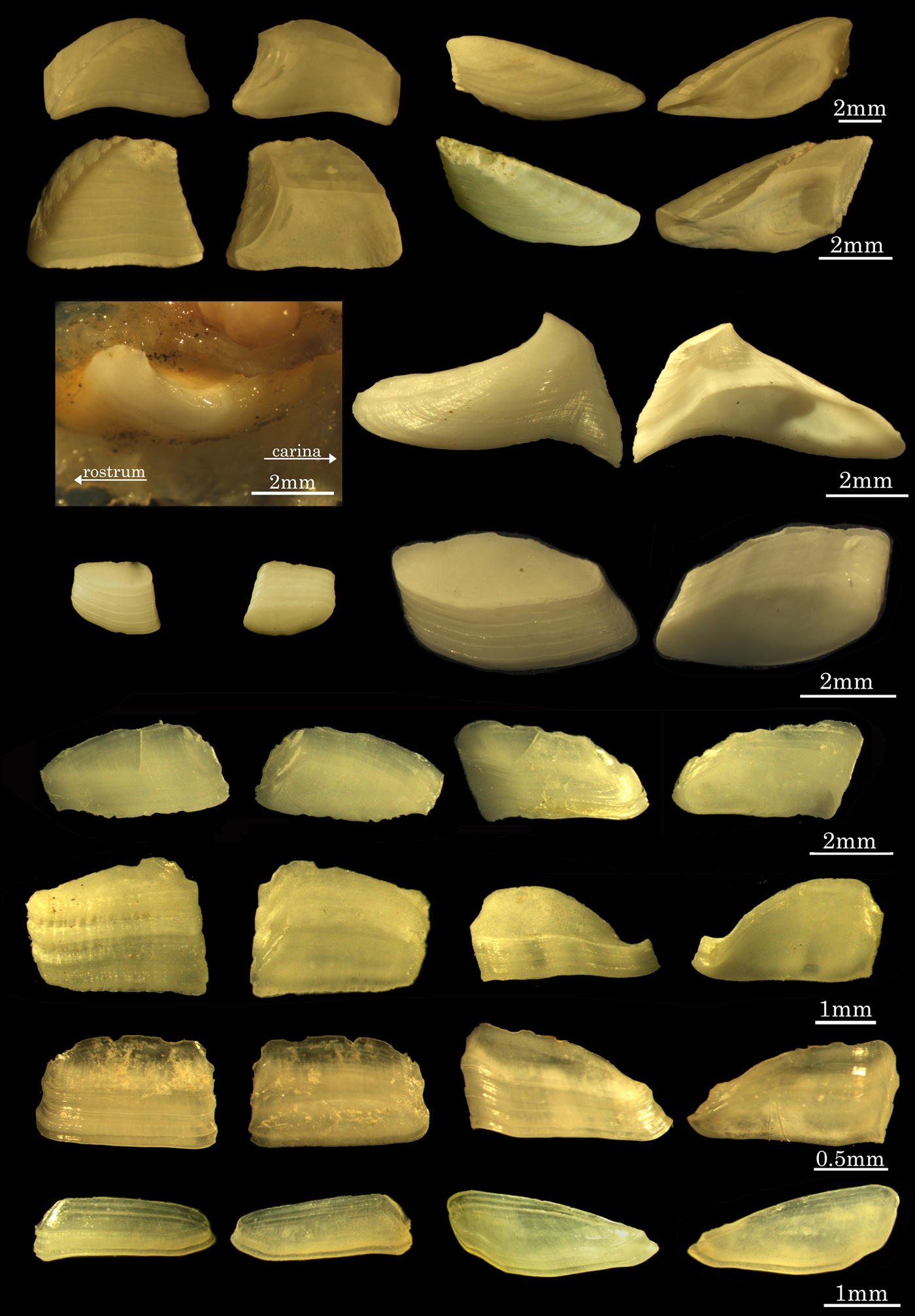
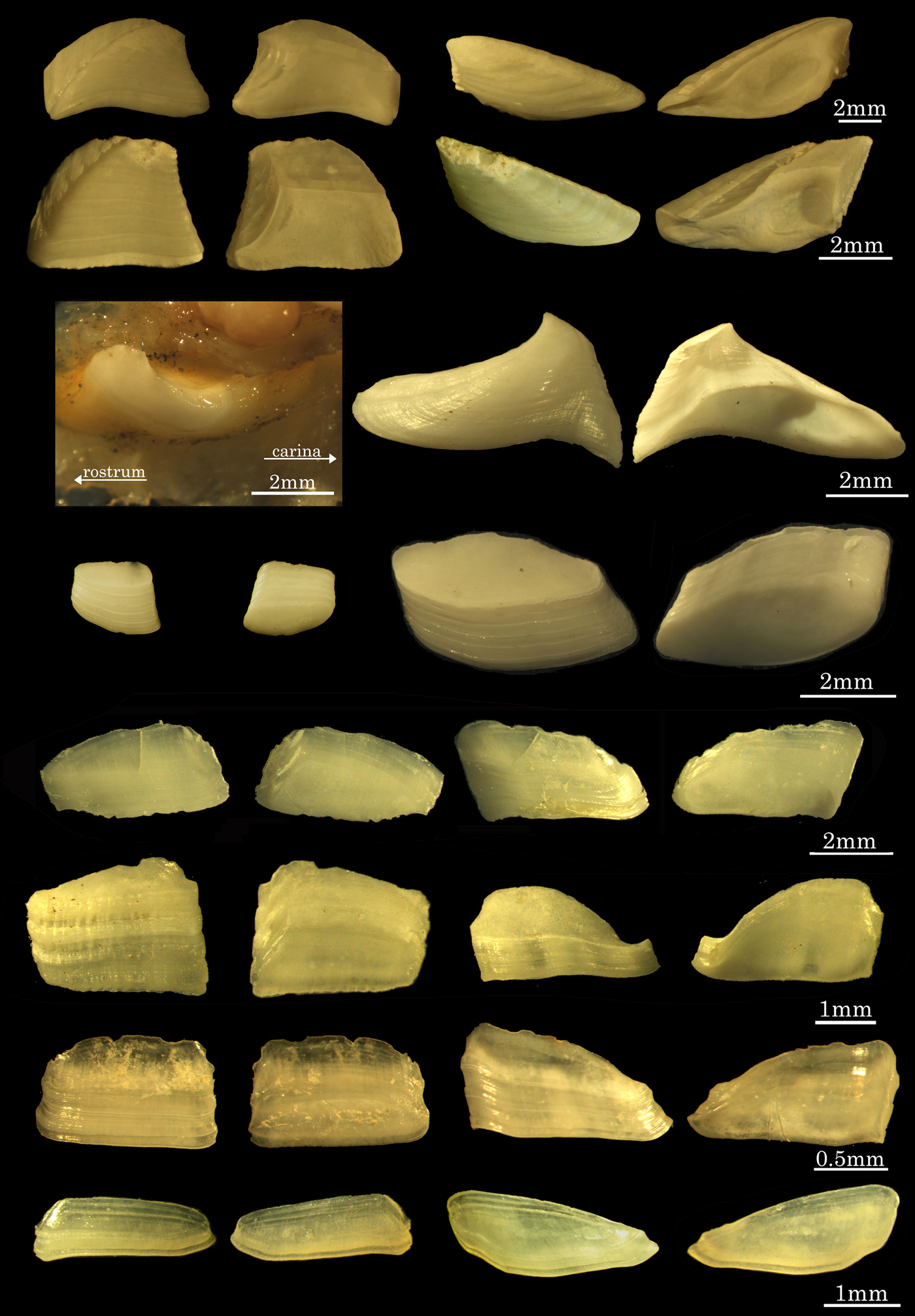
Opercular plates of Coronuloidea (surface of tergum, back of tergum, surface of scutum, back of scutum): (A) Chelonibia testudinaria; (B) C. caretta; (C) Coronula diadema (tergum absent); (D) Cryptolepas rhachianecti; (E) Tubicinella cheloniae; (F) Cylindrolepas darwiniana; (G) C. sinica; (H) Stomatolepas praegustator.
MATERIALS EXAMINED
Otsuchi, Iwate, 24 August 2006, from loggerhead turtle (SCL 71.4 cm, SCW 58.8 cm, Tag No. 47117 and 47118) (RUMF-ZC-02035); Otsuchi, Iwate, 24 August 2006, from loggerhead turtle (SCL 70.7 cm, SCW 60.0 cm, Tag No. 47125, 47126) (RUMF-ZC-02036); (RUMF-ZC-02037). New form: Ishigakijima Island, Okinawa, 3 August 2004, from green sea turtle (RUMF-ZC-02038); Ginoza, Okinawa, 18 November 2002, from green sea turtle (RUMF-ZC-02039; 02043); Ginoza, Okinawa, from green sea turtle (RUMF-ZC-02044).
DESCRIPTION
In the case of penetrated into a scale (type-A): shell almost circular in horizontal section; shaped like deep bowl in lateral view; orifice larger than the basis; compartments slightly convex, with strong projections; parietes with a medial smooth area; basal margin of parietes smooth; sheath reaches the basis and has a few fine growth lines on the surface; scutum and tergum narrow.
In the case of embedded between scales (type-B): shell small, and oval in horizontal section; shaped like shallow bowl in lateral view; orifice larger than the basis; compartments convex, with bifid projections that join adjacent plates and grab to the host skin; parietes without a medial smooth area but smooth at basal margin; sheath reaches the basis and has a few fine growth lines on the surface; scutum and tergum narrow.
Cirrus I with unequal ramus; cirrus III resembling cirrus II. Labrum with a notch in the middle; having a few teeth on each side. Palps club-shaped, covered with fine setae. Mandible with 4 or 5 main teeth; no secondary tooth between 2nd and 3rd; 2nd and 3rd bifid; inferior tooth pectinated and covered with fine setae. Maxilla I having a notch on cutting edge; two strong spines above a notch and shorter spines below; upper and inferior edge covered with fine setae. Maxilla II globular and covered with fine setae.
NEW OCCURRENCES ON JAPANESE COAST
Ginoza, Okinawa (on Chelonia mydas and Caretta caretta); Ohgimi, Okinawa (C. mydas); Yomitan, Okinawa (on C. mydas and Eretmochelys imbricata); Yakushima Island, Kagoshima (on C. caretta); Otsuchi, Iwate (on C. caretta); Hahajima Island, Ogasawara, Tokyo (on C. mydas).
PREVIOUS RECORDS ON JAPANESE COAST
Yomitan, Okinawa (on C. caretta, Hayashi, Reference Hayashi2009).
HABITAT
Penetrated into a scale or embedded between scales on front flippers of sea turtles.
REMARKS
The parietes of type-B have a bifid projection in the longitudinal medial area, whereas those of type-A lack bifid projection and have the parallel projections and medial area smooth (Figure 14D). The new form was found on only green sea turtles, embedded between epidermal scales and plastrons and not penetrating the epidermal scales, while S. muricata was also found on loggerhead and hawksbill turtles, attached on the front flippers and penetrating the epidermal scales. Morphology of soft parts is almost the same on each form.
DISCUSSION
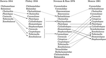
In the 19th Century, Charles Darwin classified the turtle barnacles into subfamily Balaninae (Darwin, 1854). Pilsbry (1916) divided the turtle barnacles into two subfamilies (Chelonibiinae and Coronulinae) and he regarded Coronulinae as formed by two series (the coronulid and platylepadid series) due to the presence of a median sulcus, midrib or a vestige on parietes and found on cetacea or not. Ross & Newman (Reference Ross and Newman1967) constituted a subfamily (Emersoniinae) that embraces the fossil species Emersonius cybosyrinx. Newman & Ross (Reference Newman and Ross1976) upgraded Pilsbry's two series to subfamilial level (Coronulinae and Platylepadinae) and included these subfamilies into the family Coronulidae with Chelonibiinae and Emersoniinae. Coronulidae was included in superfamily Coronuloidea with Tatraclitidae and Bathylasmatidae (Newman (Reference Newman and Forest1996) raised Newman & Ross (1976) subfamilies to familial level and organized the tetraclitids and bathylasmatids to Tetraclitoidea). Monroe (Reference Monroe1981) rejected the Platylepadinae and resurrected the subfamily Xenobalaninae Gruvel 1903, as Pilsbry's definitions of platylepadid series for shell morphology are clearly lacking in Stomatolepas and Stephanolepas. Monroe included Stomatolepas and Stephanolepas in Xenobalaninae with Xenobalanus and Tubicinella, while Platylepas and Cylindrolepas were included in Coronulinae with Coronula, Cetopirus and Cryptolepas (Figure 16). After a few decades, Ross & Frick (Reference Ross and Frick2007) rearranged the systematics of Coronuloidea in ERC (2007), however, they do not represent a published work according to the International Code of Zoological Nomenclature 4th edition (see Article 9), and hence the nomenclatural acts contained in the electronic version are not available under the Code from the work (Cylindrolepadinae: Ross & Frick, Reference Ross and Frick2007; Stomatolepadinae: Ross & Frick, Reference Ross and Frick2007; Chelolepadinae: Ross & Frick, Reference Ross and Frick2007; Cryptolepadinae: Ross & Frick, Reference Ross and Frick2007; Tubicinellinae: Ross & Frick, Reference Ross and Frick2007 are the void nomenclatures). Furthermore, barnacles are known to be phenotypically plastic in their shell form based on their substrate, habitat and density (Anderson, Reference Anderson1994; Lively et al., Reference Lively, Hazel, Schellenberger and Michelson2000), the importance of the soft parts morphology should be attended to for existing barnacle taxonomy (without fossil species), however, its description is currently insufficient in ERC (2007). In this study, the lack of information of the soft part morphology of Coronuloidea is resolved. The barnacles attached on Cetacea (Coronula, Cryptolepas and Xenobalanus) have short and thick cirri. It should be an adaptation for activity of host animals (e.g. high swimming speed, bleaching and leaping of Cetacea). Also, whale barnacles have the reduced opercular plates. Presumably, there is no need to guard themselves from predators on whale body. On sea turtle body, many other ectoparasites are recorded (e.g. molluscs (Frazier et al., Reference Frazier, Margaritoulis, Muldoon, Potter, Rosewater, Ruckdeschel and Salas1985); tunicates (Frazier et al., Reference Frazier, Goodbody and Ruckdeschel1991); bryozoa (Frazier et al., Reference Frazier, Winston and Ruckdeschel1992); copepods (Lazo-Wasem et al., Reference Lazo-Wasem, Pinou, Niz, Salgado and Schenker2007); marine leeches (Choy et al., Reference Choy, Balazs and Dailey1989; Greenblatt et al., Reference Greenblatt, Work, Balazs, Sutton, Casey and James2004); crabs (Davenport, Reference Davenport1994; Dellinger et al., Reference Dellinger, Davenport and Wirtz1997)). Particularly, crabs behave as predator for barnacles (Brousse & Baglivo, Reference Brousse and Baglivo2005; Brousse & Goldberg, Reference Brousse and Goldberg2007). While, the whale louse are recorded as ectoparasites on Cetacea, they eat the host's skin (Rowntree, Reference Rowntree1996).
Historical accounts of superfamily Coronuloidea.

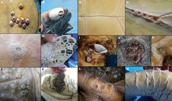
In the present study 13 species of the superfamily Coronuloidea were discovered, four of these being newly reported in Japanese waters (Chelonibia caretta, Platylepas decorata, C. darwiniana and Tubicinella cheloniae). On sea turtles, C. testudinaria attached to hard parts areas (carapace and plastron; Figure 17A), and a few individuals attached onto the posterior flippers (Figure 17B). Platylepas hexastylos attached on all over the turtle body (Figure 17C, D), while P. decorata attached onto the soft skin around the neck, tail and flippers (Figure 17E). Platylepas decorata frequently occurs with Cylindrolepas sinica (Figure 17F). Numerous S. transversa and C. sinica were found in the sulcus of green sea turtle plastron (Figure 17G, H). Cylindrolepas darwiniana is rare in Japanese waters (found only on one loggerhead sea turtle), and was found burrowed into the marginal plastron (Figure 17I). Tubicinella cheloniae burrowed into the carapace and plastron (Figure 17J, K). Stephanolepas muricata (type-A) penetrated into the scales of front flippers (Figure 17L), while type-B embedded in the sulcus of the scales (Figure 17M). Here I report the epibiont barnacles on three species of sea turtles (Caretta caretta, Chelonia mydas and Eretmochelys imbricata) that are known to migrate and lay eggs on the Japanese beaches. The leatherback turtle (Dermochelys coriacea) and olive ridley turtle (Lepidochelys olivacea) are also known to migrate around Japanese waters, without landing to reproduce (Abe & Minami, Reference Abe and Minami2008). If these turtles could be investigated, additional species will be discovered as the special parasites on leatherback turtle (D. coriacea), such as Platylepas coriacea and Stomatolepas dermochelys (Monroe & Limpus, Reference Monroe and Limpus1979).
(A) Chelonibia testudinaria on plastron of green sea turtle; (B) C. testudinaria on posterior flipper of green sea turtle; (C) Platylepas hexastylos on plastron of loggerhead sea turtle; (D) P. hexastylos on posterior flipper of green sea turtle; (E) P. decorata on soft skin around posterior flipper of green sea turtle; (F) P. decorata and Cylindrolepas sinica on tail of green sea turtle; (G) Stomatolepas transversa (left) and Cylindrolepas sinica (right) in median sulcus of green sea turtle's plastron; (H) S. transversa in the sulcus of scales on green sea turtle's plastron; (I) Cylindrolepas darwiniana occurred with Lepas spp.; found on marginal plastron of loggerhead seaturtle; (J) Tubicinella cheloniae burrowing into the carapace of loggerhead sea turtle; (K) Stephanolepas muricata (type-A) burrowed into the front flipper's scale of loggerhead sea turtle; (M) S. muricata (type-B) embedded between the front flipper's scales of green sea turtle.

Previous studies have suggested the utility of epibiont organisms of sea turtles as a tool for revealing their migratory history (Killingley & Lutcavage, Reference Killingley and Lutcavage1983; Caine, Reference Caine1986; Hayashi & Tsuji, Reference Hayashi and Tsuji2008; Hayashi, Reference Hayashi2009; Fuller et al., Reference Fuller, Broderick, Enever, Thorne and Godley2010). In Japanese waters, dugongs, some species of sea snakes and cetaceans are also known to distribute. Thorough research on barnacles and other organisms will form a useful knowledge base about marine vertebrate biology and ecology.
ACKNOWLEDGEMENTS
I thank the many people who generously helped me in various ways during the course of this study. These include the fishermen of Kanna and Toya fishery port in Okinawa, the Hahajima fishery port in Tokyo and fishermen in Iwate prefecture (Fisheries Cooperative Association of Funakoshi Bay, Hiroto, Miyako, Okirai, Omoe and Sanriku-Yamodo) for providing the live sea turtles. The Sea Turtle Association of Japan, the Nippon-Koei Co., Ltd. Okinawa office, the Entomological laboratory and Fujukan of University of Ryukyus, Ever Lasting Nature of Asia, the club NOAH Hahajima, Ogasawara Marine Center, Chura-Umi Aquarium of Okinawa, Echizen Matsushima Aquarium, Seikai National Fishery Research Institute of Ishigakijima Island, National Museum of Nature and Science Tokyo, Institute of Cetacean Research of Japan, Tomaya of Naha, Seto Marine Biological Laboratory of Kyoto University, Aquatic Conservation Laboratory and International Coastal Research Center of University of Tokyo, Historical Geology and Palaeontology laboratory of Chiba University, Whale Watching Tour of Zamamijma Island and the numerous volunteer staff of Yakushima Umigame-kan who aided RH in both field work and observations. My additional thanks Kunio Komesu, Muneyuki Kayo, Kouichi Hirate, Motoki Wakatsuki, Kazuki Tsuji, Hidetoshi Ota, Mamoru Toda, Hiroyuki Suganuma, Shinichi Tanaka, Koji Narushima, Mutsumi Kaneko, Manami Yamaguchi, Osamu Abe, Kunihiro Watanabe, Shunichi Takuma, Tomoko Narazaki, Katsufumi Sato, Shingo Kimura, Junichi Okuyama, Mitsuru Moriguchi, Haruka Sato, Nao Ozaki, Youichi Kitaji, Mari Nishizawa, Yukiko Yoshita, Shinnosuke Teruya, Akiko Fujiwara, Mitsuhiko Asakawa, Masanori Shinohara, Chisato Nakatsuka, Fujimi Fukuhara, Yukihisa Omuta, Tadasu K. Yamada, Yuko Tajima, Akiko Yatabe, Kenichiro Fujita, Masayuki Ishii, Masahiro Aizawa, Takeharu Bando, Nana Takanawa, Kazunobu Kogi, Akio Yamamoto, Yukiko Inoue, Naoko Takashima, Tadashi Yonamine, Yoshihiro Hayashi, Yuzo Ota, Ren Hirayama, Hideaki Nishizawa, Mari Kobayashi, Yoshiyuki Hisatsune, Takao Momosaki, Kotoe Sasomori, Hisashi Yamamoto, Mitsuharu Kume, Yukiko Fujiwara, Ken Okamoto, Sachi Yamaguchi, Megumi Ishizuna, Yoshie Yoshioka, Urara Kuratani, Sakana-kun and Masahito, Prince of Japan for aiding this research and providing study equipments and materials. Professor William A. Newman of Scripps Institution of Oceanography, Dr Hirotaka Tanaka of University of Ryukyus, Dr Romanus E. Prabowo of University of Jenderal Soedirman, Dr Benny K.K. Chan of Academia Sinica of Taiwan and Takuho Shuto of Atomosphere and Ocean Research Institute of University of Tokyo for their valuable advice and kindness.