Introduction
Stereotactic radiosurgery (SRS) has become an effective tool in the treatment of intracranial lesions with brain metastases being the most common indication. Reference Sheehan, Yen, Lee and Loeffler1 Techniques utilising Gamma Knife (Elekta AB, Stockholm, Sweden), CyberKnife (Accuray, Sunnyvale, CA) and linac-based approaches have all been shown to be effective. Reference Lippitz, Lindquist, Paddick, Peterson, O’Neill and Beaney2 SRS delivers high, conformal, ablative doses combined with a sharp dose fall-off to precisely target the tumour using image guidance. Due to the high precision of the delivery techniques and small target size, doses can be delivered in a single fraction. Reference De Salles, Gorgulho, Pereira and McLaughlin3 However, single fraction high dose treatments can lead to prolonged treatment times which may increase discomfort for patients and the potential for movement during treatment. Reference Hoogeman, Nuyttens, Levendag and Heijmen4 Modern linacs are equipped with flattening filter free (FFF) modes which enables a much higher dose rate compared to conventional beams with a flattening filter (FF) allowing faster beam delivery. The primary function of the FF is to produce a homogeneous dose profile. However, due to the small field sizes associated with SRS and improved treatment planning system modelling, this is less important. In addition, removing the FF may produce dosimetric advantages due to decreased scatter, less head leakage and less multi-leaf collimator (MLC) transmission all of which contribute to peripheral dose. Reference Cashmore5–Reference Fogliata, Fleckenstein and Schneider8
There is a clear interest to move towards using FFF for SRS. Reference Dimitriadis, Kirkby, Nisbet and Clark9 A recent UK audit of 21 centres providing intracranial SRS showed that 8 centres were using linac-based methods with only one centre currently using FFF (10 MV FFF) but with three of the eight centres planning to use FFF within 2 years. Reference Dimitriadis, Kirkby, Nisbet and Clark9 Several authors have investigated the use of 6 MV FFF for SRS treatments mostly using volumetric arc therapy (VMAT) and have shown that a significant reduction in beam-on-time (BOT) and improved dose gradients are achievable with comparable conformity. Reference Rieber, Tonndorf-Martini and Schramm10–Reference Stieler, Fleckenstein, Simeonova, Wenz and Lohr12 Abacioglu et al. have compared 10 MV FFF non-coplanar VMAT for a Varian TrueBeam linac to GammaKnife treatments for 12 patients with either vestibular schwannoma or cavernous sinus meningioma. The results showed that 10 MV FFF VMAT was comparable in terms of coverage, homogeneity and organ at risk (OAR) sparing with some gains in treatment efficiency. Reference Abacioglu, Ozen and Yilmaz13 Abacioglu et al. state that dosimetrically equivalent plans could have been obtained using 6 MV FFF compared to 10 MV FFF; however, no evidence is presented to support this.
Gasic et al. compared 6 MV FF to 6 MV FFF and 10 MV FF to 10 MV FFF using VMAT delivered on Varian TrueBeam for a range of tumour sites, 20 of which included intracranial single fraction metastases. They showed no statistical difference in conformity or OAR dose between FF and FFF for both 6 MV and 10 MV. However, a statistically significant reduction in the volume of healthy brain tissue irradiated for 10 MV FFF compared to 10 MV FF was noted although this was less than 0·1%. Reference Gasic, Ohlhues and Brodin14 Laoui et al. evaluated plan quality for multi-target SRS between 10 MV FFF and 6 MV FFF VMAT and found that 6 MV FFF provided a steeper dose gradient and less normal brain tissue volume irradiation. Reference Minniti, Capone and Alongi15 None of the studies specifically investigating intracranial lesions directly compare 10 MV FFF with 6 MV and 6 MV FFF with the same modality highlighting a paucity in the literature comparing the three most viable beam energies for linac-based intracranial SRS. Reference Dimitriadis, Kirkby, Nisbet and Clark9
This study investigates the use of 6 MV non-coplanar dynamic conformal arcs to treat intracranial brain metastases using a Varian TrueBeam STx (Varian Medical Systems, Palo Alto, CA). Beam data for 6 MV FFF and 10 MV FFF has been commissioned which would allow an increase in maximum dose rate compared to the clinically used 6 MV FF. A planning study was conducted to assess the plan quality and delivery efficiency of 6 MV FFF and 10 MV FFF compared to 6 MV FF for intracranial brain metastases single fraction SRS.
Materials and Methods
Patients
In total 16 patients, combining 21 intracranial metastases were treated with a single fraction prescription dose between 15 and 21 Gy prescribed to the 80% isodose. Of the total number of patients, 12 had a single metastasis, 3 had two metastases and one had three metastases, the multiple metastatic treatments had an individual plan for each metastasis. These plans were selected based on patients randomly selected from a retrospective cohort (Rec Ref: 20/HSC/0002). All patients had a 1·5T T1 weighted post gadolinium contrast enhanced magnetic resonance imaging (MRI) (Philips Healthcare, Amsterdam, Netherlands), slice thickness of 2 mm reconstructed to 1 mm and a planning computed tomography (CT) (GE Healthcare, Chicago, Illinois) with slice thickness of 1·25 mm. The MRI was fused to the CT within the iPlan (BrainLab, Munich, Germany) treatment planning system (TPS) to aide tumour and OAR delineation. Patients were immobilised in a custom fitted BrainLab frameless SRS mask (BrainLab, Munich, Germany) for both the planning CT and treatment.
Treatment planning
A planning target volume (PTV) was created for each tumour by symmetrically expanding the gross tumour volume (GTV) with a 1 mm margin Reference Kirkpatrick, Wang and Sampson16 giving a mean PTV volume and SD of 3·91 ± 4·07 cm3. All plans were created using non-coplanar dynamic conformal arcs for a Varian TrueBeam STx (Varian Medical Systems, Palo Alto, CA) linac equipped with the HD Millennium MLC with 120 leaves (0·25 cm leaf width for the inner 20 and 0·5 cm leaf width for the outer 2 × 20). All plans consisted of 3–5 non-coplanar arcs arranged to avoid critical structures if possible. For multi-target cases, beams were arranged to avoid other PTVs. Optimisation was performed through arc length, collimator angle and MLC margin adjustments to obtain PTV coverage dose to 99% of the volume (D99%) greater than the prescription dose while maintaining all OAR constraints Reference Lawrence, Li and Naqa el17–Reference Mayo, Yorke and Merchant19 and plan quality metrics.
The dose conformity to the PTV and the dose gradient of the plans were expressed through the following indices: dose conformity index (CI) = 1 + VNT(PD)/VPTV(PD). VNT(PD) is the volume of normal tissue receiving the prescription dose and VPTV(PD) is the volume of the PTV receiving the prescription dose. The local CI tolerance is 1–1·45 (PTV volume > 4 cm3) and 1–1·79 (PTV volume < 4 cm3). The Ian Paddick conformity index (IPCI) Reference Paddick20 was also used as a measure of dose conformity to the PTV and is expressed as: IPCI = V2 PTV(PD)/(VPTV*VTot(PD)) where V2 PTV(PD) is the square of the volume of PTV receiving the prescription dose, VPTV is the PTV volume and VTot(PD) is the total volume receiving the prescription dose. The local IPCI tolerance is 0·69–1 (PTV volume > 4 cm3) and 0·48–1 (PTV volume < 4 cm3). The dose gradient of the plans was expressed in terms of the gradient index (GI) Reference Paddick and Lippitz21 : GI = V50%(PD)/VTot(PD) where V50%(PD) is the volume receiving 50% of the prescription dose and VTot(PD) is the total volume receiving the prescription dose. The local GI tolerance is 5. Furthermore, the volume receiving 12 Gy (V12 Gy) for normal brain (brain-GTV) was assessed for each plan with local optimal tolerance of 10 cm3. For multiple metastatic cases the plan metrics were evaluated independently of the other metastases.
All plans were originally generated for 6 MV FF with a maximum dose rate of 600 MU/minute and were clinically acceptable. Retrospectively, each plan was re-optimised with the 6 MV FFF and 10 MV FFF dataset which allowed an increase in the maximum dose rate to 1,400 MU/minute and 2,400 MU/minute respectively. For the PTVs the dose received by 99% of the volume (D99%) and the maximum dose within the PTV (Dmax) were evaluated as a percentage of the prescription dose (80% isodose). The CI, IPCI, GI and V12 Gy were also evaluated for each plan to assess plan quality while the monitor units (MU) and BOT obtained from the TPS were evaluated to assess delivery efficiency.
It was recognised that BOT is only one part of the overall treatment process and therefore the treatment time, including verification imaging and gantry rotation between arcs, for each patient was evaluated by assessing the time of the first verification image and end of the last treatment beam where available.
All 6 MV FF plans went through the pre-treatment patient specific quality assurance (QA) process and were delivered clinically. In order to ensure deliverability for the 6 MV FFF and 10 MV FFF, a test plan was calculated for each beam energy on to the CIRS STEEV (CIRS Inc., Norfolk, VA) pre-treatment QA phantom. Each plan was delivered to the phantom with EBT XD Gafchromic film (Ashland Inc., Wayne, NJ) placed in the isocentre plane to assess the delivered isocentre absolute dose. Dose distribution analysis was also performed using the FilmQAPro (Ashland, Inc., Wayne, NJ) software using a local gamma criterion of 3%/2 mm with a 20% threshold. Reference Dang, Peters, Hickey and Semciw22 All films were scanned 24 hours after irradiation using the Epson Expression 10000XL flatbed colour scanner (Seiko Epson Corp., Nagano, Japan) in transmission mode, 48 bits RGB and 150 dpi resolution without any colour corrections. Film was calibrated using the TrueBeam photon beam which has been calibrated traceable to the UK primary standard (National Physics Laboratory—Teddington, UK). Each calibration film was positioned at the centre of the field of 10 × 10 cm at the depth of 5 cm in a WTe solid water phantom (Barts and The London NHS Trust, London, UK) with 100 cm source to surface distance. The overall uncertainty in terms of absolute dose for the film measurement was calculated as the square root of the sum of the squares of all the individual uncertainties. These include: calibration of linear accelerator output for film calibration, calibration of linear accelerator output for film plan irradiation, film calibration fit function for creation of dose map in FilmQA Pro software, TPS calculation, positional uncertainty and scanning of film. The overall uncertainty (coverage factor K = 1) was estimated to be 3·3%. Reference Mohamed Yoosuf, Jeevandram, Whitten, Workman and McGarry23
The evaluated data are presented as median values and ranges with statistical analysis conducted in MATLAB (v9·2-R2017b) (Mathworks Inc., Massachusetts, USA) using the two-sided paired-sample Wilcoxon signed rank test with p values < 0·05 considered significant.
Results
Table 1 shows all the metrics used for evaluation with statistical comparison made between 6 MV FF and both FFF plans as well as a comparison between the two FFF plans. Statistical analysis shows a significant difference in PTV dose coverage when compared to the 6 MV FF plans; however, all plans were deemed acceptable in terms of coverage and the difference was not deemed clinically significant. 10 MV FFF shows an increase in the median maximum dose when compared to both 6 MV FF and 6 MV FFF with 6 MV FF and 6 MV FFF being comparable. Figure 1a shows the distribution for PTV coverage assessed by D99% for each beam energy highlighting that while the median values are similar there was a greater spread for 6 MV FF. Figure 1b shows that the increase in Dmax was observed throughout the distribution for 10 MV FFF but all plans were within the local tolerance of 105%. Both conformity indices, CI and IPCI, show no difference between 6 MV, 6 MV FFF and 10 MV FFF.
Median and range of dosimetric parameters for treatment plans using 6MV, 6MV FFF and 10MV FFF.

Abbreviations: FFF, flattening filter free; D99%, dose as a percentage of prescription (80%) covering at least 99% of PTV; Dmax, maximum dose in PTV as percentage of prescription (80%); CI, Conformity index; IPCI, Ian Paddick Conformity index; GI, Gradient index; V12Gy, Volume of normal brain receiving 12Gy; MU/Gy, monitor units per Gray; BOT, Beam-on-time.
(a) D99% comparison between energies. D99%: Dose to 99% of PTV volume as a percentage of prescription showing the median, 25th and 75th percentile and the whiskers which extend to the most extreme data points which are considered non-outliers. Black dots indicate outliers in the 5/95th percentile. (b) Dmax comparison between energies. Dmax: Maximum dose in PTV as a percentage of prescription showing the median, 25th and 75th percentile and the whiskers which extend to the most extreme data points which are considered non-outliers. Black dots indicate outliers in the 5/95th percentile.

Table 1 shows comparable gradient index values between 6 MV FFF and 6 MV FF. 10 MV FFF shows an increase in median dose gradient when compared to 6 MV (2·74 to 3·05; +11·3%; p = 0·001) and 6 MV FFF (2·67 to 3·05; +14·2%; p < 0·001). Figure 2a highlights the trends in the distribution between the energies. The variation in dose gradient between the energies manifests itself in the v12 Gy constraint for Brain-GTV with similar trends observed as shown in Figure 2b. There is an approximately 0·5 cm3 (11·4%; p < 0·001) increase in the median volume of normal tissue receiving 12 Gy for 10 MV FFF compared to both 6 MV FF and 6 MV FFF.
(a) GI comparison between energies. GI: Gradient index showing the median, 25th and 75th percentile and the whiskers which extend to the most extreme data points which are considered non-outliers. Black dots indicate outliers in the 5/95th percentile. (b) V12Gy comparison between energies. V12Gy: Volume of normal brain (brain-GTV) receiving 12Gy showing the median, 25th and 75th percentile and the whiskers which extend to the most extreme data points which are considered non-outliers. Black dots indicate outliers in the 5/95th percentile.

BOT for each met when using 6 MV FF, 6 MV FFF and 10 MV FFF.

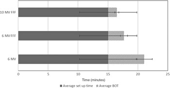
Average total treatment time when using 6 MV FF, 6 MV FFF and 10 MV FFF showing break down between set up time and BOT.

Similarly, no significant difference was observed between 6 MV FF and 6 MV FFF in the number of monitor units per gray required with a decrease in median value of 18 MU/Gy observed for 10 MV FFF plans. FFF significantly reduces the median BOT by 3·7 minutes (57·9%; p < 0·001) for 6 MV FFF and 4·9 minutes (76·3%; p < 0·001) for 10 MV FFF (Figure 3). In total there were 80 arcs, all 6 MV and 6 MV FFF arcs utilised the maximum dose rate available. For 10 MV FFF, 28 of the arcs did not use the full 2,400 MU/minute dose rate as they were limited by the gantry rotation speed of 1 minute/360°.
Data for 18 of the 21 treatments were available to assess the overall treatment time. Figure 4 shows that the average total treatment time for 6 MV treatments from first verification image to the end of the last beam was 21·0 ± 5·1 minutes. This equates to an estimated average reduction in overall treatment for 6 MV FFF of 16·8% and 21·5% for 10 MV FFF compared to conventional 6 MV beams.
The absolute measured isocentre dose was compared to the TPS calculated isocentre dose with the percentage difference being less than 1% for both plans. The dose distribution in the isocentre plane was assessed using gamma analysis with both plans showing greater than 95% of pixels passing at 3%/2 mm with a 20% threshold.
Discussion
This study aimed to build on the work by Lai et al. Reference Youqun, Shanyu and Changdong11 , Rieber et al. Reference Rieber, Tonndorf-Martini and Schramm10 and Laoui et al. Reference Minniti, Capone and Alongi15 by evaluating 10 MV FFF alongside 6 MV FFF for single fraction intracranial SRS specifically using non-coplanar dynamic conformal arcs compared to a 6 MV flattened beam. The FFF option on modern linacs is appealing to use for high dose single fraction treatments due to the increased dose rate which can reduce treatment times. FFF treatments are already in widespread use for extracranial stereotactic radiotherapy. Reference Fogliata, Fleckenstein and Schneider8,Reference Gasic, Ohlhues and Brodin14,Reference Lang, Shrestha and Graydon24,Reference Hsu, Lai, Jeng and Tseng25
All plans were individually optimised and evaluated against standard constraints. In practice, for a dynamic conformal arc method, this means optimising gantry arcs, collimator angles, couch angles and MLC positions. This is in contrast to a full inversely optimised intensity modulated approach in which the set optimisation objectives influence the dose distribution which may to some degree mitigate against the dosimetric effects of the different beam energies. For cases with more than one metastases the plans were optimised on an individual basis with the added constraint of having no beams passing through any of the other metastases. This may have an effect when comparing with other studies in which only one metastases is planned as some beam angles may be compromised but will still allow for direct comparison between energies within this study as the same data set is used.
Statistically the D99% PTV coverage of the FFF plans were different compared to the 6 MV FF plans; however, when evaluating the boxplots in Figure 1a it is clear that the median values are similar with differences observed in the range of results. This may be explained when evaluated in conjunction with the results for the maximum dose in Figure 1b. The local protocol was to keep the maximum dose less than 105% which proved to be more difficult with 10 MV FFF as is noted with the higher median Dmax and greater range, in order to keep the maximum value less than 105% the 80% isodose coverage was pushed down as close to tolerance as possible hence reducing the range of D99% values. Lai et al. Reference Youqun, Shanyu and Changdong11 observed a small decrease in the maximum dose for 6 MV FFF VMAT compared to 6 MV FF; however, this is most likely due to certain planning constraints applied in the inverse optimisation process which aim to keep the maximum dose at a desired level.
No significant difference was observed between the 6 MV FF plan and the 6 MV and 10 MV FFF plans for conformity. Lai et al. Reference Youqun, Shanyu and Changdong11 investigated the difference between 6 MV FF and 6 MV FFF VMAT SRS plans and also found no difference in conformity. Rieber et al. Reference Rieber, Tonndorf-Martini and Schramm10 used a mix of 3D conformal, IMRT step and shoot and VMAT techniques to investigate 6 MV FF and 6 MV FFF and noted slight superiority in conformity of 1·3% (p = 0·001) for FF plans. Laoui et al. Reference Minniti, Capone and Alongi15 investigating 10 MV FFF and 6 MV FFF VMAT plans also found the conformity to be comparable which is in agreement with the results in this study. Consistency between the plans for both PTV coverage and conformity indicates that the plans are comparable in terms of dose distribution to the target as these can often be a trade-off with organ at risk dose.
The beam quality index characterises the energy of the beam which gives an indication of how penetrating it will be. A beam with a higher quality index will be more penetrating and hence may contribute to higher peripheral dose. The gradient index is a measure of how quickly the out-of-field dose decreases away from the target edge and hence is linked to the dosimetric characteristics of the beam. Rieber et al. Reference Rieber, Tonndorf-Martini and Schramm10 using an Elekta FFF beam showed an improved dose gradient of 29·6% for 6 MV FFF when compared to 6 MV FF while Lai et al. Reference Youqun, Shanyu and Changdong11 using Varian FFF beams reported an improvement of 1·9% which was similar to the 2·6% reduction observed in this study for 6 MV FFF when compared to 6 MV FF although not statistically significant. The greater improvement observed by Rieber et al. may be related to the different treatment techniques used which included 3D conventional, step and shoot intensity modulated radiotherapy and VMAT. An improved dose gradient is indicative of less peripheral dose and should lead to improved OAR and normal brain doses. In contrast, the more penetrating beam energy of 10 MV FFF leads to an increase in the median dose gradient of 11·3% (p = 0·001) when compared with the 6 MV FF plans which may lead to an increase in peripheral dose at distance.
Abacioglu et al. Reference Abacioglu, Ozen and Yilmaz13 compared VMAT SRS using 10 MV FFF generated by a TrueBeam linac to Gammaknife and found that Gammaknife had superior dose gradient. Using a VMAT technique Abacioglu et al. were able to achieve an average gradient index of 3·8 ± 0·6 for 10 MV FFF Reference Abacioglu, Ozen and Yilmaz13 which is higher than the median dose gradient values for the 10 MV FFF plans in this study. Hsu et al. Reference Hsu, Lai, Jeng and Tseng25 evaluated the dose gradient for 6 MV cone based SRS versus 6 MV FFF VMAT delivered using Elekta linacs and found an improved dose gradient for 6 MV cone based treatment; however, they conclude that this is related to the use of cones for shaping the beam rather than an energy effect. Dang et al. Reference Dang, Peters, Hickey and Semciw22 in a review of ten studies evaluating the use of flattening filter free for stereotactic body radiotherapy reported that there were minimal differences in conformity but some improvement in dose gradient for FFF which is in agreement with much of the current literature and this study.
The importance of a steep dose gradient is to reduce the dose spillage to surrounding healthy tissues. The v12 Gy dose constraint in brain SRS treatments is used as a predictor of radiation necrosis post treatment. Reference Blonigen, Steinmetz, Levin, Lamba, Warnick and Breneman26 The inferior dose gradient observed with 10 MV FFF correlates with the inferior v12 Gy for (brain-GTV) for 10 MV FFF. Similarly the comparable dose gradient observed for 6 MV FFF compared to 6 MV FF translates to the same median V12 Gy between 6 MV and 6 MV FFF. However, both Rieber et al. Reference Rieber, Tonndorf-Martini and Schramm10 and Lai et al. Reference Youqun, Shanyu and Changdong11 observed a decrease in mean brain dose when moving to 6 MV FFF from 6 MV FF which is indicative of the improved gradient seen for 6 MV FFF although was not deemed statistical significiant in this study. Interestingly, Gasic et al. Reference Gasic, Ohlhues and Brodin14 reported a lower volume receiving 10–16 Gy using 10 MV FFF VMAT when compared to 10 MV FF VMAT for single fraction SRS brain targets delivered on Varian TrueBeam linacs; however, it was noted that the small reduction would most likely be considered as non-significant from a clinical point of view. Similarly, Laoui et al. found an improved dose gradient and normal tissue brain sparing with 6 MV FFF when compared to 10 MV FFF. The results from this study show a trend of comparable plan quality between 6 MV FF and 6 MV FFF with a reduction in quality when using 10 MV FFF in terms of dose gradient and normal brain dose which is in broad agreement with the studies mentioned. While removal of the flattening filter leads to a reduction in scatter and leakage radiation which contributes to the peripheral dose, Reference Kragl, af Wetterstedt and Knäusl27 it appears that this effect has minimal significance in this study. Due to the differences observed between the 6 MV FFF and 10 MV FFF, it is the change in beam energy spectrum which is significantly impacting the gradient index and v12 Gy normal brain doses.
A reduction in the total number of monitor units was noted for 10 MV FFF; however, in terms of beam delivery time savings, the main reduction is due to the higher dose rate available. This study has shown that BOT is significantly reduced with the use of FFF beams compared to FF by approximately 57·9 and 76·3% for 6 MV FFF and 10 MV FFF respectively. Several studies Reference Rieber, Tonndorf-Martini and Schramm10–Reference Stieler, Fleckenstein, Simeonova, Wenz and Lohr12,Reference Dzierma, Nuesken, Palm, Licht and Ruebe28,Reference Wang, Rice, Mundt, Sandhu and Murphy29 have found a reduction in BOT for 6 MV to 6 MV FFF of approximately 50% consistent to the results presented in this manuscript. Although 10 MV FFF shows a further reduction in BOT, 35% of the arcs used were limited by gantry rotation speed and hence cannot fully utilise the maximum dose rate. Lang et al. Reference Lang, Shrestha and Graydon24 investigated BOT reduction for 6 MV FFF and 10 MV FFF for extracranial lung and abdomen stereotactic radiotherapy delivered with VMAT and found that prescription doses above 4 Gy and 10 Gy per arc were required for 6 MV FFF and 10 MV FFF respectively in order to take advantage of the maximum dose rate available due to gantry speed. Gasic et al. Reference Gasic, Ohlhues and Brodin14 investigated the use of Varian TrueBeam FFF for VMAT including SRS brain mets, 20 single met plans were evaluated with a BOT reduction of 56·1 and 69·5% for 6 MV FFF and 10 MV FFF respectively again in good agreement with the results provided here.
There are a number of limitations to this study. Each of the plans were created with a single isocentre per single target (SIST). Recent literature has suggested a move towards single isocentre, multiple target (SIMT) treatments using advanced optimisation methodologies. Reference Minniti, Capone and Alongi15 SIST will utilise the centre of the beam which has the highest differential between dose-rates between FF and FFF. SIMT will sample a larger proportion of the beam which will result in areas off-axis being sampled where the dose-rate will be less. Hence, further work will analyse the impact of FFF beams on SIMT plan quality and delivery.
Clearly, significant gains in BOT can be obtained by using FFF for high dose single fraction treatments using SIST. However, the majority of time for an overall treatment is not the BOT but the time required for patient setup including verification imaging. An overall reduction in average total treatment time of 16·8 and 21·5% for 6 MV FFF and 10 MV FFF respectively was determined in this study. Lai et al. Reference Youqun, Shanyu and Changdong11 calculated mean treatment delivery time of 20·63 ± 0·83 minutes using VMAT, representing an average of 16·1% reduction for 6 MV FFF compared to 6 MV. Rieber et al. Reference Rieber, Tonndorf-Martini and Schramm10 again using VMAT observed a treatment time reduction of 27·8%. While the magnitude of the total time reduction due to the reduced BOT appears to be small it is still of clinical significance as it is during the actual beam on that unwanted motion is critical. For multiple dynamic conformal arcs verification imaging is still recommended between each arc where there is a couch movement to allow for corrections to be made sequentially throughout the treatment. Due to the fact that 6 MV FFF provided significant BOT reduction while maintaining comparable dose coverage and plan quality, we have implemented 6 MV FFF within our institution.
Conclusion
This study has shown that significant BOT improvements can be made using FFF which in turn should benefit the patient treatment. However, it was noted that despite the improved BOT reduction seen with 10 MV FFF these plans had a poorer dose gradient and normal brain tissue sparing when compared with 6 MV FF and FFF for equivalent plan parameters. 6 MV FFF provided significant BOT reduction while maintaining comparable dose coverage and plan quality.
Funding
This research did not receive any specific grant from funding agencies in the public, commercial, or not-for-profit sectors.
Acknowledgements
None.