Introduction
Wilms tumour (WT), also called nephroblastoma, is the most common renal neoplasm in the paediatric populationReference Ehrlich, Ritchey and Hamilton 1 and tends to appear between 1 and 5 years of life, with an average age of diagnosis of 44 months.Reference Dalul, Nicolás, Nicolás, Alberto, Diego and Rodolfo 2 , Reference Zapata, González and Pérez 3 In about 90% of patients presenting with WT, tumours are unilateral and 12% of these cases present with synchronous multifocal lesions and approximately 7% are bilateral.Reference Ferris, Berbel, Ortega, López, García and Balaguer 4 The incidence of WT is relatively uniform and stable in different continents, approximately 1–2 cases per year and per million inhabitants or 1/10,000 births.Reference Hernández Fernández 5
In Mexico, there is no precise data on WT incidence; however, it seems to have a higher frequency of presentation. The age of presentation ranges from 1 to 5 years, with a mean of 3·5 years; 90% occur in children under 7, it is rare in newborns, and there is no difference between gender. 6
Clinically patients typically present with a clinical presentation of an increased abdominal volume, and this may be accompanied by fever (23%), abdominal pain (37%), hypertension (25%) and haematuria (20%).Reference Beckwith and Palmer 7 – Reference Weirich, Ludwig and Graf 10
The treatment protocol is according to the National Wilms Tumor Study (NWST) and International Society of Pediatric Oncology protocol, radiotherapy is frequently delivered using conformal 3D modality with fields such as flank or hemi-abdomen and whole abdominal radiotherapy (WAR) with doses no greater than 1,080 cGy; and in cases with residual disease or thrombus in unresectable cava, the dose is increased up to 2,100 cGy.Reference D’Angio 11 – Reference De Kraker, Graf, van Tinteren, Pein, Sandstedt, Godzinski and Tournade 13
As new radiotherapy techniques become available, along with their improved technical accuracy, there is an improvement in the overall survival in WT; and given these patients are young and are still growing, it is very important to keep the dose to organs at risk (OAR) as low as possible. Keeping the dose as low as possible to the kidney, lungs and heart is going to reduce the late toxicity, renal insufficiency, ischemic cardiopathy and lung fibrosis and improve the quality of life of these patients.
The purpose of this study is to undertake a dosimetric comparison of whole abdominal treatment plans of patients diagnosed with stage III WT, to assess the benefits of treating these patients with either volumetric arch therapy (VMAT) or 3D conformal radiotherapy.
Material and methods
We realise a retrospective, cross-sectional study, including 23 patients diagnosed with WT who received treatment with WAR in the two modalities, 3D conformal or VMAT in the 2013–2017 period. The treatments were all planned using the Eclipse treatment planning system (Varian Medical Systems, Palo Alto, CA). A total of 23 patients were assessed, 14 treated with VMAT and 9 with 3D conformal radiotherapy.
Treatment plan volumes for the clinical target volume (CTV) included all the borders of peritoneum, and the planning target volume (PTV) included the CTV plus 5 mm axial and 10 mm cephalocaudal, according to the NWST. The dose for WAR was 1,080 cGy in all cases.Reference Halperin, Constine and Tarbell 14
The determination of the conformality index (CI), homogeneity index (HI) and coverage quality (CQ) was carried out according to the Radiation Therapy Oncology Group.Reference Van’t Riet, Mak and Moerland 15 , Reference Feuvret, Noël, Mazeron and Bey 16 We compared these indices for all patients treated with 3D conformal and VMAT. The statistical analysis was performed using the Mann–Whitney U test with the software GraphPad Prism 5·0.
Results
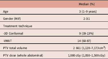
In this retrospective study, we analysed the treatment plans of 23 patients diagnosed with WT and referred for WAR due to the characteristics of the initial disease, surgical findings or characteristics of the definitive pathology report, during the period of March 2014 to March 2017. The epidemiological characteristics and treatment details of the patients are shown in Table 1.
Epidemiological and treatment features of the patients

Abbreviations: M, male; F, female; VMAT, volumetric arch therapy; PTV, planning target volume.
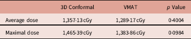
The comparison between the dosimetric parameters of 3D conformal and VMAT is shown in Table 2, using the Mann–Whitney U test, where the maximum dose to the PTV was observed to be significantly higher in the conformal 3D modality in comparison to VMAT.
Whole abdominal PTV

Abbreviations: VMAT, volumetric arch therapy; PTV, planning target volume.
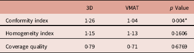
The dosimetric values of the 9 patients planned for 3D conformal techniques and 14 for VMAT are shown in Table 3, the same CQ was found, but the conformation index differed, were more favourable for the VMAT modality and both had the same HI.
Index

Abbreviation: VMAT, volumetric arch therapy.
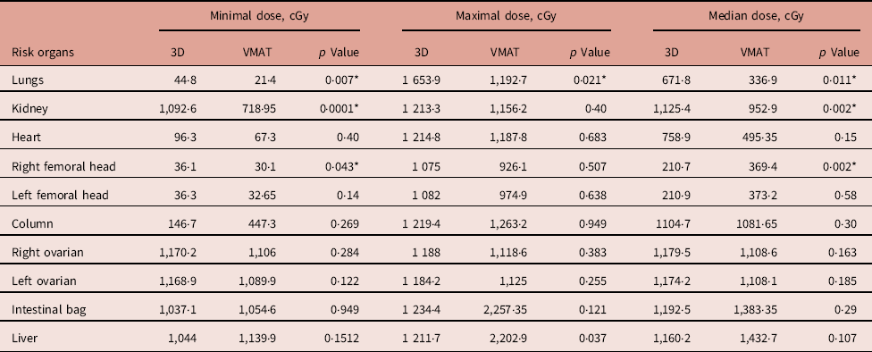
Regarding the assessment of doses to the OAR, higher values of minimum, maximum and average doses were found for the conformal 3D technique as well as a statistically significant difference between both techniques, showing an improvement in the VMAT group, as shown in Table 4.
Dose to the organ at risk

*Statistical significance.
Abbreviation: VMAT, volumetric arch therapy.
Discussion
With new radiotherapy treatment techniques such as VMAT, improved treatment plans for patients have been achieved by reducing the radiation dose to the OAR and increasing the conformation of the PTV. However, few studies have been conducted in the paediatric population to support the fact that the VMAT technique is a new standard of treatment.
In this study, the dosimetric comparison was performed in patients with WT for 3D conformal versus the VMAT technique. The dosimetric parameters for the whole abdominal PTV in the 3D technique and VMAT showed no statistical difference, showing that both treatments have the same quality coverage of the PTV and not showing statistical significance in the average doses. However, it was shown that VMAT had a statistically significant and better CI. The results show that VMAT reduces the average doses to the OAR, the lung, remaining kidney and heart, achieving a reduction in 50%, 15·32% and 34·69%, respectively.
The statistical analysis between the two techniques verified that this decrease is statistically significant for lung and kidney. No statistical significance was demonstrated for the other OAR.
This decrease in doses to OAR and improved PTV conformity, as compared with the 3D technique, has been reported in other studies in which a reduction in dose to the OAR with techniques such as VMAT or intensity modulated radiotherapy has been shown.Reference Newhauser and Durante 17 , Reference Chojnacka, Zygmuntowicz-Piętka, Semaniak, Pędziwiatr, Dąbrowski and Skowrońska-Gardas 18 Proton therapy has shown a greater advantage in decreasing the dose to the OAR; however, this modality is currently not available in developing countries. Therefore, the use of VMAT is the best available option to achieve a reduced dose to the OAR, in order to reduce late radiation toxicity and improve the quality of treatment for patients treated with WAR for a WT diagnosis.
The limitations of this study are the small number of patients included in the analysis and that it is a retrospective study.
Conclusion
This study proposes using the VMAT radiotherapy technique in the treatment of the whole abdomen in patients diagnosed with WT. The dosimetric analysis shows improved conformity, without affecting the quality of the PTV coverage compared with the 3D conformal technique. In addition, VMAT reduces the doses to OAR such as the remaining kidney and the lungs that are important to preserve to reduce the probability of radiation toxicity in these patients.
Acknowledgement
We are grateful for the collaboration of the Radiotherapy Service of the Mexican Social Insurance Institute for the support of this project. Also to Estefania Beltran and Estefania Olmos for their unconditional support.