1. Introduction
Ichthyosaurs are one of the groups of secondarily aquatic tetrapods most highly modified for an aquatic way of life. It was recognized as a unique lineage over 200 years ago, thanks in part to the collecting efforts of the Anning family (reviewed by Evans, Reference Evans, Moody, Buffetaut, Naish and Martill2010), and even the earliest interpretations of ichthyosaurian limbs regarded them as fish-like and specialized for a marine way of life (De la Beche & Conybeare, Reference De la Beche and Conybeare1821). The limbs of derived ichthyosaurs are still regarded as one of the most unique characteristics of this group, and receive a disproportionate amount of research attention from specialists for this reason (Johnson, Reference Johnson1979; Caldwell, Reference Caldwell1997a ,Reference Caldwell b ; Motani, Reference Motani1997, Reference Motani1998, Reference Motani1999a ; Maisch & Matzke, Reference Maisch and Matzke1998; Maxwell, Reference Maxwell2012). However, in addition to these papers examining morphology, homology and variation in the appendicular skeleton, the limbs of derived ichthyosaurs represent a morphological endpoint for the phenomena of hyperphalangy and hyperdactyly – that is, they represent a classic example of morphospace occupation that is dissimilar to anything naturally occurring in an extant taxon. This has led to the inclusion of this clade as a comparative point in studies on the genetics of limb patterning in extant tetrapods (e.g. Caldwell, Reference Caldwell2002; Fedak & Hall, Reference Fedak and Hall2004; Archer, Dowthwaite & Francis-West, Reference Archer, Dowthwaite, Francis-West and Hall2007; Zhu et al. Reference Zhu, Zhang, Alber and Newman2010). Relatively few studies consider the identity of the resulting elements (e.g. Wagner & Chiu, Reference Wagner and Chiu2001; Woltering & Duboule, Reference Woltering and Duboule2010). Element identity is an intriguing problem in the highly modified ichthyosaur limb, influencing both ingroup homology determination and understanding of the stability of proximodistal patterning in vertebrate limbs.
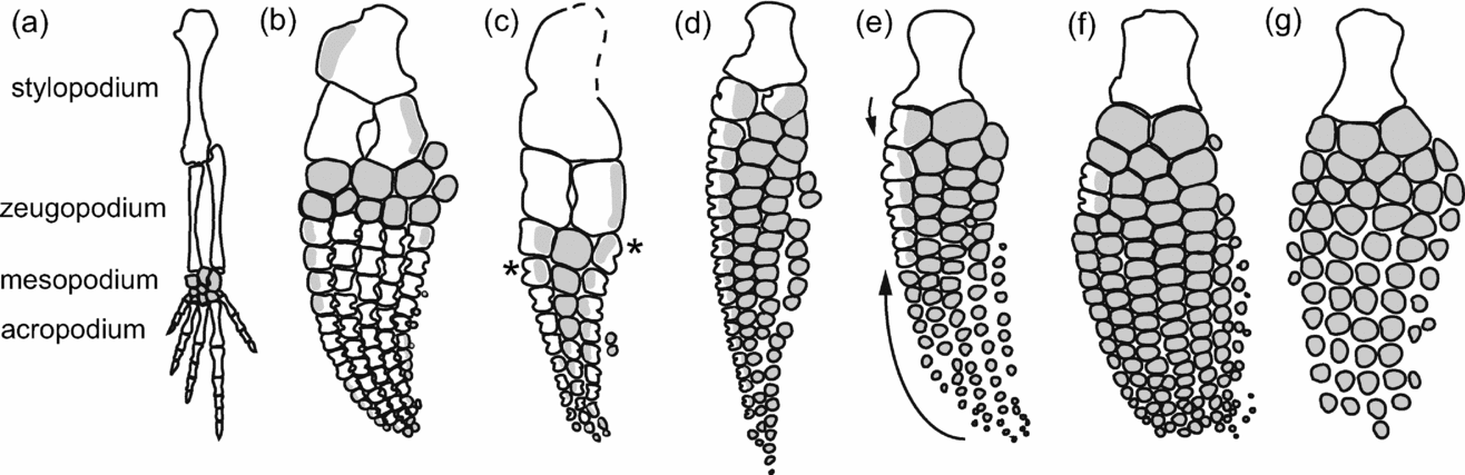
Although ichthyosaur limbs are extensively modified for aquatic locomotion, the typical tetrapod pattern is conserved in the most basal forms (Nicholls & Brinkman, Reference Nicholls, Brinkman and Sarjeant1995; Motani, Reference Motani1997). However, the proximal elements (stylopodium, zeugopodium) as well as the metapodials and phalanges are shortened and the manus is elongated through the addition of elements, as is typical for secondarily aquatic tetrapods (Thewissen & Taylor, Reference Thewissen, Taylor and Hall2007). In derived forms, these stereotypical changes are taken to an extreme, and proximodistal regionalization has become blurred: clear zeugopodial, mesopodial, metapodial and phalangeal identities are absent and elements are dorsoventrally flattened and nodular, lacking perichondral bone not only on the proximal and distal articular surfaces, but also on the anterior and posterior surfaces (Caldwell, Reference Caldwell1997a ,Reference Caldwell b ) (‘mesopodialization’: Wagner & Chiu, Reference Wagner and Chiu2001). Only some elements of the leading-edge digit retain perichondral bone on their anterior surface (digit II: Motani, Reference Motani1999a ), resulting in a distinctive notched morphology (C-shaped invagination: Caldwell, Reference Caldwell1997a ; peripheral ‘shaft’: Motani, Reference Motani1999b ). Although intraspecific and asymmetric variability is widespread (McGowan, Reference McGowan1974; Johnson, Reference Johnson1979), the proximodistal location of the notched elements can have diagnostic value (McGowan & Motani, Reference McGowan, Motani and Sues2003). For instance, the absence of notched elements is considered to be a feature uniting ophthalmosaurids, a diverse clade comprising most Middle Jurassic – Cretaceous ichthyosaurs (Fischer et al. Reference Fischer, Masure, Arkhangelsky and Godefroit2011) (Fig. 1). Although the notched morphology is distinctive, the homology and function of this character is debated (Johnson, Reference Johnson1979; Caldwell, Reference Caldwell1997a ; Motani, Reference Motani1999b ) and has implications for proximodistal identity determination within the limb.
Sequence of perichondral bone loss in forelimbs of selected ichthyosaurs. Grey shading denotes the area of the limb from which perichondral bone is absent. (a) Petrolacosaurus, representing the terrestrial diapsid condition. Modified from Reisz (Reference Reisz1981); (b) Mixosaurus cornalianus (basal grade), modified from Maxwell (Reference Maxwell2012); (c) Toretocnemus (intermediate grade), modified from McGowan & Motani (Reference McGowan, Motani and Sues2003), asterisks denote acquisition of a notched element morphology in the mesopodium. (d) Temnodontosaurus trigonodon (non-ophthalmosaurid parvipelvian grade), (SMNS 50000); (e) Stenopterygius quadriscissus (non-ophthalmosaurid parvipelvian grade), modified from Maxwell (Reference Maxwell2012), arrows denote intraspecific trends in perichondral bone loss; (f) Ichthyosaurus communis (non-ophthalmosaurid parvipelvian), modified from Maxwell (Reference Maxwell2012); (g) Ophthalmosaurus (ophthalmosaurid grade), modified from McGowan & Motani (Reference McGowan, Motani and Sues2003).

Two hypotheses have been proposed to explain the notches in ichthyosaur limb elements. Johnson (Reference Johnson1979) suggested that notches transmitted nerves or blood vessels to the Mm. extensor and flexor carpi radialis. She further speculated that the notch might form the posterior wall of a foramen completed anteriorly by fibrocartilage (Johnson, Reference Johnson1979). This speculation receives support from e.g. Shonisaurus, where the proximal and distal margins of the phalanx curl together anterior to the notch and are almost in contact (Camp, Reference Camp1980). Caldwell (Reference Caldwell1997a ) also supported notching as an adaptation protecting rerouted nerves and blood vessels following the loss of the antebrachial space. This hypothesis is not mutually exclusive with the hypothesis of homology presented below, but does not explicitly require the notches to be homologized with the shaft of long bones. As it relies on soft tissue anatomy, it cannot be directly tested.
The second hypothesis is one of homology. Huene (1922) proposed that the anterior notching of elements was a primitive feature, a retained vestige of the dumbbell shape of long bones in the terrestrial condition. This view was supported by Motani (Reference Motani1999b ), who tentatively considered the notches to be homologous to the shafts of long bones. However, notching of the carpal and tarsal elements, sometimes in the absence of notching of elements considered to be long bones (e.g. zeugopodials, metapodials, phalanges) (Motani, Reference Motani1999a ) needs to be addressed.
In this contribution, we draw on multiple lines of evidence to examine the evolution, structure and function of notching in the limb elements of derived ichthyosaurs. We summarize the taxonomic and proximodistal distribution of notches across the clade in order to infer the evolutionary history of this morphology. We treat notching of limb elements as a morphological property of the elements within an accepted positional framework (Motani, Reference Motani1999a ) rather than as a marker of element identity, which is appropriate given both the evolutionary flexibility in location and the levels of intraspecific variability reported. We also examine the histology of notched elements to assess morphological divergence in bone microstructure along the anteroposterior axis and potential functional drivers. Lastly, we identify factors influencing intraspecific variability in the distribution of notched limb elements in the Early Jurassic ichthyosaur Stenopterygius quadriscissus. Based on the combined results of the preceding three sections, we propose a developmental mechanism underlying the evolutionary changes observed.
2. Materials and methods
2.a. Institutional abbreviations
MHH – Urwelt Museum Hauff, Holzmaden, Germany; SMNS – Staatliches Museum für Naturkunde Stuttgart, Germany.
2.b. Phylogenetic variation in proximodistal regionalization
Owing to their taxonomic utility, ichthyosaur limbs are relatively well known and thoroughly described in the literature, so we can supplement the observations presented by Caldwell (Reference Caldwell1997b , Reference Caldwell2002) to gain a better understanding of the pattern of loss of perichondral bone (and thus notching) in the digits and the gain of perichondral bone in the mesopodial region. We frame the evolutionary pattern as a transition series rather than as an optimized character, owing in part to controversies regarding the interrelationships of Triassic ichthyosaurs (e.g. Sander et al. Reference Sander, Chen, Cheng and Wang2011; Thorne, Ruta & Benton, Reference Thorne, Ruta and Benton2011), but also because of the suspected influence of individual age and degree of ossification (Johnson, Reference Johnson1977) on the pattern observed. We divided ichthyosaurs into four grades: basal (e.g. grippidians, mixosaurids), intermediate (more derived than mixosaurids, but basal to parvipelvians sensu Sander et al. Reference Sander, Chen, Cheng and Wang2011), non-ophthalmosaurid parvipelvians and ophthalmosaurids. The terms ‘peripheral’ and ‘contiguous’ are used in the sense of Motani (Reference Motani1999b ) to refer to the morphological state observed along the anterior and posterior margins of the flipper and the centre, respectively.
2.c. Histology and microstructure of long bones with partial perichondral reduction
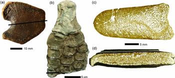
We conducted a preliminary analysis to examine whether differences in microstructure exist between the periosteum of the notched edge and the periosteum of the dorsal and ventral surfaces of mesopodialized elements. We sampled a tibia (SMNS 51130) from the Posidonia Shale referable to Stenopterygius cf. uniter (Fig. 2a). In addition, we sampled a metacarpal II (mcII) referable to Temnodontosaurus cf. trigonodon (SMNS 50329) (Fig. 2b). Both elements externally exhibited notching of the anterior surface.
(a) Tibia of SMNS 51130, Stenopterygius cf. uniter, and (b) flipper of SMNS 50329, Temnodontosaurus cf. trigonodon prior to sectioning, with the plane of sectioning indicated. (c) Microstructural overview of tibia (SMNS 51130) and (d) metacarpal II (SMNS 50329); anterior is to the left.

Although several ichthyosaur limb elements have been examined histologically, none of these exhibited notching in the sense discussed here. Comparative histological information is available for humeri (Kiprijanoff, Reference Kiprijanoff1881; de Buffrénil & Mazin, Reference de Buffrénil and Mazin1990; Canoville & Laurin, Reference Canoville and Laurin2010; Kolb, Sánchez-Villagra & Scheyer, Reference Kolb, Sánchez-Villagra and Scheyer2011), the tibia of a Triassic intermediate-grade taxon retaining both the contiguous and peripheral shaft (de Buffrénil & Mazin, Reference de Buffrénil and Mazin1990), as well as zeugopodial and more distal limb elements of ophthalmosaurids (Kiprijanoff, Reference Kiprijanoff1881; Lopuchowycz & Massare, Reference Lopuchowycz and Massare2002).
The sampling of the bones followed standard petrographic thin-sectioning procedures, as were outlined by Chinsamy & Raath (Reference Chinsamy and Raath1992). Both the tibia and the mcII were sectioned parallel to the anteroposterior axis, through the mid-point of the anteriorly directed notch to elucidate the structure and distribution of periosteal versus endosteal bone (Fig. 2a, b). The thin-sections were analyzed using a composite polarizing microscope Leica DM 2500 M equipped with a Leica DFC420 C digital camera.
2.d. Intraspecific variation in Stenopterygius quadriscissus
In order to investigate the relationship between function, ontogeny and the notched morphology of limb elements, we examined correlation patterns between the number of notches and predictor variables (size, number of phalanges in the longest digit, stratigraphy and number of notches in the serial homologue) in the fore- and hind limbs of the parvipelvian ichthyosaur Stenopterygius quadriscissus, from the Lower Jurassic Posidonia Shale of Baden-Württemberg, Germany. See Table S1 in the online Supplementary Material at http://journals.cambridge.org/geo for a list of specimens examined. We selected these variables based on previous studies indicating that they influenced aspects of limb morphology in secondarily aquatic tetrapods more generally and Stenopterygius in particular (Johnson, Reference Johnson1977; Dawson, Reference Dawson2003; Maxwell, Reference Maxwell and Dececchi2012; Maxwell, Fernández & Schoch, Reference Maxwell2012; Maxwell & Dececchi, Reference Maxwell, Fernández and Schoch2012).
We evaluated the relationship between number of notches, number of phalanges in digit II and size (using length of the lower jaw as a proxy; McGowan, Reference McGowan1973) for the fore- and hind limbs separately to maximize sample size, and then together to test the effect of serial homology. Because of uncertainty regarding independence of the variables (stage of ontogeny and digital length appear to be slightly correlated: Maxwell, Reference Maxwell2012), we examined relationships using non-parametric partial correlation (Kendall's tau) implemented in R (R Development Core Team, 2011). Sequential Bonferroni correction for multiple comparisons was applied as per Quinn & Keough (Reference Quinn and Keough2002). When the number of notches or phalanges varied within an individual, the average was used for statistical purposes. Stratigraphy was not included in the correlation analysis because examination of the data suggested that although the number of notched elements was greatest in the stratigraphically youngest specimens (falciferum Zone, late elegans Subzone) there was not a linear increase through the sampled interval.
3. Results
3.a. Phylogenetic variation in proximodistal regionalization
Concerning our proposed transition series (Fig. 1), we suggest that the first transition involved a reduction in perichondral bone in digits I and V; the second involved reduction of perichondral bone in the central digits in a proximal to distal sequence and in the marginal digits in a contiguous to peripheral sequence (data are presented in Table S2 in the online Supplementary Material at http://journals.cambridge.org/geo). Concurrent with the latter stage, the peripheral surface of the anterior mesopodials became notched, likely following a distal to proximal sequence. The next major change was the dissociation between the fore- and hind limbs, with the evolutionary loss of perichondral bone in the hind limb delayed relative to its loss in the forelimb. The dissociation between the forelimb and hind limb coincides approximately with the first appearance of the clade Parvipelvia, but the timing could be an artefact of a poor Middle–Late Triassic record: there is some evidence that extent of notching in the forelimb was reduced relative to the hind limb in the intermediate-grade genus Besanosaurus (E. Maxwell, pers. obs.; Dal Sasso & Pinna, Reference Dal Sasso and Pinna1996). The decoupling between the pattern in the fore- and hind limbs is followed by the loss of the contiguous shaft of the radius (Fig. 1d), then that of the ulna (Fig. 1e). Subsequent trends in perichondral bone loss in Early Jurassic ichthyosaurs are less clearly directional.
All Early Jurassic genera show variation in the number of notches between species. Two patterns of reduction in the number of notches are observed, but the data are not easily interpreted in a phylogenetic context. The first pattern is more widespread and involves a progressive reduction of notching of the leading-edge digit in a distal to proximal direction (e.g. Temnodontosaurus platyodon, Hauffiopteryx, Leptonectes tenuirostris, Excalibosaurus and Eurhinosaurus). The second pattern is similar, but involves the loss of notches not only from distal to proximal in the phalanges, but also from proximal to distal in the radius and proximal carpals. This second trend is seen in Stenopterygius (Fig. 1e; except in S. aaleniensis) and Ichthyosaurus (Fig. 1f; except in the hind limb of I. conybeari) (Table S2 in the online Supplementary Material at http://journals.cambridge.org/geo).
3.b. Histology and microstructure of long bones with partial perichondral reduction
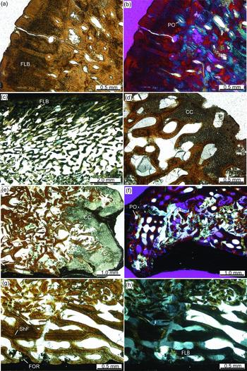
In the thin-section of the Stenopterygius tibia, periosteal bone is present along the anterior (Fig. 3a–c), dorsal and ventral surfaces of the element. The posterior surface, the margin that articulates with the fibula, lacks a periosteal layer but is instead covered by a layer of articular calcified cartilage (Fig. 3d). The periosteal layer is thickest towards the anterior end of the element and thins posteriorly. In the mcII of Temnodontosaurus, the articular cartilage is also seen along the posterior margin in the thin-section in the region that would have articulated with mcIII (Fig. 3e), whereas the dorsal, ventral and the anterior notched regions (Fig. 3f) of the element show periosteal bone composed of fibrolamellar bone (Fig. 3g–h). The internal part of both elements is composed of secondary trabeculae and the articular surfaces are slightly concave. Furthermore, the periosteal layers taper off just before the posterior articular surface, so that the adjacent dorsal and ventral surface areas are also composed of calcified cartilage (Fig. 2c–d). The microstructure of the dorsal and ventral periosteal layer is identical to the periosteal layer of the notched region, and both the fibrolamellar bone matrix and the vascularization pattern can be continuously traced through the sections. Sharpey's fibres are present throughout the periosteal bone.
(a–d) Tibia of SMNS 51130, Stenopterygius cf. uniter; (e–h) SMNS 50329, Temnodontosaurus cf. trigonodon metacarpal II. Images in (a), (d), (e) and (g) are in normal transmitted light, (c) and (h) in cross-polarized light, and (b) and (f) in cross-polarized light using a lambda compensator. (a, b) Close-up of the cortical bone in the notched area. (c) Close-up of external (dorsal) cortical bone and adjacent interior cancellous bone. (d) Close-up of calcified cartilage cap and adjacent interior trabecular bone. (e) Close-up of the calcified cartilage cap and collapsed interior trabecular framework. (f) Close-up of the notched cortex. Note dorsoventral crushing of element. (g, h) Close-up of the internal (ventral) cortical bone and adjacent interior collapsed trabecular framework. Abbreviations: CC – calcified cartilage; FLB – fibrolamellar bone; FOR – foramen; PO – primary osteon; ShF – Sharpey's fibres.

Both elements experienced dorsoventral compaction. In the mcII, crushing is much stronger, leading to the collapse of internal trabecular structures and a displacement of the anterior periosteal bone (Fig. 2d). In the tibia, crushing is minimal, restricted to a thin area within the cancellous bone (visible as a meandering thin band of trabecular ‘debris’) (Fig. 2c). The concavity of the articular (posterior) margin of the tibia is thus genuine and not a taphonomic artefact. Owing to the stronger degree of structural collapse, this feature is not so clear in the mcII. Although the latter is generally thinner than the tibia (taking into account the crushing the elements experienced), the cortical bone is more strongly vascularized throughout, presenting the ‘osteoporotic’ appearance typical of derived ichthyosaur bones (e.g. de Buffrénil & Mazin, Reference de Buffrénil and Mazin1990; Kolb, Sánchez-Villagra & Scheyer, Reference Kolb, Sánchez-Villagra and Scheyer2011). In the tibia, the periosteal layer is less vascularized. Starting from around the centre of the elements, the orientation of the vascular cavities and the cortical canal system change from being arranged sub-parallel and perpendicular to the outer bone surface, respectively, to being angled towards the articular surface opposite to the notch. Sharpey's fibres (Fig. 3g–h), which are present in both sections throughout the periosteal bone, generally follow the vascular arrangement, changing from a perpendicular arrangement in the notch area to more oblique angles towards the opposite articular surface.
3.c. Intraspecific variability in Stenopterygius quadriscissus
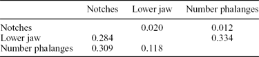
Notching is present on the anterior edge of digit II in both the fore- and hind limbs of all specimens of Stenopterygius quadriscissus studied. In the forelimb, notching develops in a roughly proximal to distal sequence, radiale > (distal carpal (dc) II < > radius) > mcII > phalanges (digit II), with the terminal events in the series occurring variably in adults. Up to the second phalanx may be notched in S. quadriscissus; however, in most specimens the distal-most notched element is dcII (Table 1). Left–right asymmetry in the number of notches is very common: out of 13 specimens with both forelimbs adequately preserved, 7/13 (54%) showed asymmetry. The within-individual difference in the number of notches on the right and left sides is never greater than one. The number and identity of notched elements is size dependent (Table 2). Notching is occasionally absent in the radius but present in more distal elements (e.g. radiale, dcII). This is ontogenetically dependent, found only in embryos and small juveniles, confirming the results of a previous interspecific study (Johnson, Reference Johnson1977). However, the radial notch is not restricted to the postnatal stages, sometimes developing prior to birth (e.g. SMNS 7402).
Descriptive statistics: number of notched elements, Stenopterygius quadriscissus

*sd – standard deviation. Values in bold were calculated excluding juveniles from the sample.
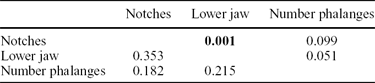
Partial correlations (forelimb), p-values above the diagonal, partial correlations below

N = 41. Values in bold were significant following Bonferroni correction.
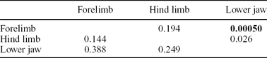
Notching is more variable in the hind limb than in the forelimb. The tibia is more frequently unnotched than the radius, and the two morphologies occur independently. When an ossification is present anterior to the tibia (Fraas, Reference Fraas1891; Maxwell, Reference Maxwell2012) both it and the tibia are unnotched. As few as three notches are present in the hind limb (MHH 1981/13) (Table 1); this is weakly correlated with size but the relationship is not significant (Table 3). In most specimens, the most distal notched element is the proximal phalanx of digit II, but up to the sixth phalanx may be notched (Table 1). Of the 13 specimens with hind limbs adequately preserved, 6/13 (46%) show left–right asymmetry, but as in the forelimb, the within-individual difference is no greater than one. The hind limb has on average one notched element more than the forelimb.
Partial correlations (hind limb)

No values were significant following Bonferroni correction. N = 34.
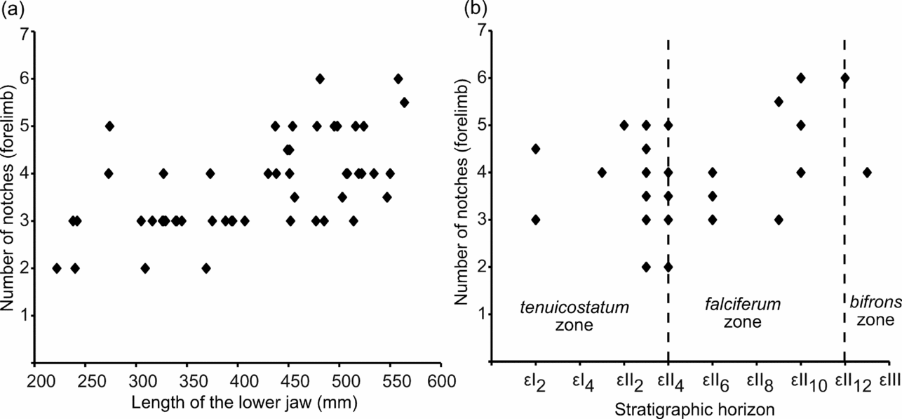
In both the statistical analysis of the forelimb alone and that including the hind limb (Tables 2, 4), body size was the only significant predictor of the number of notched forelimb elements. The relationship between the variables was positive (larger specimens possessed a greater number of notched elements) (Fig. 4a). No variables were significant in predicting the number of notches in the hind limb (Table 3). The total number of phalanges explained the largest fraction of observed variance, and, as in the forelimb, the relationship was positive.
Correlation between serial homologues, at constant body size

Values in bold were significant following Bonferroni correction. N = 40.
Number of notches in the forelimb of Stenopterygius quadriscissus (a) plotted against lower jaw length, a proxy for body size, and (b) number of notches in the forelimb plotted against stratigraphic provenance. The numbering of the beds follows a local scheme for the Posidonia Shale in southwestern Germany (see Urlichs, Wild & Ziegler, Reference Urlichs, Wild and Ziegler1979). The radius is always counted as notched, even when variable, such that the number of notches reflects the identity of the most distal notched element.

The significance of the observed increase in number of notches in the stratigraphically youngest specimens (Fig. 4b) was evaluated by calculating the probability of the increase observed in the late elegans Subzone being attributable to chance. Resampling the data to mimic the number of adult specimens in the best-sampled time interval (late semicelatum Subzone; N = 11) suggested that the observed distribution pattern of specimens with the most notches in the forelimb is not different from that expected by chance.
4. Discussion
4.a. Implications of histological, phylogenetic and intraspecific data
Caldwell (Reference Caldwell1997b ) challenged the hypothesis of homology of notches in the leading-edge elements of ichthyosaur limbs to the shafts of long bones, noting the onset of perichondral ossification was delayed along the anterior margin of the notched elements. The dorsal and ventral periostea ossified separately and later fused anteriorly (Caldwell, Reference Caldwell1997b ), in contrast to the early ossification of the perichondrium in the long bones of most extant diapsids. It has been argued that changes in element shape are related to the timing and nature of ossification (Blanco, Misof & Wagner, Reference Blanco, Misof and Wagner1998). In extant cetaceans with proximodistally shortened acropodial elements, phalangeal ossification proceeds through the formation of an initial ovoid ossification centre, followed by delayed perichondral ossification at the anterior or posterior margin (delta-shaped bone: Dawson, Reference Dawson2003), and lastly by ossification of the opposite margin. In some species, ossification remains arrested at the ovoid state in the distal phalanges, and ossification of the anterior and posterior perichondrium never occurs (Mellor et al. Reference Mellor, Cooper, Torre and Brownell2009). A heterochronic delay in the ossification of the phalangeal margins is consistent with the size-related increase in the number of notches seen in Stenopterygius quadriscissus (this study; Johnson, Reference Johnson1977). Although bone histology shows no indication for a fusion of separate dorsal and ventral periosteal layers or later development of the anterior phalangeal edge, we cannot rule out the possibility that these events occurred early in development, but that their traces were subsequently removed by internal bone remodelling. Thus, although the change in mode of ossification in the notched limb elements of derived ichthyosaurs appears to be mediated through heterochronic processes, examination of the histology of a growth series is needed to test this hypothesis.
The distribution of Sharpey's fibres throughout the periosteal layer of the Stenopterygius tibia indicates a close interface between the bone and surrounding connective tissue within the flipper, as expected. It is noteworthy that the thickness of the periosteal layer in the notched region of this element is well developed and the vascularity is reduced. These microstructures are in accordance with ossification induced by leading-edge stresses on the anterior digit, suggesting the notches were not foramina surrounded by fibrocartilage anteriorly (as proposed by Johnson, Reference Johnson1979). Cetaceans show proximodistal ankylosis of phalanges along the leading edge of the flipper in response to hydrodynamic stresses (Cooper & Dawson, Reference Cooper and Dawson2009), which are greatest along the anterior surface (Cooper et al. Reference Cooper, Berta, Dawson and Reidenberg2007a ,Reference Cooper, Dawson, Reidenberg and Berta b ). The reduced porosity of the anterior periosteal layer of the tibia relative to the posterior margin implies that the hind limb of Stenopterygius may also experience leading-edge forces. The tibia of the intermediate-grade ichthyosaur examined by de Buffrénil & Mazin (Reference de Buffrénil and Mazin1990) did not show this anteroposterior gradient in periosteal porosity, being osteoporotic throughout. The correlation between morphological changes related to thunniform swimming and microstructure of the limb elements promises to be an intriguing direction of future investigation into ichthyosaurian functional morphology.
Perichondral bone on the anterior surface of ichthyosaur limb elements is found in most Triassic and Early Jurassic genera. An almost continuous occurrence throughout the early record of ichthyosaurs, following a trend towards reduction and loss, suggests that the notches are phylogenetically conserved structures, homologous across the clade and, by extension, to the shaft of the long bone in terrestrial tetrapods (von Huene, Reference von Huene1922). However, the position of notched elements varies extensively along the proximodistal axis of the limb and also between the fore- and hind limbs, and this creates problems if a notch is considered a property linked with a given element. For instance, the anterior surface of dcII is a novel expression domain for a long bone-like peripheral shaft; likewise, the first phalanx of the leading-edge digit loses and re-evolves a notched morphology multiple times. While the latter pattern can be explained through differences in timing of ossification, the former can only be reconciled with homology if the notched morphology is viewed to be the result of an identity shift within a series of structures. This is analogous to qualitative and quantitative changes in the number and identity of elements in the vertebral column, where boundaries between elements showing a given morphology shift along the anterior-posterior axis over evolutionary time (e.g. Buchholtz, Reference Buchholtz2007; Müller et al. Reference Müller, Scheyer, Head, Barrett, Werneburg, Ericson, Pol and Sánchez-Villagra2010). We discuss a hypothesis based on molecular developmental mechanisms in the following section that elucidates how this type of change may have arisen.
4.b. Possible mechanistic underpinnings of mesopodialization in ichthyosaurs
Developmental biology can provide a complementary perspective on how the patterning of the ancestral tetrapod limb could have been modified to give rise to the remarkably divergent ichthyosaurian limb. Limb patterning is understood through the study of two model systems: the house mouse, Mus musculus, and the domestic chicken, Gallus gallus (Tabin & Wolpert, Reference Tabin and Wolpert2007; Towers & Tickle, Reference Towers and Tickle2009; Zeller, López-Ríos & Zuniga, Reference Zeller, López-Ríos and Zuniga2009; Woltering & Duboule, Reference Woltering and Duboule2010). Despite large morphological differences between the forelimbs of these two models, they show remarkable similarity in the genetic networks underlying limb development, strongly suggesting tetrapod limb diversity is achieved through modifications to existing networks and known genes, and not through the activity of genes unique to limb development in that species or clade (Woltering & Duboule, Reference Woltering and Duboule2010; Young et al. Reference Young, Bever, Wang and Wagner2011). The relationship between progressive mesopodialization of the acropodium and zeugopodium and acquisition of a notched morphology in the mesopodium in ichthyosaur limb evolution are areas in which developmental biology can provide information on potential pathways of character evolution. In particular, mesopodialization is a process for which a great deal of information is available.
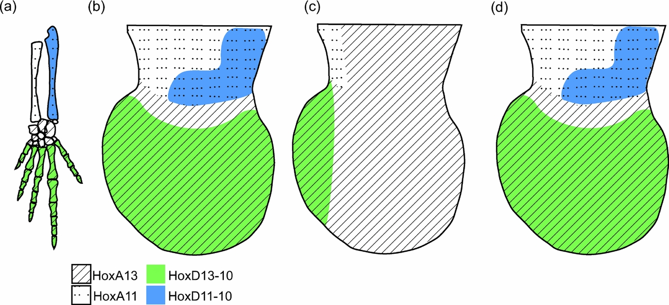
Hypotheses put forward to explain mesopodialization of the ichthyosaur acropodium centre around the Hox family of genes. The transcription of Hox genes in specific areas of the developing embryo is closely associated with acquisition of the appropriate positional identity of resulting structures (Swalla, Reference Swalla2006). This is also true in the limb, in which a specific pattern of Hox expression dictates element identity along the proximodistal axis prior to the differentiation of skeletal tissues (Woltering & Duboule, Reference Woltering and Duboule2010). The expression of a combination of HoxA11 and HoxD10–HoxD12 indicates zeugopodial identity; HoxA13 and little to no expression of HoxD genes characterize the mesopodium; HoxA13 and HoxD13 are expressed throughout the acropodium, while all but the region producing the most anterior digit express HoxD10–HoxD12 (Woltering & Duboule, Reference Woltering and Duboule2010) (Fig. 5a–b). Theoretically, changing the proximodistal location of a given Hox expression pattern should result in a predictable proximodistal transformation of element identity along the limb axis (i.e. a homeotic transformation). In derived ichthyosaurs, the mesopodialization of the acropodium could be owing to a distal shift of mesopodial Hox gene expression into the acropodium. Similarly, a proximal shift in the autopodial profile of Hox expression could produce zeugopodial elements with mesopodial characteristics (Wagner & Chiu, Reference Wagner and Chiu2001) (Fig. 5c). Unfortunately, experiments examining the effect of modifications to Hox gene expression in mice argue against this hypothesis. Although reduced acropodial expression of HoxD genes should theoretically induce a homeotic transformation resulting in the mesopodialization of the metapodials and phalanges, in reality these elements become shorter but retain their long bone morphology (Reno et al. Reference Reno, McCollum, Cohn, Meindl, Hamrick and Lovejoy2008; Montavon et al. Reference Montavon, Soshnikova, Mascrez, Joye, Thevenet, Splinter, de Laat, Spitz and Duboule2011).
Regionalization of Hox gene expression (a) correlated to adult limb regions in a terrestrial diapsid; (b) in the limb bud of a mouse; (c) expression profile expected in the ichthyosaur limb bud based on morphological change driven by transcriptional change in Hox expression; and (d) expression profile expected in the ichthyosaur limb bud if changes in Hox protein structure are driving morphological change. Anterior is to the left.

The second hypothesis involves changes to the Hox gene products themselves (Fig 5d). A HoxD13 mutant, synpolydactyly homologue (spdh), exists that results in a homeotic-like transformation of metapodial elements to mesopodialized elements in humans and mice (Muragaki et al. Reference Muragaki, Mundlos, Upton and Olsen1996; Horsnell et al. Reference Horsnell, Ali, Malik, Wilson, Hall, Debeer and Crow2006; Cocquempot et al. Reference Cocquempot, Brault, Babinet and Herault2009). Instead of modifying the transcription of HoxD13, the spdh mutation changes the protein coding sequence by increasing the number of alanine residues within the polyalanine tract of the protein (Bruneau et al. Reference Bruneau, Johnson, Yamamoto, Kuroiwa and Duboule2001; Villavicencio-Lorini et al. Reference Villavicencio-Lorini, Kuss, Friedrich, Haupt, Farooq, Türkmen, Duboule, Hecht and Mundlos2010). The mutant phenotype exhibits proximal acropodial elements that resemble those in the mesopodium, becoming shorter, with reduced perichondral bone (Muragaki et al. Reference Muragaki, Mundlos, Upton and Olsen1996; Horsnell et al. Reference Horsnell, Ali, Malik, Wilson, Hall, Debeer and Crow2006). Increasing the number of alanine repeats increases the severity of the mutant phenotype (Goodman et al. Reference Goodman, Mundlos, Muragaki, Donnai, Giovannucci-Uzielli, Lapi, Majewski, McGaughran, McKeown, Reardon, Upton, Winter, Olsen and Scambler1997; Villavicencio-Lorini et al. Reference Villavicencio-Lorini, Kuss, Friedrich, Haupt, Farooq, Türkmen, Duboule, Hecht and Mundlos2010). The reduction of perichondral bone observed in the mutant phenotype is driven by the interaction between the HoxD13 protein and Runx2, a protein that regulates the ossification of the perichondrium (Villavicencio-Lorini et al. Reference Villavicencio-Lorini, Kuss, Friedrich, Haupt, Farooq, Türkmen, Duboule, Hecht and Mundlos2010). HoxD13 and Runx2 are produced in the cytoplasm but act in the nucleus, and the expansion of the polyalanine tract of HoxD13 prevents this migration. Increasing the number of alanine residues proportionally increases the retention of HoxD13 and Runx2 proteins outside the nucleus, where they are inactive (Villavicencio-Lorini et al. Reference Villavicencio-Lorini, Kuss, Friedrich, Haupt, Farooq, Türkmen, Duboule, Hecht and Mundlos2010). With the Runx2 protein sequestered, the ossification of the perichondrium is delayed, resulting in shorter, more nodular bones (Villavicencio-Lorini et al. Reference Villavicencio-Lorini, Kuss, Friedrich, Haupt, Farooq, Türkmen, Duboule, Hecht and Mundlos2010).
The polyalanine tract of HoxD13 is not found in all tetrapods but is thought to have arisen prior to the origin of amniotes (Anan et al. Reference Anan, Yoshida, Kataoka, Sato, Ichise, Nasu and Ueda2007), and thus its presence can be inferred in ichthyosaurs. Natural variation in the length of the tract results in predictable changes similar to those described based on the spdh phenotype. Within mammals, an expansion of the polyalanine tract occurred during the evolution of the cetacean HoxD13 gene (Wang et al. Reference Wang, Yuan, Rossiter, Zuo, Ru, Zhong, Han, Jones, Jepson and Zhang2008). It is reasonable to suggest that an increase in the number of alanine residues in the cetacean HoxD13 could result in the initiation of mesopodialization of the cetacean acropodium, including shorter, more nodular phalanges, and delayed or absent perichondral ossification (Dawson, Reference Dawson2003; Mellor et al. Reference Mellor, Cooper, Torre and Brownell2009).
The gradual mesopodialization of the limb in ichthyosaurs would have been associated with proximodistal homogenization of Hox activity, either through altered expression domains or through altered protein structure. The notches along the margins of the limb could result from either reduced penetrance of the expanded polyalanine phenotype along the periphery of the limb, or through dilute expression of zeugopodial and acropodial cues. These may have been conserved via interaction between proximodistal and anteroposterior axis specification mechanisms, effected through signalling centres on the distal and posterior margins of the limb (apical ectodermal ridge and the zone of polarizing activity) (Zeller, López-Ríos & Zuniga, Reference Zeller, López-Ríos and Zuniga2009). In contrast, a strictly mechanical hypothesis for the notching of the anterior digit, for instance the presence of dorsoventral nerves and/or blood vessels creating the notching, is doubtful: notches are more numerous in the hind limb than the forelimb in most ichthyosaurs, but at the same time the hind limb is reduced both in size, number of elements, and theoretically also in function (Buchholtz, Reference Buchholtz2001).
Although the above discussion is speculative, it investigates the existing developmental data in light of the phenotypes seen in ichthyosaurs. Further developmental research is needed on the relationship between the transcription of genes relevant to limb development, their molecular biology and their effects on phenotype, especially the mesopodial region. The structure of the HoxD13 polyalanine tract over a broader range of diapsids would also provide information on its conservation and morphogenetic role in non-mammalian systems. Improved understanding of the morphology and evolution of highly derived phenotypes, such as the ichthyosaur limb, can provide a new perspective on the morphogenesis of less divergent forms.
5. Conclusions
We find strong support for the opinion of earlier workers that the notched morphology seen in the phalangeal elements of Early Jurassic ichthyosaurs is homologous to the shaft of the long bones in terrestrial tetrapods (von Huene, Reference von Huene1922; Motani, Reference Motani1999b ) using phylogenetic arguments. However, because the presence of notches on the anterior edge of limb elements does not appear to be irreversibly associated with element identity based on proximodistal position, further explanations are required. Therefore, we propose a morphogenetic hypothesis that might underlie the loss of proximodistal identity (mesopodialization) of ichthyosaurian limbs. Intraspecific data from Stenopterygius quadriscissus is consistent with these conclusions: although the number of notches varies within this species, the significant correlation with body size suggests that this morphology is caused by a delay in perichondral ossification, as has been reported in extant cetaceans. Although the examination of the histology and microstructure of notched elements could not confirm the observation of delayed perichondral ossification along the anterior phalangeal margin in Stenopterygius (Caldwell, Reference Caldwell1997b ), it did not suggest that the periosteum lining the notch was qualitatively different from that on the dorsal and ventral phalangeal surfaces.
Drawing on multiple lines of evidence to understand the evolution and function of unique morphologies is relatively new to studies of marine reptile palaeontology. However, as demonstrated here using phylogenetic, intraspecific and histological data, this approach holds great promise as a way to understand trends in the evolution and development of adaptations to a secondarily aquatic way of life. In the future, such techniques could be used to identify different mechanistic underpinnings of convergence between independent marine radiations.
Acknowledgements
Thanks to R. Schoch, M. Sánchez-Villagra and H. Larsson for discussion, R. Schoch and J. Müller for logistical support, P. Havlik (Geologisch-Paläontologisches Institut und Museum, Universität Tübingen), R. Hauff (Urwelt Museum Hauff), R. Brocke (Senckenberg Forschungsinstitut und Naturmuseum Frankfurt), J. Ebbestad (Evolutionsmuseet, Uppsala Universitet) and U. Göhlich (Naturhistorisches Museum Wien) for collections access, and to B. Kear for coordinating this special issue. The comments of two anonymous reviewers significantly improved this manuscript. This project was funded through Natural Sciences and Engineering Research Council of Canada (NSERC) and Alexander von Humboldt postdoctoral fellowships (E. M.), and the Swiss National Science Foundation (T. M. S., 31003A 127053).