Kaolinite, with a structural formula Al2Si2O5(OH)4, is a naturally abundant 1:1 aluminosilicate composed of a tetrahedral silica sheet and an octahedral alumina sheet, held together by hydrogen bonds, dipole–dipole interactions and van der Waals forces (De Paiva et al., Reference De Paiva, Morales and Valenzuela2008; Yuan et al., Reference Yuan, Tan, Annabi-Bergaya, Yan, Liu and Liu2013; He et al., Reference He, Ma, Zhu, Frost, Theng and Bergaya2014). Due to its strong interlayer cohesion and low cation-exchange capacity, pristine kaolinite is only weakly reactive towards most organic molecules, resulting in a low degree of surface functionalization and limited application potential in advanced materials fields (Tunney & Detellier, Reference Tunney and Detellier1993; Komori et al., Reference Komori, Enoto, Takenawa, Hayashi, Sugahara and Kuroda2000; Yuan et al., Reference Yuan, Tan, Annabi-Bergaya, Yan, Liu and Liu2013). Consequently, developing effective strategies to modify the kaolinite interlayer space remains an important topic for further research.
Organic intercalation has long been recognized as an effective method to activate kaolinite by expanding its interlayer space and weakening the intrinsic hydrogen bonding (Bergaya & Lagaly, Reference Bergaya and Lagaly2013; Dedzo & Detellier, Reference Dedzo and Detellier2016, Reference Dedzo and Detellier2018). The aluminol groups confined in the interlayer space of kaolinite could react with organic compounds, and a limited number of highly polar molecules or hydrogen bond donors, such as hydrazine, urea, formamide and dimethyl sulfoxide (DMSO; Johnston et al., Reference Johnston, Sposito, Bocian and Birge1984; Frost et al., Reference Frost, Kristof, Paroz and Kloprogge1998, Reference Frost, Kristof, Horvath and Kloprogge2000; Dedzo et al., Reference Dedzo, Letaief and Detellier2012; Cheng et al., Reference Cheng, Zhang, Liu, Li and Frost2015b; Zhang et al., Reference Zhang, Liu, Cheng and Zeng2015; Qiao et al., Reference Qiao, Shen, Wu, Wang, Pei, Liu and Ren2020), can be directly intercalated. Additionally, the alkali salts of short-chain fatty acids (acetates, propionates, butyrates and isovalerates) can also be directly intercalated, such as potassium acetate (Bergaya & Lagaly, Reference Bergaya and Lagaly2013). Among them, intercalation using polar molecules such as DMSO is a crucial pretreatment step that expands the interlayer space and increases the accessibility of internal hydroxyl groups. However, the disadvantage of intercalation is that most intercalated guest compounds are susceptible to desorption by washing with water or by heating (Dedzo & Detellier, Reference Dedzo and Detellier2016; Detellier, Reference Detellier2018).
By contrast, products obtained from grafting, whereby organic molecules form covalent bonds with kaolinite hydroxyl groups, demonstrated more functional groups, greater stability under certain conditions and a wider range of applications (Lagaly, Reference Lagaly1981; Komori et al., Reference Komori, Sugahara and Kuroda1999, Reference Komori, Enoto, Takenawa, Hayashi, Sugahara and Kuroda2000; Janek et al., Reference Janek, Emmerich, Heissler and Nüesch2007; De Faria et al., Reference De Faria, Ciuffi, Nassar, Vicente, Trujillano and Calefi2010; Kuroda et al., Reference Kuroda, Ito, Itabashi and Kuroda2011; Dedzo et al., Reference Dedzo, Letaief and Detellier2012; Qiao et al., Reference Qiao, Yang, Liu, Zhao and Ren2014). The first interlamellar grafting of organic compounds in the kaolinite interlayer was reported by Tunney & Detellier (Reference Tunney and Detellier1993, Reference Tunney and Detellier1994), in which DMSO- or N-methyl formamide-intercalated kaolinite was used as an intermediate and ethylene glycol and a series of diols were grafted. The resulting grafted structures typically involve Al–O–C, Al–O–Si and Al–O–CO linkages, leading to increased thermal stability and functional versatility (Letaief & Detellier, Reference Letaief and Detellier2007; Avila et al., Reference Avila, de Faria, Ciuffi, Nassar, Calefi, Vicente and Trujillano2010; De Faria et al., Reference De Faria, Ciuffi, Nassar, Vicente, Trujillano and Calefi2010; Dedzo et al., Reference Dedzo, Letaief and Detellier2012; Hou et al., Reference Hou, Li, Li and He2014; Qiao et al., Reference Qiao, Ding, Zhao, Li, Liu and Ren2017; Dedzo & Detellier, Reference Detellier2018).
Alcohols, such as methanol and ethylene glycol, have been widely used as grafting agents. Methoxy grafting, first achieved by Tunney & Detellier (Reference Tunney and Detellier1996) under extreme conditions and later reported at room temperature by Komori et al. (Reference Komori, Enoto, Takenawa, Hayashi, Sugahara and Kuroda2000), produces a grafted layer with a stoichiometry of Al2Si2O5(OH)3.64·(OCH3)0.36·0.58H2O for the dried product. Subsequently, methoxy-modified kaolinite serves as an important intermediate for various guest species, such as alkylamines and quaternary ammonium salts (Gardolinski & Lagaly, Reference Gardolinski and Lagaly2005; Yuan et al., Reference Yuan, Tan, Annabi-Bergaya, Yan, Liu and Liu2013). The ethylene glycol-grafted kaolinite was first reported by Tunney & Detellier (Reference Tunney and Detellier1993), who described the covalently bonded 9.4 Å phase. Subsequently, two distinct derivatives with basal spacings of 10.8 and 9.4 Å were systematically prepared and characterized, and the critical role of water in controlling phase selectivity was elucidated (Tunney & Detellier, Reference Tunney and Detellier1994). The 9.4 Å grafted phase, formed under anhydrous conditions, resists hydrolysis and withstands temperatures up to 350–400°C. Hirsemann et al. (Reference Hirsemann, Köster, Wack, Wüllen, Breu and Senker2011) subsequently provided nuclear magnetic resonance (NMR) evidence for covalent Al–O–C bond formation, quantifying a grafting density of 0.6 ethylene glycol molecules per formula unit, corresponding to a composition of Al2Si2O5(OH)3.4·(OC2H4OH)0.6. Furthermore, the distance of 13C–27Al between the bonded carbon atom and the octahedral aluminium was determined to be 3.1 Å (Hirsemann et al., Reference Hirsemann, Köster, Wack, Wüllen, Breu and Senker2011). The differences between the methanol- and ethylene glycol-grafted kaolinite thus lie not in the presence of covalent Al–O–C bonds but in the structural characteristics and stability of the resulting products. However, a direct comparative understanding of the two grafted systems remains lacking, which motivated the present study.
From a molecular perspective, the molecular size, hydroxyl functionality and hydrogen-bonding capability of methanol and ethylene glycol may lead to distinct interlayer coordination modes when grafted onto kaolinite. In particular, the role of the interlayer water generated during grafting reactions has been overlooked, despite its potential influence on molecular coordination and structural stability. A systematic understanding of how the molecular structure and interlayer water jointly control the formation, ordering and stability of alcohol-grafted kaolinite is still lacking.
In this study, methanol- and ethylene glycol-grafted kaolinite were synthesized using DMSO-intercalated kaolinite as a precursor. A comprehensive suite of characterization techniques, including X-ray diffraction (XRD), Fourier-transform infrared (FTIR) spectroscopy, solid-state magic-angle spinning nuclear magnetic resonance (MAS NMR), elemental analysis (CHNS) and thermogravimetry–differential scanning calorimetry (TG–DSC), was employed to elucidate the structural transformation, interlayer environment and thermal behaviour of the two grafting products. This work aims to clarify the role of alcohol functionality in controlling interlayer structure and stability and to provide new insights into the mechanism of alcohol grafting on kaolinite.
Materials
Well-crystallized kaolinite, with a Hinckley index of 1.31, was obtained from Hebei Province, China. The chemical composition, expressed as percentage by mass, is presented in Table 1. Analytical-grade ethanol, DMSO, methanol and ethylene glycol were purchased from Sigma Aldrich and used as received.
The chemical composition by percentage mass of the original kaolinite.

Table 1 Long description
Mass percentages are listed for oxides SiO2, TiO2, Al2O3, Fe2O3, FeO, MnO, MgO, CaO, Na2O, K2O, P2O5, and LOI. Values are SiO2 46.01, TiO2 0.91, Al2O3 35.63, Fe2O3 0.55, FeO 0.08, and MnO 0.01. The second set lists MgO 0.45, CaO 0.32, Na2O 0.18, K2O 0.15, P2O5 0.15, and LOI 15.3. Among the minor oxides, TiO2 is the highest at 0.91, followed by Fe2O3 at 0.55 and MgO at 0.45. K2O and P2O5 share the same value at 0.15, and MnO is the smallest at 0.01.
LOI = loss on ignition.
Experimental
Sample preparation
The pre-intercalates, DMSO-intercalated kaolinite (Kaol-DMSO), were prepared by dispersing 2 g of kaolinite in a mixture of 18 mL DMSO and 2 mL deionized water at 90°C for 2 h. The obtained Kaol-DMSO compounds were centrifuged, washed with methanol and dried.
The synthesis of methanol- and ethylene glycol-grafted kaolinite was accomplished using the guest displacement method following literature procedures (Komori et al., Reference Komori, Enoto, Takenawa, Hayashi, Sugahara and Kuroda2000; Gardolinski & Lagaly, Reference Gardolinski and Lagaly2005): 1 g of Kaol-DMSO was refluxed in 10 mL of methanol or ethylene glycol at boiling temperature for 24 h. The obtained powder was filtered, washed three times with ethanol and dried at 60°C for 12 h. The products were denoted as Kaol-MeOH and Kaol-EG, respectively.
Characterization
XRD analysis was performed using a Rigaku D/MAX 2500 PC diffractometer with Cu-Kα radiation operating at 45 kV and 100 mA. The samples were scanned at 2°2θ min–1 in the range of 3°–60°. FTIR spectra of KBr pellets of samples were recorded on a Thermo Fisher Nicolet 6700 spectrometer (4000–600 cm−1, 32 scans at 4 cm−1 resolution). Solid-state C and H NMR spectra were obtained under MAS conditions using an Agilent 600 DD2 NMR spectrometer.
Elemental CHNS analysis of grafted kaolinite was performed using an Elementar UNICUBE CHNS analyser (Germany) operating under dynamic combustion conditions. Each sample was analysed twice, and the average value was reported. The precision of duplicate measurements, expressed as relative standard deviation (RSD), was typically better than 1%.
TG–DSC was carried out using a simultaneous Netzsch STA 449 F3 thermal analyser. Measurements were taken at 30–1100°C with a heating rate of 10°C min–1 under a flowing nitrogen atmosphere (100 mL min–1).
Results and discussion
Interlayer structural transformation revealed by XRD
The structural transitions occurring during the transformation from pristine kaolinite to the grafted products were recorded with XRD (Fig. 1). Raw kaolinite showed a diffraction maximum at 0.72 nm, corresponding to the typical (001) basal spacing of kaolinite layers. Upon DMSO intercalation, the (001) reflection shifted to a smaller angle with a basal spacing of 1.12 nm, confirming the intercalation of DMSO molecules into interlayer space of kaolinite (Johnston et al., Reference Johnston, Sposito, Bocian and Birge1984; Zhang et al., Reference Zhang, Liu, Cheng and Zeng2015).
XRD traces of kaolinite, Kaol-DMSO, Kaol-MeOH and Kaol-EG: (a) full-range XRD traces and (b) enlarged (001) region.

Figure 1 Long description
A showing four stacked line graphs labeled Kaol-EG, Kaol-MeOH, Kaol-DMSO and kaolinite. The horizontal axis is labeled 2 theta, with tick labels from 5 to 60. The vertical axis label is not shown. Each trace has multiple narrow peaks. Text labels near the left side of each trace read 0.94 nanometer for Kaol-EG, 0.86 nanometer for Kaol-MeOH, 1.12 nanometer for Kaol-DMSO and 0.72 nanometer for kaolinite. B showing a line graph with four overlaid traces matching the four samples. The x axis is labeled 2 theta, with tick labels from 6 to 15 at increments of 1. The y axis is labeled Intensity, a u, with tick labels from 0 to 25000 at increments of 5000. Four main narrow peaks are labeled at their tops: 1.12 nanometer on the leftmost peak near 2 theta about 7, 0.94 nanometer on the next peak near 2 theta about 9, 0.86 nanometer on a smaller peak near 2 theta about 10 and 0.72 nanometer on the rightmost tall peak near 2 theta about 12.
When DMSO was replaced by alcohols, two distinct structural responses were observed. Subsequent grafting led to the disappearance of the DMSO-related (001) reflection and partial contraction of the interlayer space to 0.86 nm for methoxy-modified kaolinite and to 0.94 nm for ethylene glycol-grafted kaolinite. According to the calculation method proposed by Lagaly (2006; see also Tunney & Detellier, Reference Tunney and Detellier1993, Reference Tunney and Detellier1994), the intercalation ratios of methanol- and ethylene glycol-grafted kaolinite were determined to be 68.7% and 87.0%, respectively. In addition, the Hinkley index values of the two grafted products were evaluated, yielding values of 0.82 for methoxy-modified kaolinite and 1.23 for ethylene glycol-grafted kaolinite. This indicated that structural differences were formed by the two alcohol molecules within the interlayers of kaolinite.
Compared with methoxy-modified kaolinite, the ethylene glycol-grafted kaolinite displayed a more ordered interlayer structure. Methoxy-modified kaolinite displayed broader reflections with reduced intensity (Fig. 1b). By contrast, the ethylene glycol-grafted sample exhibited a sharper and more symmetric (001) reflection, indicating increased structural coherence and a more uniform interlayer environment. The full width at half maximum (FWHM) values, calculated in terms of 2θ, were 0.41 for methoxy-modified kaolinite and 0.29 for ethylene glycol-grafted kaolinite. These results indicated that, although both alcohols successfully replaced DMSO, the bifunctional characteristic of ethylene glycol promoted a more ordered interlayer structure.
Hydrogen bonding and chemical grafting evidenced by FTIR spectroscopy and MAS NMR
FTIR spectra confirmed the intercalation of DMSO into the interlayers of kaolinite (Fig. 2a). The FTIR spectra of pristine kaolinite displayed well-defined inner-surface O–H stretching bands and an inner-layer OH band (Fig. 2a). These features reflected the highly ordered hydrogen bonding framework inherent to the 1:1 layered structure. Upon DMSO intercalation, new bands emerged at 3538 and 3502 cm–1, typical of S=O···H–O hydrogen bonds between DMSO molecules and the inner-surface hydroxyls of kaolinite (Johnston et al., Reference Johnston, Sposito, Bocian and Birge1984; Zhang et al., Reference Zhang, Liu, Cheng and Zeng2015). Moreover, new bands appeared at 3021 and 2936 cm−1, corresponding to the bands of antisymmetric C–H stretching and symmetric C–H stretching, supporting the interlayer incorporation of DMSO (Madejová & Komadel, Reference Madejová and Komadel2001; Cheng et al., Reference Cheng, Hou, Liu, Li and R.L2015a). The broad and less intense band at 1637 cm–1 was ascribed to the water molecules adsorbed on the external surface of kaolinite (Komori et al., Reference Komori, Sugahara and Kuroda1999, Reference Komori, Enoto, Takenawa, Hayashi, Sugahara and Kuroda2000). These results confirmed that strong host–guest interactions were formed within the interlayer and that some amount of water physically adsorbed on the external surface of Kaol-DMSO.
FTIR spectra of kaolinite, Kaol-DMSO, Kaol-MeOH and Kaol-EG in the regions of (a) 3800–2800 cm–1 and (b) 1800–800 cm–1.

Figure 2 Long description
Four stacked FTIR spectra labeled, Kaol-EG, Kaol-MeOH, Kaol-DMSO and kaolinite. The x-axis is labeled, Wavenumbers, with unit, centimeter superscript minus 1, ranging from 3800 to 2800. The y-axis has no visible label and no visible unit. Kaol-EG has labeled peaks at 3575 and 3387 and additional labeled peaks at 2969, 2943 and 2894. Kaol-MeOH has labeled peaks at 3543 and 2942. Kaol-DMSO has labeled peaks at 3538 and 3502 and additional labeled peaks at 3021 and 2936. Kaolinite has labeled peaks at 3694 and 3620. The image B showing four stacked FTIR spectra labeled, Kaol-EG, Kaol-MeOH, Kaol-DMSO and kaolinite. The x-axis is labeled, Wavenumbers, with unit, centimeter superscript minus 1, ranging from 1800 to 800. The y-axis has no visible label and no visible unit. Kaol-EG has labeled peaks at 1462 and 1383 and labeled peaks at 1323 and 1236 and a labeled peak at 1091. Kaol-MeOH has a labeled peak at 1653. Kaol-DMSO has a labeled peak at 1637. Kaolinite has labeled peaks at 1030 and 1008 and a labeled peak at 912.
Following alcohol grafting, significant transformations in the C–H and O–H stretching regions were observed (Fig. 2a). Both grafted samples demonstrated that the O–H stretching bands of kaolinite were perturbed by the guest species, but the band due to the O–H stretching frequency of the inner hydroxyls at 3620 cm–1 remained unchanged. For the methanol-modified product, a broader and less intense O–H stretching region relative to pristine kaolinite was observed. As indicated by the significantly weakened bands at 3538 and 3502 cm–1, DMSO molecules in the interlayer space of kaolinite were substituted by methanol. The band at 1653 cm–1 was assigned to H–O–H bending, indicating that some amount of water was present in the methoxy-modified kaolinite. The band at 3550 cm–1 was attributed to the hydrated kaolinite, indicating that hydrogen bonds were present between the OH groups of kaolinite and the interlayer water molecules. Due to factors such as hydrogen bond competition, steric hindrance coupled with interlayer space constraints and solvation effects leading to hydrogen bond network reorganization, this part of the water may serve as a significant inhibitor of the direct hydrogen bonding between kaolinite and methanol.
By contrast to the methoxy-modified kaolinite product, distinct differences were observed in the C–H and O–H stretching regions of the ethylene glycol-modified sample. The band at 3575 cm–1 was assigned to the hydrogen bonded component of the interlamellar surface hydroxyls of kaolinite, together with a hydrogen bonded O–H stretching frequency at 3387 cm–1 for the ethylene glycol moiety. Three distinct C–H stretching bands at 2894, 2943 and 2969 cm–1 were observed, different from that observed for neat ethylene glycol (Tunney & Detellier, Reference Tunney and Detellier1994). In the region of the CH2 deformation, four very weak bands at 1236, 1323, 1383 and 1462 cm–1 could be identified (Fig. 2b), for which the presence of the band at 1323 cm–1 was attributed to the trans conformation of the ethyleneoxy groups. The presence of C–O and C–H absorptions verified successful grafting through Al–O–C linkages, replacing the hydrogen bonded DMSO intercalate. The ethylene glycol molecules adopt a trans configuration in the interlayers of kaolinite, with the grafted product hosting a minute fraction of non-bonded excess ethylene glycol and a measurable amount of water molecules. In contrast to the methanol-grafted product, although water may perturb the optimal Al–OH···ethylene glycol hydrogen bonds, ethylene glycol’s structural advantages allow the system to accommodate and reorganize, potentially leading to a water-inclusive interlayer hydrogen bond network with retained stability.
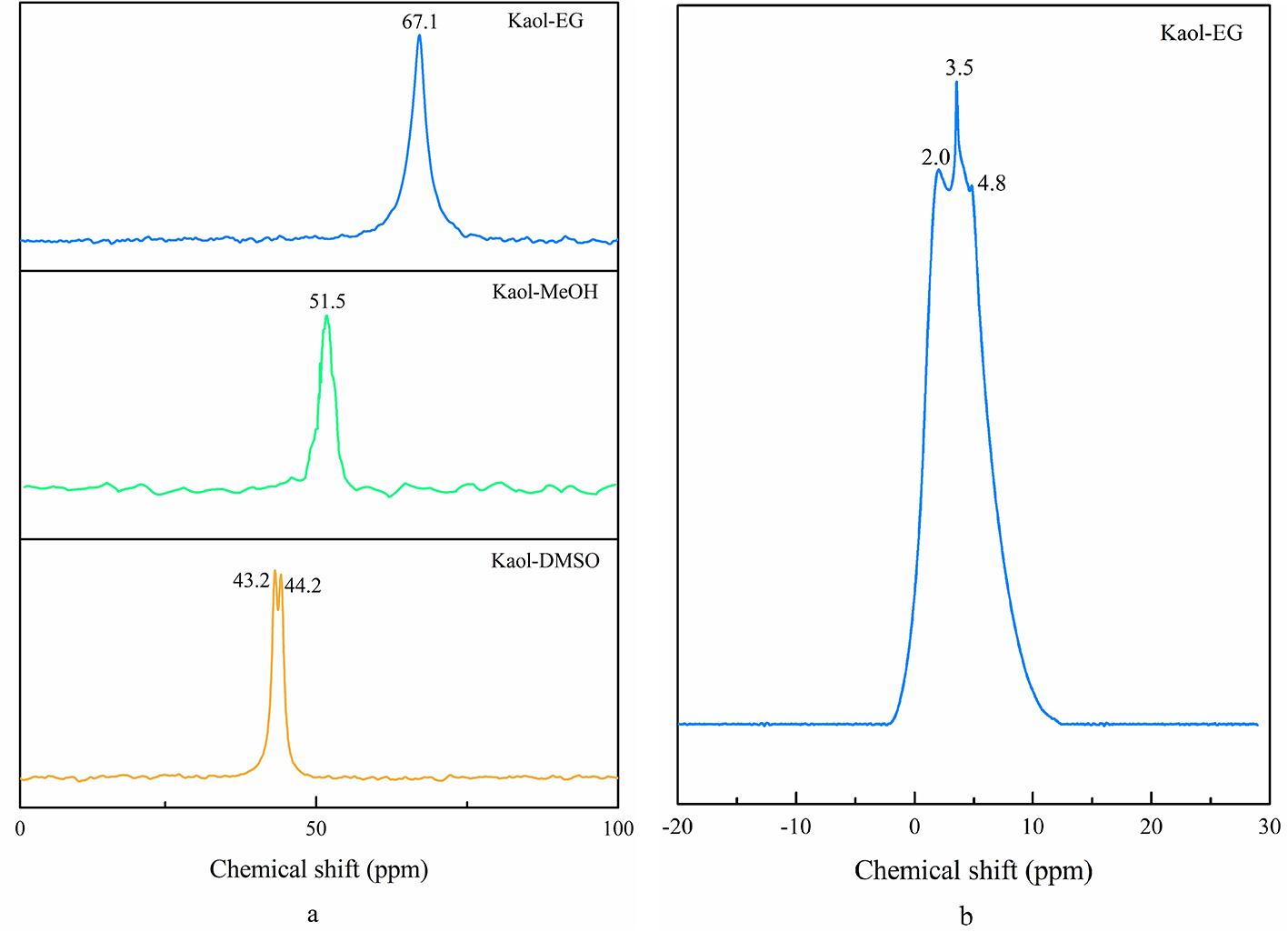
The solid-state 13C MAS NMR spectra revealed distinct bonding environments and interlayer structures among the Kaol-DMSO, methanol- and ethylene glycol-grafted samples (Fig. 3a). In general, the spectra transformed from a single DMSO-derived resonance to more shifted and defined alcohol-related signals, indicating transformation from hydrogen-bonded to covalently grafted configurations. Specifically, the Kaol-DMSO displayed a main resonance with slight splitting, consistent with previous work for confined DMSO, where two methyl groups experience non-equivalent local environments due to asymmetric hydrogen bonding (Thompson, Reference J.G1985; Duer et al., Reference Duer, Rocha and Klinowski1992). The methoxy-modified kaolinite showed a resonance at 51.5 ppm, representing the methyl carbon of the grafted methanol molecules. By contrast, the ethylene glycol-grafted sample exhibited a single sharp signal centred at 67.1 ppm, evidencing a uniform Al–O–C linkage (Tunney & Detellier, Reference Tunney and Detellier1994) and a more compact, homogeneous interlayer arrangement of organic units.
(a) 13C MAS NMR spectra of Kaol-DMSO, Kaol-MeOH and Kaol-EG and (b) 1H MAS NMR spectrum of Kaol-EG.

The 1H NMR spectra showed that water molecules are present in the Kaol-EG (0.94 nm; Fig. 3b). For ethylene glycol-grafted kaolinite, a weak but distinct resonance appears at 3.5 ppm, accompanied by broad shoulders near 2.0 and 4.8 ppm. The weak resonance at 3.5 ppm originated from –CH2– and –OH protons of grafted ethylene glycol, whereas the shoulders correspond, respectively, to residual surface hydroxyls and hydrogen-bonded water molecules confined in the interlayer. Compared with the spectra of DMSO and methanol derivatives, this profile highlights stronger hydrogen-bonding interactions and the retention of interlayer water in the ethylene glycol system, confirming the presence of a stabilized hydrogen bond network and a denser interlayer configuration (Hirsemann et al., Reference Hirsemann, Köster, Wack, Wüllen, Breu and Senker2011).
Organic loading and water retention: insights from CHNS and TG–DSC analyses
The carbon, hydrogen and sulfur contents provide information regarding the chemical composition of the interlayer space of kaolinite, which was expected to change significantly upon intercalation of DMSO. Significant increases in carbon and sulfur contents were detected after DMSO intercalation (Table 2), consistent with the presence of the inserted DMSO molecules within the interlayer space. On the basis of sulfur content (7.53 mass%), the number of S=O groups (x) per Al2Si2O5(OH)(4 – x) was calculated to be 0.69 (for details, see the Supplementary Material), yielding a structural formula for the DMSO intercalated kaolinite Al2Si2O5(OH)3.31⋅(C2H6S)0.69. The calculated structural formula was used to validate the contents of carbon and hydrogen, yielding values of 5.73% and 2.60%, respectively, which were in good agreement with the listed experimental data.
Elemental contents (carbon, hydrogen, sulfur) of Kaol-DMSO, Kaol-MeOH and Kaol-EG and the calculated chemical formulas.
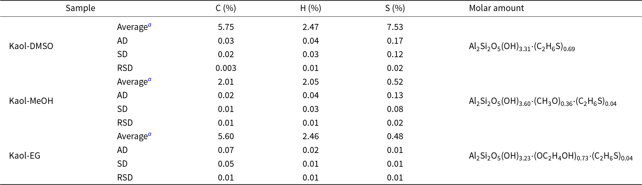
Table 2 Long description
Elemental percentages for carbon, hydrogen, and sulfur are listed for three samples, Kaol- DMSO, Kaol-MeOH, and Kaol-EG, along with a molar amount entry for each sample. For Kaol- DMSO, the Averagea row reports carbon 5.75 percent, hydrogen 2.47 percent, and sulfur 7.53 percent, followed by a molar amount entry. For Kaol-DMSO, AD is 0.03 for carbon, 0.04 for hydrogen, and 0.17 for sulfur; SD is 0.02, 0.03, and 0.12; RSD is 0.003, 0.01, and 0.02. For Kaol- MeOH, the Averagea row reports carbon 2.01 percent, hydrogen 2.05 percent, and sulfur 0.52 percent, followed by a molar amount entry. For Kaol-MeOH, AD is 0.02 for carbon, 0.04 for hydrogen, and 0.13 for sulfur; SD is 0.01, 0.03, and 0.08; RSD is 0.01, 0.01, and 0.02. For Kaol-EG, the Averagea row reports carbon 5.60 percent, hydrogen 2.46 percent, and sulfur 0.48 percent, followed by a molar amount entry. For Kaol-EG, AD is 0.07 for carbon, 0.02 for hydrogen, and 0.01 for sulfur; SD is 0.05, 0.01, and 0.01; RSD is 0.01, 0.01, and 0.01.
a All results were derived from duplicate experiments.
AD = absolute difference between the two measurements; SD = standard deviation.
The structural formulae of methanol- and ethylene glycol-grafted kaolinite were also calculated based on the carbon and sulfur elemental contents, and the hydrogen elemental content was used for validation. As demonstrated earlier, residual sulfur in both grafted products indicated incomplete but substantial removal of DMSO. Consequently, the structural formulae derived from the carbon and sulfur contents were Al2Si2O5(OH)3.60⋅(CH3O)0.36⋅(C2H6S)0.04 for methoxy-modified kaolinite and Al2Si2O5(OH)3.23⋅(OC2H4OH)0.73⋅(C2H6S)0.04 for ethylene glycol-grafted kaolinite.
As revealed by the structural formulae, the ethylene glycol grafting reaction consumed more hydroxyl groups, leading to organic loading of ethylene glycol molecules within the kaolinite interlayers that was approximately double that for methoxy-modified kaolinite. However, the calculated hydrogen content was smaller than the experiment data, indicating the presence of additional hydrogen sources, which originated from the condensation reaction between the Al–OH groups of kaolinite and the hydroxyl groups of the alcohols.
Figure 4 presents the TG and DSC curves of kaolinite, Kaol-DMSO, Kaol-MeOH and Kaol-EG under nitrogen. Pristine kaolinite exhibited a characteristic single-step mass loss (13.12%) associated with dehydroxylation that ranged from 400°C to 700℃, corresponding to the DSC peak centred at ∼524°C. In contrast, two mass loss steps were observed in the TG curve of Kaol-DMSO: decomposition of DMSO molecules below 200°C and dehydroxylation of kaolinite near 480°C. The mass losses associated with these effects were calculated to 17.46% and 10.67%, respectively.
(a) TG and (b) DSC curves of kaolinite, Kaol-DMSO, Kaol-MeOH and Kaol-EG.

Figure 4 Long description
Panel A shows TG (mass change) curves for kaolinite, Kaol-DMSO, Kaol-MeOH and Kaol-EG. The x-axis is labeled Temperature (degrees Celsius), ranging from 30 to 1100. The y-axis likely represents mass change, but units are not legible. All curves show a decrease with temperature. Kaol-EG has mass loss steps at 1.71 percent, 8.71 percent and 9.35 percent. Kaol-MeOH shows mass loss at 1.31 percent and 13.01 percent. Kaol-DMSO exhibits mass loss at 17.46 percent and 10.67 percent, with a low-temperature event below 200 degrees Celsius. Kaolinite shows a single mass loss of 13.12 percent. Panel B displays DSC (heat-flow) curves for the same samples. The x-axis is labeled Temperature (degrees Celsius), ranging from 30 to 1100. The y-axis likely represents heat flow, but units are not legible. Kaol-EG shows a heat-flow event at 450 degrees Celsius. Kaol-MeOH has an event at 519 degrees Celsius. Kaol-DMSO shows events at 194 degrees Celsius and 518 degrees Celsius. Kaolinite exhibits events at 524 degrees Celsius and a high-temperature event at 998 degrees Celsius. The plots compare thermal decomposition and dehydroxylation behavior of intercalated kaolinite versus pristine kaolinite.
The grafted samples showed distinctly different thermodynamic responses. The methoxy-modified kaolinite exhibited a low-temperature (115–210°C) mass loss associated with water molecules, followed by a higher-temperature mass loss corresponding to dehydroxylation of the kaolinite from 360°C to 850°C. By contrast, three major mass loss steps were recorded in the TG curve of ethylene glycol-grafted kaolinite. The first mass loss step was clearly identified, but the second and third step partially overlapped. The first mass loss step at 100–200°C, corresponding to the DSC peak centred at ∼160°C, was ascribed to the removal of water molecules. The second mass loss at 250–460°C, corresponding to an 8.71% mass loss, was attributed to the removal of ethylene glycol molecules. The third 9.35% mass loss at 460–640°C was attributed to the dehydroxylation of kaolinite. Compared with the methoxy-modified kaolinite, a higher onset temperature for dehydroxylation was observed, reflecting the stronger interlayer binding and increased thermal stability of ethylene glycol-grafted kaolinite.
Formation mechanism of methanol- and ethylene glycol-grafted kaolinite
The transformation sequence is summarized schematically in Fig. 5. In this stepwise intercalation–grafting reaction, DMSO molecules, possessing a high dipole moment (4.0 Debye) and strong S=O···H–O bonding capability, intercalated directly into the interlayer space of kaolinite and acted as an essential ‘interlayer template’, expanding the basal spacing and weakening the Al–OH···O–Si network. DMSO is a strong hydrogen bond acceptor, enabling it to form strong hydrogen bonds with the inner-surface Al–OH groups and create Al–O–S bonds. Subsequent treatment with methanol or ethylene glycol led to the substitution of relatively weak Al–O–S interactions by stronger covalent Al–O–C bonds. The overall displacement reaction was thermodynamically spontaneous due to this exchange being towards a higher bond strength and a more stable grafted structure.
Schematic representation of the transformation from kaolinite to Kaol-DMSO, Kaol-MeOH and Kaol-EG. Al = dark grey; C = black; H = white; O = red; Si = yellow.

Figure 5 Long description
The schematic shows the transformation of kaolinite into Kaol-DMSO, Kaol-MeOH and Kaol-EG. The initial structure, labeled as kaolinite, has a basal spacing of 0.72 nanometers. DMSO molecules intercalate into the kaolinite structure, expanding the basal spacing to 1.12 nanometers, forming Kaol-DMSO. Subsequent treatment with methanol reduces the spacing to 0.98 nanometers, resulting in Kaol-MeOH. Alternatively, treatment with ethylene glycol reduces the spacing to 0.94 nanometers, forming Kaol-EG. The atoms are color-coded: aluminum is dark grey, carbon is black, hydrogen is white, oxygen is red and silicon is yellow. Arrows indicate the transformation pathways involving DMSO, methanol and ethylene glycol.
The water molecules generated by a condensation reaction may have a significant impact on the arrangement of alcohols and the stability of grafting products. For methanol-grafted products, water molecules may have competed with methanol for the Al–OH groups of kaolinite, forming hydrogen bonds that block grafting. In addition, the presence of water also introduced steric constraints that prevented methanol from achieving a favourable orientation. Consequently, some methanol molecules may have interacted indirectly with Al–OH via water bridges (Al–OH···OH2···HOCH3), forming a weaker, reorganized hydrogen bond network and so producing a disordered and poorly crystalline methoxy-modified kaolinite.
By contrast with methanol, ethylene glycol functions as a bidentate ligand, offering more binding sites and granting ethylene glycol greater competitiveness, enabling it to partially displace or coexist with water molecules at the interface. Spatially, the ethylene glycol-grafted kaolinite provided more space to accommodate water, thereby reducing steric hindrance and facilitating water molecules to integrate into the complex hydrogen bond network. Therefore, water was not only a disruptor but also could be incorporated into a hybrid network. As a result, the interlayer structure, organic loading and thermal stability collectively demonstrated that the ethylene glycol-grafted kaolinite exhibited a more stable, chemically integrated grafted configuration than its methanol counterpart, reflecting a balance between steric accessibility and hydrogen bond-mediated cohesion. Thus, the interlayer structure and bonding mechanism of grafting were presumably influenced by the molecular polarity, size, hydrogen-bonding capacity and steric hindrance of the substituent alcohols, as well as by the presence of confined water.
Conclusions
The ethylene glycol-grafted kaolinite exhibited a more ordered and compact grafting structure than its methoxy-modified counterpart, and water molecules coexisted with ethylene glycol in the interlayer space. This difference was controlled by a combination of molecular properties (polarity, size, hydrogen-bonding capability) and their synergy with confined water molecules. Notably, water molecules appeared to play an adverse role in methanol and ethylene glycol grafting.
Supplementary material
The supplementary material for this article can be found at https://doi.org/10.1180/clm.2026.10061.
Financial support
This study received financial support from multiple sources, including the National Natural Science Foundation of China (No. 52201178), the National Project of Undergraduate Innovation and Entrepreneurship (202510077013), the Doctoral Research Start-up Fund of Hebei GEO University (H2025093), and the Open Foundation of Hebei Key Laboratory of Green Development of Rock and Mineral Materials (RM2025003).
Competing interests
The authors declare that they have no financial or personal relationships with other people or organizations that can inappropriately influence their work described in this paper. There are no professional or other personal interests of any nature or kind in any product, service and/or company that could be construed as influencing the position presented in the enclosed article.