Tetralogy of Fallot, pulmonary atresia, and major aortopulmonary collaterals (TOF/MAPCAs) is a form of TOF characterised by extreme heterogeneity of pulmonary blood supply, which can be difficult to manage due to the complex anatomy of the pulmonary circulation.Reference Jefferson, Rees and Somerville 1 – Reference Rossi, Hislop and Anderson 3 Our programmatic approach to this condition emphasises single-stage unifocalisation and extensive reconstruction of the proximal and distal pulmonary vasculature, with complete intra-cardiac repair during the same procedure in most patients.Reference Bauser-Heaton, Borquez and Han 4 – Reference McElhinney, Reddy and Hanley 7 Using this surgical approach, extensive dissection into the parenchyma and around the trachea and distal airways is common, which can have implications for the proximal and distal airways.Reference Sidell, Koth and Bauser-Heaton 8 As documented previously, respiratory factors are one of the primary sources of peri-operative morbidity after complete repair of TOF/MAPCAs using our approach.Reference Asija, Hanley and Roth 9 , Reference Asija, Roth and Hanley 10 Respiratory morbidity is likely multi-factorial, but native and iatrogenic airway abnormalities may play a role in some cases.Reference Perri, Albanese and Carotti 11 – Reference Carotti, Digilio and Piacentini 13 For example, proximal and distal airway compression as a result of the variable anatomy has been noted prior to repair.Reference Mainwaring, Patrick and Carrillo 14 Also, there is a high incidence of chromosome 22q11 microdeletion in patients with TOF/MAPCAs, anywhere from 17% to 40%.Reference Bauser-Heaton, Borquez and Han 4 , Reference Mercer-Rosa, Pinto and Yang 15 , Reference O’Byrne, Yang and Mercer-Rosa 16 Associated clinical problems in patients with this genetic anomaly include airway abnormalities.Reference Huang and Shapiro 17 In a series of 35 patients with a chromosome 22q11 microdeletion, Leopold et al defined a 14% incidence of airway abnormalities, including laryngeal web, sub-glottic stenosis, and laryngomalacia.Reference Leopold, De Barros and Cellier 18 While peri-operative survival after TOF/MAPCAs surgery at our Institution is excellent, prevention of respiratory failure in these patients has been a major focus in our continued efforts at process and outcome improvement.Reference Li, Zhang, Li and Wang 19 , Reference Davis, Tucker and Russo 20 A prior pilot study did not show chromosome 22q11 deletion to be associated with respiratory failure,Reference Asija, Hanley and Roth 9 but post-operative bronchoscopy has provided more insight into potential airway abnormalities in TOF/MAPCAs patients, many with 22q11 microdeletion.Reference Sidell, Koth and Bauser-Heaton 8
Pre-operative computed tomography (CT) angiography is frequently performed in TOF/MAPCAs patients at our institution to provide three-dimensional definition of the pulmonary vascular anatomy. Although specific detection of airway abnormalities is not usually the primary focus of these exams, identification of airway anomalies can be an ancillary benefit. Post-operative CT imaging is more often performed to further characterise potential abnormalities, such as extrinsic compression of the airway that might contribute to prolonged mechanical ventilation or other clinical findings. Based on previously reported bronchoscopic findings in TOF/MAPCAs patients with post-operative respiratory failure, pre-operative bronchoscopy, particularly in patients with a chromosome 22q11 deletion, has been adopted as a standard of care since 2017. Laryngoscopy and direct airway evaluation during anaesthesia for various imaging, catheterisation, and surgical procedures has also been performed in these patients, allowing characterisation of patients with a difficult airway or to identify intrinsic or extrinsic laryngeal, tracheal, or bronchial pathology. However, the association between difficult airway, chromosome 22q11 microdeletion, and airway abnormalities on CT and bronchoscopy has not been previously assessed in our series of over 500 patients with TOF/MAPCAs.
Materials and methods
Patients
After Stanford University Institutional Board approval, we identified patients with TOF/MAPCAS who underwent unifocalisation or pulmonary artery reconstruction surgery at Lucile Packard Children’s Hospital/Stanford Children’s Healthcare from March, 2002 to June, 2018 using our electronic database within the Betty Moore Children’s Heart Center. Airway evaluation either by direct laryngoscopy, chest CT, and/or bronchoscopy was described in relation to the date of surgery. Patients with multiple surgeries and multiple airway evaluations by direct laryngoscopy, chest CT, and/or bronchoscopy were also included in the analysis. The diagnosis of a chromosome 22q11 microdeletion was confirmed if determined by fluorescence in situ hybridization.
Difficult airway
Anaesthetic records for all patients with TOF/MAPCAS who underwent unifocalisation or pulmonary artery reconstruction surgery were reviewed for difficulty associated with direct laryngoscopy and intubation. The medical records were further reviewed for any association with difficult airway evaluation by anaesthesia for general surgical procedures and other services upon direct laryngoscopy and intubation.
Difficult airway can be defined according to standard ASA external and internal airway classification. External classification is predictive for a potentially difficult airway and accounts for mouth opening, thyromental distance, and neck mobility/range of motion. Internal classification is based on the ability to view the vocal cords upon direct laryngoscopy and to pass the endotracheal tube through the vocal cords. In this study, difficult airway refers to the latter assessment, namely, difficulty in endotracheal intubation upon direct laryngoscopy. At our institution, this is noted and highlighted in the electronic medical record as would be a drug allergy. It alerts caregivers that a given patient would benefit from an individual skilled in airway management and endotracheal intubation, such as an anaesthesiologist or otolaryngologist, if the need for endotracheal intubation should arise.
Chest CT
Chest CT angiography is performed during pre-operative assessment in many patients with TOF/MAPCAS, regardless of airway concerns, at our institution. Chest CT reports, collected from the electronic medical record, were reviewed for evidence of airway (trachea and bronchus) abnormality and specifically defined as “airway” in the report. Reports for chest CT performed at outside institutions were located in the electronic medical record. Patients in whom the chest CT report could not be located were defined as missing data and not assumed to have a normal airway.
Bronchoscopy
Indications for bronchoscopy were not systematically defined in this patient population until recently, and most bronchoscopy studies were performed for intra-operative and post-operative respiratory management, that is, clearance of mucous plug or clot, or for specific peri-operative management indications. Some were performed out of concern for complete tracheal rings as determined by pre-operative chest CT or upon direct laryngoscopy and intubation at the time of the surgical procedure. Due to more recent concernsReference Sidell, Koth and Bauser-Heaton 8 that post-operative abnormal bronchoscopy findings are manifestations of abnormal pre-surgical anatomy, particularly in patients with a chromosome 22q11 microdeletion, pre-operative bronchoscopy is now performed in this subset of TOF/MAPCAs patients or others with pre-operative symptoms.
Data analysis
Categorical data were reported as number (N) of patients (%). Numerical data were reported as median (range) for skewed data and mean (standard deviation) for normally distributed data.
Results
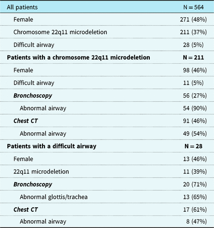
From March, 2002 to June, 2018, 564 patients with TOF/MAPCAs underwent unifocalisation or pulmonary artery reconstruction surgery at our centre and had records available for review. Selected demographic and diagnostic data are presented in Table 1. In total, 28 patients (5%) had a difficult airway/intubation at the time of surgery.
Characteristics of patients with TOF/PA/MAPCAs

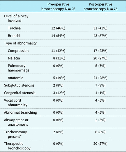
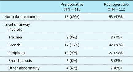
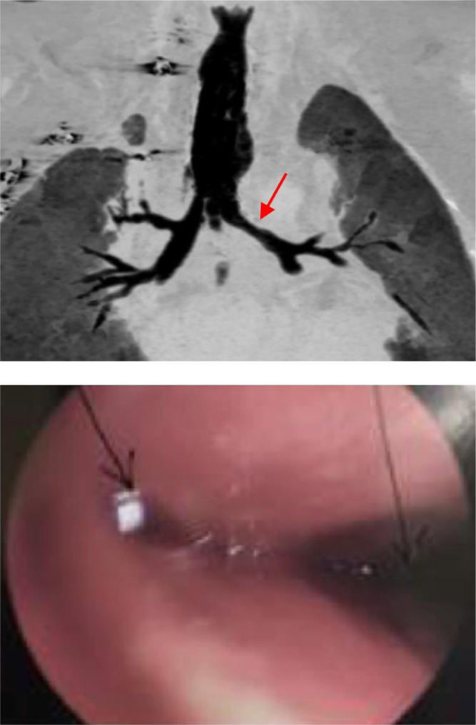
Reports were available for chest CT and/or peri-operative bronchoscopy performed at our centre in 234 (41%) of these patients. Bronchoscopy was performed in 101 patients, 26 pre-operatively and 75 intra- and/or post-operatively. Of these, 54 (53%) had a 22q11 microdeletion and 17 (17%) had a difficult airway. Upon examination, 13 (13%) had no abnormality or intervention, while the rest had at least one abnormal finding, as detailed in Table 2. Chest CT was performed in 199 patients, 110 pre-operatively and 112 post-operatively. Of these, 82 (41%) had a chromosome 22q11 microdeletion and 15 (8%) had a difficult airway. Airway/lung abnormalities were reported in 31% and 53% of patients studied with CT pre- and post-operatively, respectively, as summarised in Table 3. Almost all of the abnormal CT findings related to the trachea and bronchi were narrowing, generally indicative of compression or malacia. Of these patients, 35 had both CT and bronchoscopy within 3 months of each other: in 32 cases, the findings on CT and bronchoscopy were concordant, and in 3 they were partially concordant (the bronchoscopy report did not include all of the abnormal findings reported from the CT) (Figure 1).
Findings on pre- and post-operative bronchoscopy

* Tracheostomy is also included as an anatomic abnormality.
Findings on pre- and post-operative CT imaging

An example of CT and bronchoscopy findings in a patient with TOF/MAPCAS and left mainstem bronchus compression. (Top) CT and (bottom) bronchoscopy. Arrow(s) indicate airway abnormality.

Of the 28 patients labelled as having a difficult airway or intubation, 20 underwent bronchoscopy, 13 of whom (65%) had an abnormal larynx/trachea, and 17 had a CT scan, which was abnormal in 8 (47%), including 4 (24%) with tracheal findings. Among 56 patients with a documented chromosome 22q11 deletion, 11 (5%) had a difficult airway/intubation, 49 (23%) had an abnormal CT, and 52 (90%) had abnormal bronchoscopy findings (Table 1).
Discussion
In this report based on management of more than 500 patients with TOF/PA/MAPCAs at a single institution, we provide a descriptive experience of airway abnormalities documented by direct laryngoscopy, chest CT, and/or bronchoscopy. In this complex population, there are numerous risk factors for airway abnormalities, and respiratory issues are the most comment source of peri-operative morbidity. This characterisation of airway findings both pre-operatively and after surgical repair can be useful information for not only understanding but also caring for this population.
The association of airway abnormalities in children with congenital heart disease has been described. The largest series, based on diagnostic codes without confirmation by direct laryngoscopy, chest CT, and/or bronchoscopy, reported an incidence of 4%.Reference Lee, Jeng and Tsao 21 In a few smaller series describing findings of direct laryngoscopy, an incidence of 11% was reported in patients with congenital heart diseaseReference Kazim, Berdon and Montoya 22 and TOF.Reference Kazim, Quaegebeur and Sun 23 Our 5% incidence of airway abnormalities identified by direct laryngoscopy in patients with TOF/PA/MAPCAs is not unexpected.
Of patients with congenital heart disease 30% have a genetic syndrome.Reference Ferencz, Correa-Villasenot and Loffredo 24 , Reference Harris, Cronk and Cassidy 25 Microdeletion of chromosome 22q11 is one of most common and important genetic syndromes in patients with congenital heart disease and is highly prevalent in patients with various anomalies, including TOF 10%,Reference Marino, Digilio and Grazioli 26 pulmonary atresia with ventricular septal defect (VSD) (16–40%),Reference Anacierio, Marino and Carotti 27 truncus arteriosus (30–35%),Reference McElhinney, Driscoll and Emanuel 28 interrupted aortic arch (40–50%),Reference Marino, Digilio and Toscano 29 other arch anomalies (24%),Reference McElhinney, Clark and Weinberg 30 and VSD (10–50%).Reference Toscano, Anacierio and Digilio 31 An incidence of 22q11 microdeletion of 29%Reference Mercer-Rosa, Elci and Pinto 32 has been observed in patients with TOF/PA/MAPCAs.
Airway abnormalities were identified in 71% of patients with a chromosome 22q11 microdeletion as determined by microlaryngoscopy and bronchoscopy, including bronchomalacia, laryngeal web, and tendency towards airway bleeding.Reference Sacca, Zur and Crowley 33 Most patients in that series had an associated cardiac defect. The incidence of airway abnormalities in this selected cohort of patients with TOF/MAPCAs was 87% by bronchoscopy, with confirmation by chest CT in 32% and direct laryngoscopy in 71%.
Tracheostomy after cardiac surgery has an overall low incidence (1.3%)Reference Cotts, Hirsch and Thorne 34 and the number of patients in our series was similar at 1.4% (8 patients). Indications for tracheostomy can be multi-factorial (37%), but the majority of cases are related to trachea- or bronchomalacia (24%). It has been shown that hospital (75%) and long-term survival (53%) are lower in cardiac patients after tracheostomy than in patients without tracheostomy. Thus, improved awareness of airway abnormalities by pre-operative bronchoscopic evaluation may serve as means to improve overall pulmonary status in a population at risk for respiratory failure, including patients with TOF/MAPCAs, in whom a chromosome 22q11 deletion alone is not predictive of post-operative respiratory failure.Reference Asija, Hanley and Roth 9
In a series of 45 children, low-dose multi-detector CT with virtual tracheobronchoscopy was compared to flexible tracheobronchoscopy for the evaluation of airway stenosis or suspected airway abnormality. Though CT with virtual tracheobronchoscopy was 87% sensitive and 86% specific with an accuracy of 87% in detecting most airway abnormalities, the ability to detect tracheobronchomalacia was limited as compared to flexible tracheobronchoscopy.Reference Heyer, Nuesslein and Jung 35 These findings are similar to the 87% incidence of abnormalities seen with bronchoscopy and 31% pre-operative and 53% post-operative incidence of airway abnormality with CT. Though we report a high correlation of concordant findings between chest CT and bronchoscopy, a similar limitation of tracheobronchomalacia was observed. Some have suggested the use of dynamic pulmonary CT angiography to assess airway abnormalities in children with congenital heart disease due to improved ability to detect both trachea- and lobar-bronchomalacia, which led to a change in medical or surgical management in 70% of the patients in a prior study.Reference Greenberg and Dyamenahalli 36 Despite advanced dynamic imaging modalities, there remain significant limitations in the ability to characterise or grade malacia of the airways.
The limitations of the study include the retrospective analysis of the data, more importantly the review of previously documented clinical direct laryngoscopy, chest CT, and bronchoscopy reports. In this large surgical series, only 41% of the 564 patients had chest CT or bronchoscopy, but all had at least one direct laryngoscopy by an experienced paediatric cardiac anaesthesiologist. Direct laryngoscopy only provides upper airway evaluation, whereas chest CT and bronchoscopy allow for lower airway evaluation. In addition, bronchoscopy can give additional information pertaining to laryngeal webs and laryngeal clefts. Laryngeal clefts and webs can be the most frequently missed airway abnormalities and are not routinely seen by direct laryngoscopy or chest CT. The focus of this study was to report findings related to the airway in this population and not to evaluate outcomes related to or management of the abnormalities detected, so specific clinical recommendations or conclusions cannot be drawn from our findings. These findings should be considered preliminary and hypothesis generating.
In summary, this is the largest series of detailed airway findings (direct laryngoscopy, CT, and bronchoscopy) in TOF/MAPCAs patients. Although these findings are specific to an at-risk population for airway abnormalities, patients with TOF/MAPCAs, they support the utility of CT and /or bronchoscopy in detecting airway abnormalities in patients with TOF/MAPCAs.
Acknowledgments
The authors wish to acknowledge the Pediatric Cardiac Anesthesia Division at Stanford University/Lucile Packard Children’s Hospital: Drs. Gail Boltz, Komal Kamra, Calvin Kuan, Manchula Navaratnam, Chandra Ramamoorthy and Jumbo Williams.
Financial Support
This study was supported by a grant from The Gerber Foundation.
Conflicts of Interest
None.
Ethical Standards
The authors confirm that all procedures contributing to this work comply with the ethical standards of the relevant national guidelines on human experimentation, retrospective chart review, and with the Helsinki Declaration of 1975, as revised in 2008, and that the study was approved by the Stanford University Institutional Review Committee.