Although syncope is a frequent and generally benign event in children, it may present as a symptom of potentially lethal underlying disease. Diagnosis of the underlying disease is important for stratification of risk and provision of appropriate therapy. This is particularly the case for patients subsequent to surgical correction of congenital cardiac malformations, of for those primary electrical abnormalities, since arrhythmias are a common cause of syncope. The aetiology of recurrent but infrequent syncope often remains unexplained, despite extensive investigations. The major explanation for the low diagnostic yield of most diagnostic approaches is the sporadic and unpredictable nature of the underlying pathophysiologic mechanism.
The optimal diagnostic tool in recurrent syncope must offer long-term monitoring in order to obtain a correlation of symptoms with cardiac rhythm in patients who suffer infrequent events. After detecting an arrhythmia, the monitor should autoactivate recording of the electrocardiogram. Recent developments in the technology of implantable loop recorders now allow such long-term electrocardiographic monitoring, with either automatic or patient-activation of recording. The compelling data relative to safety and efficacy from several large trials have led to increasing use of these devices in adults with recurrent unexplained syncope.1–4 Experience in children with implantable loop recorder, in contrast, is still limited.5–8 The purpose of our retrospective study, therefore, was to report our experience with these devices in children and adolescents.
Patients and methods
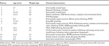
Between January 1999 and August 2005, we implanted a loop recorder in 12 children, using either the Reveal™ or Reveal™ Plus as manufactured by Medtronic Inc. The mean age at implantation was 10.9 years, with a range from 2 to 17 years. Of the patients, 6 had structural disease, 3 had primary electrical abnormalities, and 3 had no history of cardiovascular disease. The clinical characteristics of the patients are noted in Table 1.
Table 1. Clinical characteristics of patients.

In all cases the indication for implantation of a loop recorder was recurrent syncope, or near syncope, that remained undiagnosed after standard diagnostic tests. The preceding diagnostic tests included the surface electrocardiogram, echocardiography, and repeated 24-hour Holter monitor in all 12 patients, exercise testing in 7, tilt-table testing in 3, electroencephalographic recordings in 8, cardiac catheterization in 4, electrophysiological studies in 5, and external loop recording in another 5 patients. If possible, we always first tried to use an external loop recorder, but toddlers, and patients with disorders like Down's syndrome, often do not tolerate the external device.
The loop recorders are capable of recording a single-lead electrocardiogram using two sensing electrodes on the surface of the device (Fig. 1). The storage capacity is 42 minutes. The life of the battery ends 14 months after implantation. All devices were implanted subcutaneously in the left prepectoral region. The surgical procedure, along with a postoperative chest X-ray, are shown in Figure 2. Recording of the cardiac rhythm can be started by the patient, or automatically if the heart rate exceeds a preset upper or lower limit.

Figure 1. Sensing electrodes of the implantable loop recorder Reveal™ from Medtronic Inc.

Figure 2. Subcutanous implantation (a) of the device in the left prepectoral region, and the chest X-ray of a 10-year-old female (b) after implantation.
All patients had regular follow-up after each symptomatic event to analyze the recorded contents, or at 2-monthly intervals if no event occurred. The electrocardiograms recorded during events were transferred to the analyzing unit Programmer 9790 from Medtronic Inc. After analysis, the electrocardiographic data were stored electronically on disc. The device was either explanted after the diagnosis was established, or after 14 months if syncope did not recur.
Results
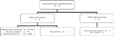
Resyncope occurred in 9 of 12 patients. Of these 9, 6 suffered events in the first six weeks after implantation. A definitive diagnosis of an arrhythmic cause of the syncope was made in 4 of these 9 patients (44%). Definitive treatment could be rendered to all 4 patients with arrhythmic diagnosis (Fig. 3).

Figure 3. Outcomes for our patients.
There were no complications due to bleeding, no skin erosions, and no infections related to implantation of the recorder, and no instance of malfunction of the device. An acceptable electrogram was obtained in all patients. The mean duration until explantation was 8.3 months, with a range from 7 weeks to 14 months.
Patients with resyncope
Of the 12 patients, 9 developed syncope or presyncope after implantation of the recorder, at a mean interval of 6.8 weeks, with a range from 3 weeks to 4 months. Syncope was arrhythmogenic in 4 of these 9 patients. The second patient, a two year old boy, had undergone corrective surgery for tetralogy of Fallot at the age of 3 months. The records obtained from this patient revealed intermittent asystole of 5 seconds duration during syncope (Fig. 4). Following implantation of a VVI-pacemaker, syncope did not recur. The fourth patient developed complete atrioventricular block following surgical correction of a ventricular septal defect, and had also been implanted with a VVI-pacemaker. This girl, aged 14 years, had recurrent syncopes despite normal function of the pacemaker. The recorder showed an interference of atrial contractions and pacemaker rhythm, the so-called “pacemaker syndrome”, during syncope. The lack of normal atrioventricular synchrony results in decreased cardiac output and increased atrial pressure, which elicits a systemic hypotensive reflex response.9, 10 The VVI pacemaker was upgraded to a DDD-pacemaker, with no recurrence of syncope. The tenth patient had Wolff-Parkinson-White-syndrome, with recurrent supraventricular tachycardias and syncopes. Despite successful radiofrequency ablation of an accessory muscular atrioventricular connection, syncopes recurred 6 weeks after implantation of the recorder, which revealed idiopathic ventricular fibrillation with subsequent asystole of 8 seconds duration (Fig. 5). Antiarrhythmic therapy was instituted with Metoprolol, and in addition the girl underwent implantation of an implantable cardioverter-defibrillator (VVI-ICD) at the age of ten years. During a period of five years, the implanted cardioverter delivered 4 adequate shocks for recurrent episodes of ventricular fibrillation. The twelfth patient is a boy aged 8 years with recurrent syncopal events. Echocardiography was able to exclude structural cardiac disease. Holter monitoring showed isolated monotopic ventricular extrasystoles. The impantable loop recorder allowed correlation of symptoms with the prevailing rhythm, revealing the presence of ventricular fibrillation during syncope. Antiarrhythmic therapy with Metoprolol was started, and a VVI-ICD was inserted 4 months after implantation of the loop recorder.

Figure 4. Intermittent asystole with a duration of 5 seconds documented with implantable loop recorder during syncope in a 2 years old male patient with surgically repaired tetralogy of Fallot.

Figure 5. Fast monomorphic ventricular Tachycardia with subsequent asystole of 8 seconds duration documented with an implantable loop recorder during syncope in a 10-year-old female with Wolff-Parkinson-Whitesyndrome following radiofrequency ablation of an accessory muscular atrioventricular connection.
Syncope was nonarrhythmogenic in 5 of these 9 patients. The third patient, without structural cardiac disease, was in normal sinus rhythm during a syncopal event 4 months after implantation of the recorder. The fifth patient, with mitral atresia treated by creation of a total cavopulmonary connection, had experienced recurrent syncope and near syncope with increased cyanosis. Recordings obtained 3 weeks after implantation demonstrated regular sinus rhythm during presyncope. The ninth patient had undergone replacement of the aortic valve with a homograft because of aortic stenosis at the age of 15 years. Holter monitoring performed after presyncope showed nonsustained ventricular tachycardia. Nonsustained fast monomorphic ventricular tachycardia was inducible during electrophysiologic testing. The recorder, however, demonstrated sinus rhythm during presyncope, and excluded malignant arrhythmias. Medical treatment was instituted with Metoprolol. The sixth patient, a boy of 11 years with long QT syndrome, with maximal QTc duration of 0.55 seconds, had syncopal events despite medication of Metoprolol. Based on loop recording, it was possible to exclude ventricular arrhythmias such as torsades de pointes as the cause of several syncopal events. Medical therapy was continued with Metoprolol. The eleventh patient, a 3-year-old boy with family history of sudden death, had recurrent syncopal events. Electrocardiographic findings suggested Brugada-syndrome. Loop recordings excluded arrhythmias during several syncopes. Ajmaline-testing was not done in our institution because of the poor diagnostic and prognostic value of the testing, in addition to the risk of prolonged resuscitation.11, 12 During follow-up, the electrocardiographic findings normalized, and no specific therapy was instituted. Hence, implantable loop recordings in these 5 patients allowed exclusion of malignant arrhythmias, and avoided unnecessary and potentially harmful escalation of antiarrhythmic therapy.
Patients with no resyncope
In 3 of the 12 patients, there was neither presyncope or syncope, nor recorded malignant arrhythmia. After 14 months, the recorders were explanted in all these patients. The seventh and the eighth patient had undergone surgical procedures for correction of complex congenital cardiac malformations, whilst the other patient, our first, had a structurally normal heart. All patients remained asymptomatic during follow-up without the need for antiarrhythmic therapy.
Discussion
Syncope is a frequent event in children. In the majority of cases, it is not associated with structural cardiac disease, but is rather due to mechanisms such as vasovagal syncope.13, 14 In the majority of patients, the diagnosis can be established based on a careful history and physical examination as well as an electrocardiogram.15 Arrhythmias, however, are a common cause of syncope in patients who have undergone surgical correction of congenital cardiac malformations, or in those with primary electrical abnormalities, such as long QT syndrome.16–19 In these patients, caution should be used in making the diagnosis of vasovagal syncope without excluding potentially life-threatening arrhythmias.
Cardiovascular testing typically involves an electrocardiogram, echocardiography, short-term monitoring, followed in some cases by provocative tilt and electrophysiological testing. Tilt testing is reported to have a sensitivity of 67% to 83%, and a specifity of about 90%.20, 21 This estimate, however, includes patients with a clinical presentation that is likely to represent neurally mediated syncope. Electrophysiological testing is more likely to detect abnormalities in patients with structural cardiac disease.22, 23 The diagnostic yield of Holter monitoring in the evaluation of syncope is disappointing.24 A prolonged strategy for monitoring with external loop recorders is difficult to apply in patients with rare events, due to waning compliance and motivation of the patients, and the relatively short memory capabilities of these devices.25,26
Despite a wide array of diagnostic tools, the cause of syncope is not determined yet after initial investigations in from one-third to half of all patients.27 Even after inclusion of tilt table and electrophysiological testing, up to one-quarter of patients will still remain undiagnosed.28–30 Recent advances in technology have led to the development of an implantable loop recorder (ILR RevealR Plus, Medtronic Inc.) that is active for at least 14 months. This device incorporates continous recording of the cardiac rhythm that is stored when the device is activated by the patient, parent, or surrogate. There is also an autoactivation mode that allows the device to record rhythms automatically if the heart rate exceeds a preset upper or lower limit.
Use of this technology in children certainly increases the probability of establishing an accurate diagnosis. In adults with recurrent undiagnosed syncope, this technology allowed for correlation of symptoms to rhythm in well over nine-tenths of patients.1, 30
Use of implantable loop recorders in children and adolescents
Experience with implantable loop recorder is still limited in children. This is certainly due to its invasive nature. Sanatani et al.7 reported on 4 patients, 2 with congenital disease and 2 with long QT syndrome. All of them experienced repeat symptoms after implantation, and activated the device appropriately at a median of 86 days, with a range from 46 to 102 days. Malignant arrhythmias were excluded in all, and a likely cause of symptoms was identified in 2 of them. According to the experience of these authors, avoidance of escalation of therapy in patients at high risk, as well as reassurance of the families, were the major benefits of implantation.
The largest number of children receiving recorders was published by Rossano et al.6 who described 21 patients from 3 centres. In their study, 5 of the 21 patients had structural disease, and 3 had primary electrical abnormalities, while the remaining 13 patients had no cardiovascular disease. Correlations were achieved in all 14 patients who continued to have symptoms after implantation, and definitive diagnosis, with subsequent treatment, was established in 11 of them. This applied to 2 of 5 children with structural disease, 2 of 3 children with QT prolongation, and 7 of 13 children without cardiovascular disease.
Our data confirms previous findings that implantation of the loop recorder represents an effective approach in selected children when conventional testing is inconclusive. The majority of our patients had either congenital cardiac disease or primary electrical abnormalities. Correlation of symptoms with abnormal rhythms was possible in all 9 patients with recurrent events. Potentially life-threatening arrhythmias were found in 3 of 4 patients with arrhythmic cause of the syncope, resulting in implantation of a pacemaker in one, and of a cardioverter-defibrillator in the remaining 2 patients. Malignant arrhythmias as a possible cause of syncope were excluded in a further 5 patients. In these, as well in those patients who did not experience a further event, implantation resulted in reassurance of the patient and the family.
Complications related to implantation have not been encountered either in our study or in the preceeding ones. Due to its invasive nature, nonetheless, the indication for implantation should be made carefully, and should be restricted to a very limited number of patients with suspicion of potentially life-threatening arrhythmias. This applies preferentially for children with complex congenital cardiac disease following surgery, as well as for children with primary electrical disorders. In patients with long QT syndrome, syncopes have to be considered to result from torsade de pointes unless proven otherwise. Patients with documented torsade de pointes despite effective medical treatment with beta-adrenergic blocking agents have to be considered for implantation of a cardioverter-defibrillator.31 Although such implantation can be performed in children and adolescents, it would be preferable that this invasive therapy is instituted after nonarrhythmic cause of the syncope has been excluded.
Based on our own experience, and the data from the literature, we conclude that not only children with congenital cardiac disease and/or electric cardiomyopathy profit from adequate diagnosis of syncope. Even children with structural normal hearts and repeated syncope need a fast answer to rule out life-threatening arrhythmias. If the diagnosis cannot be made by standard diagnostic testing, and autonomic dysfunction as cause of orthostatic hypotension is unlikely, implantation of a loop recorder is the most effective tool to uncover an underlying arrhythmia.