Introduction
In vitro embryo culture is an important step to achieving success in reproductive technology and is challenging due to multiple factors. The development potential of embryos obtained using in vitro fertilization (IVF) is related to the composition of the culture medium (Paternot et al., Reference Paternot, Debrock, D’Hooghe and Spiessens2010; Sameni et al., Reference Sameni, Javadinia, Safari, Tabrizi Amjad, Khanmohammadi, Parsaie and Zarbakhsh2018). Even though many advances have been made in cell culture techniques, the proportion of embryos that reach the blastocyst stage remains variable (Sepúlveda et al., Reference Sepúlveda, Garcia, Arriaga, Diaz, Noriega-Portella and Noriega-Hoces2009; Sills and Palermo, Reference Sills and Palermo2010; Yu et al., Reference Yu, Long, Lyu, Zhang, Yan, Liang, Chai, Yan, Kuang and Qi2014). It is well known that one of the factors that negatively affects embryonic development is oxidative stress and therefore the oxygen (O2) concentration used in the incubation system emerges as a crucial factor to be considered. Some studies have reported that in vivo oxygen tension in the female reproductive tract of mammals ranges from 2% to 8% (Bishop, Reference Bishop1957; Mastroianni Jr and Jones, Reference Mastroianni and Jones1965; Morin, Reference Morin2017) and that oxygen tension decreases around the time of blastocyst formation (Fischer and Bavister, Reference Fischer and Bavister1993; Morin, Reference Morin2017). Also, improved in vitro development of mammalian embryos under physiological oxygen tension has been reported (Whitten, Reference Whitten1969; Morin, Reference Morin2017) and because the oxygen tension in the uterus is indeed lower than in the oviduct (Bishop, Reference Bishop1957; Steptoe et al., Reference Steptoe, Edwards and Purdy1971; Morin Reference Morin2017), a dynamic embryo culture system that adjusts oxygen concentration could represent the most physiological system.
Low O2 tension triggers a wide range of cellular events in embryonic development, focused on the regulation of hypoxia-inducible factors (HIF) (Webster and Abela, Reference Webster and Abela2007; Mantikou et al., Reference Mantikou, Bontekoe, van Wely, Seshadri, Repping and Mastenbroek2013; Ma et al., Reference Ma, Chen and Tzeng2017). HIFs are transcriptional regulators of hypoxic cellular responses and consist of two basic helix–loop–helix protein subunits, HIF-1α and HIF-1β. The α subunit is activated and stable only in cells under conditions of low O2 content (Wang et al., Reference Wang, Jiang, Rue and Semenza1995; Dunwoodie, Reference Dunwoodie2009), producing the activation of a wide range of genes (Caramelo et al., Reference Caramelo, Peña Deudero, Castilla, Justo, De Solis, Neria, Peñate and Gonzalez-Pacheco2006), although the expression and function of HIF-1α in embryogenesis is still unclear (Yoon et al., Reference Yoon, Juhn, Ko, Yoon, Ko, Lee and Lim2013). Conversely, it has been shown that the expression of HIF-1α protein can be induced using a variety of stimuli other than hypoxia, such as hormones, cytokines, growth factors (Carroll and Ashcroft, Reference Carroll and Ashcroft2006; Pringle et al., Reference Pringle, Kind, Sferruzzi-Perri, Thompson and Roberts2010; Yoon et al., Reference Yoon, Juhn, Ko, Yoon, Ko, Lee and Lim2013), and flavonoids (Bogacz et al., Reference Bogacz, Mikołajczak, Wolek, Górska, Szulc, Ożarowski, Kujawski, Czerny, Wolski, Karpiński and Seremak-Mrozikiewicz2021).
In contrast, high levels of atmospheric O2 favour a greater production of reactive oxygen species (ROS), which are highly harmful and can cause cell damage to oocytes and embryos (Guérin et al., Reference Guérin, El Mouatassim and Ménézo2001; Martín-Romero et al., Reference Martín-Romero, Miguel-Lasobras, Domínguez-Arroyo, González-Carrera and Álvarez2008; Yu et al., Reference Yu, Long, Lyu, Zhang, Yan, Liang, Chai, Yan, Kuang and Qi2014; Sovernigo et al., Reference Sovernigo, Adona, Monzani, Guemra, Barros, Lopes and Leal2017) being an important cause of low efficiency in oocyte maturation and embryo development in several species (Luberda, Reference Luberda2005). The female reproductive system is rich in antioxidants, such as catalase and glutathione, among others (Carbone et al., Reference Carbone, Tatone, Delle Monache, Marci, Caserta, Colonna and Amicarelli2003) and it has been widely proposed that the use of substances with antioxidant properties could counteract the increase in ROS and improve embryo production (De Matos et al., Reference De Matos, Gasparrini, Pasqualini and Thompson2002; Kere et al., Reference Kere, Siriboon, Lo, Nguyen and Ju2013). Therefore, culture medium often contains a wide variety of antioxidants (Kere et al., Reference Kere, Siriboon, Lo, Nguyen and Ju2013; Mishra et al., Reference Mishra, Reddy, Gupta and Mondal2016; Rocha-Frigoni et al., Reference Rocha-Frigoni, Leão, Dall’Acqua and Mingoti2016) to reduce ROS that is produced by the medium during in vitro embryo culture (Martín-Romero et al., Reference Martín-Romero, Miguel-Lasobras, Domínguez-Arroyo, González-Carrera and Álvarez2008).
Flavonoids are phytophenolic compounds with a strong antioxidant effect that work as free radical scavengers. Within the flavonoid family, quercetin is the most potent scavenger of ROS (Cushnie and Lamb, Reference Cushnie and Lamb2005; Boots et al., Reference Boots, Haenen and Bast2008) and nitrogen species (Heijnen et al., Reference Heijnen, Haenen, Oostveen, Stalpers and Bast2002). These antioxidant properties could be attributed to its ability to chelate metals (Ferrali et al., Reference Ferrali, Signorini, Caciotti, Sugherini, Ciccoli, Giachetti and Comporti1997; Aherne and O’Brien, Reference Aherne and O’Brien2000), for radical scavenging (Aherne and O’Brien, Reference Aherne and O’Brien2000), enzymatic inhibition (Nagao et al., Reference Nagao, Seki and Kobayashi1999), and/or induction of repair mechanisms (Myhrstad et al., Reference Myhrstad, Carlsen, Nordström, Blomhoff and Moskaug2002). Quercetin is very abundant in fruits and vegetables such as apple, strawberry, onion, tea and broccoli (Kazuo et al., Reference Kazuo, Taijiro, Takashi, Tadashi, Machiko, Yoichi and Shozo1977; Nakayama et al., Reference Nakayama, Yamada, Osawa and Kawakishi1993; Mlcek et al., Reference Mlcek, Jurikova, Skrovankova and Sochor2016). The effect of this compound has been extensively studied by researchers over the past 35 years in hamster embryonic cells (Kazuo et al., Reference Kazuo, Taijiro, Takashi, Tadashi, Machiko, Yoichi and Shozo1977), V79 Chinese hamster cells (Nakayama et al., Reference Nakayama, Yamada, Osawa and Kawakishi1993), human sperm (Khanduja et al., Reference Khanduja, Verma and Bhardwaj2001), hepatocytes human HepG2 (Dong et al., Reference Dong, Kim, Cho, Shin and Lee2006), and porcine oocytes (Kang et al., Reference Kang, Moon, Choi, Park, Kim, Saadeldin and Lee2016). It has been reported to have antioxidant, antimutagenic, and anti-inflammatory activities, thereby reducing abnormalities in rat embryos, decreasing neural tube defects in diabetic mouse embryos, and reducing the damage to hair cells in zebrafish embryos (Karampour et al., Reference Karampour, Arzi, Varzi, Mohammadian and Rezaei2014; Lee et al., Reference Lee, Oh, Chung, Park, Lee, Kwon and Choi2015; Kang et al., Reference Kang, Moon, Choi, Park, Kim, Saadeldin and Lee2016).
In contrast, there have been several reports that indicated a possible toxic effect of quercetin in in vitro obtained mouse embryos through unknown mechanisms and, therefore, there is still controversy about its possible utilization in IVF (Pérez-Pastén et al., Reference Pérez-Pastén, Martínez-Galero and Chamorro-Cevallos2010; Hashemzaei et al., Reference Hashemzaei, Delarami Far, Yari, Heravi, Tabrizian, Taghdisi, Sadegh, Tsarouhas, Kouretas, Tzanakakis, Nikitovic, Anisimov, Spandidos, Tsatsakis and Rezaee2017). Consequently, the objective of this study was to gain insight into the effect of quercetin on pre-implantation mouse embryos obtained using IVF, especially during the passage from morula to blastocyst. Furthermore, we studied whether quercetin influenced embryonic development by stimulating the expression of HIF-1α.
Materials and methods
Animals
Female B6D2F1/J (C57BL/6JOlaHsd × DBA/2OlaHsd, Envigo RMS, The Netherlands) hybrid mice were used as oocyte and embryos donors, and male B6D2F1/J hybrid mice were used as semen donors. All mice were 8–12 weeks old. Animals were housed in the animalarium of the Jesús Usón minimally invasive surgery centre (certified by ES100370001499) under controlled conditions, 12 h light:12 h dark, 20–25°C temperature and 40–70% relative humidity, with food and water ad libitum. Males were housed individually in cages, whereas females were housed in groups of two or four per cage, and were controlled by the veterinarian throughout the entire realization of this work. Confounders were not controlled. The number of animals used for the experiments was divided as follows: 60 females were used to obtain embryos through IVF and treated with quercetin for long periods of time to study the development potential; 11 females were used to obtain embryos using IVF and treat with quercetin for short periods of time and to analyze the development potential, the cell number, the apoptosis rate, and the expression of HIF-1α; 11 females were used to obtain embryos in utero and treat them with quercetin for short periods of time and analyze the same parameters as before. This means, in total, 82 females were used in this work, along with, in total, 40 males. All experiments were approved by the Ethics Committee of the University of Extremadura according to RD 53/2013 1 February, and all procedures followed the guiding principles for research involving animals in accordance with the Institute for Laboratory Animal Research (ILAR) Guide for Care and Use of Laboratory Animals, and the Spanish Regulation (RD1201/2005).
Collection of in utero embryos and in vitro (IVF) obtained embryos
Embryos developed under physiological conditions
Mature female mice were stimulated with 8 IU equine chorionic gonadotropin (eCG; Syncrostim, CEVA Animal Health, S.A. Barcelona, Spain) and 48 h later, with 8 IU human chorionic gonadotropin (hCG; Veterin Corion, Divisa Farmavic, S.A. Barcelona, Spain). Females were mated with fertile males of the same age and strain to induce pregnancy (1♂:1♀) overnight. Pregnancy was confirmed by visualization of the vaginal plug on day 1 of embryo development. Females were sacrificed 72 h and 96 h post-hCG injection by cervical dislocation and developing embryos were recovered in potassium simplex optimized medium (KSOM; Merck-Millipore, Madrid, Spain) after flushing the uterus with M2 medium (Sigma-Aldrich, Barcelona, Spain).
Embryos obtained using IVF
Mature female mice were stimulated with 8 IU of eCG and 48 h later, with 8 IU of hCG. To perform IVF, male mice were euthanized by cervical dislocation and sperm was obtained from the caudal epididymides and then incubated in human tubal fluid (HTF) medium to allow capacitation. Mature oocytes were recovered from the ampulla of the oviduct of females sacrificed by cervical dislocation 12 h post-hCG injection and placed in HTF medium. Insemination was performed with capacitated spermatozoa (3 × 10⁶) in 600 µl of HTF medium containing the oocytes. Fertilized oocytes were washed and cultured in KSOM medium at 37°C and atmospheric oxygen concentration (21%) until required for the experiments.
Quercetin treatment and low-oxygen concentration culture
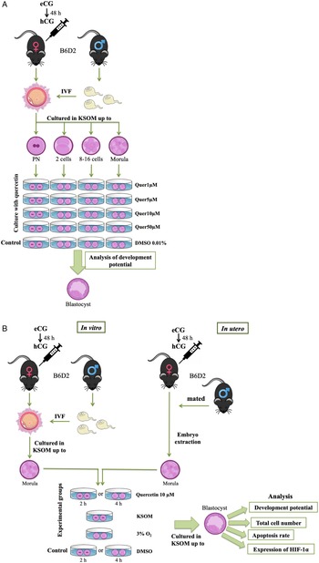
Quercetin (Sigma-Aldrich, Barcelona, Spain) diluted in dimethyl sulphoxide (DMSO; 0.01%) (Sigma-Aldrich, Barcelona, Spain) at different concentrations (1 µM, 5 µM, 10 µM, or 50 µM), was added to the culture medium at different stages of development [pronucleus (PN) stage, 2-cell, 8–16-cell and morulae], and then embryos were cultured until the blastocyst stage, at 37°C. To study the effect of the vehicle DMSO, a set of embryos was cultured only with DMSO (0.01%), and all the results were compared with embryos cultured in KSOM medium throughout the entire development (Figure 1A).
Diagram of the workflow of the treatment of the embryos with quercetin. (A) Treatment of the embryos during long periods of time. The top of the image shows how the embryos were obtained by in vitro fertilization and then cultured up to different stages of development. Embryos at different days of development were treated up to the blastocyst stage with different concentrations of quercetin, and then the development potential was studied (bottom part). (B) Treatment of the embryos for short periods of time. The top of the image shows obtaining the embryos for the experiments, by in vitro fertilization (left side) or by extraction from the uterus at the morula stage (right side). The bottom of the image explains how the embryos were treated with quercetin, or DMSO for the control, and what parameters were then analyzed.

Based on the results obtained, we performed new experiments exposing embryos to quercetin at 10 µM for short periods of time. We added this flavonoid to the culture medium of embryos at the morula stage for 2 h or 4 h. These embryos were both obtained using IVF or developed in utero (IU) (Figure 1B). Then, embryos were cultured again in conventional medium up to the blastocyst stage. For comparison, two other sets of embryos, both obtained using IVF and IU, were cultured either in KSOM medium or under 3% O2. Finally, once the embryos reached day 4.5 of development, the percentage of embryonic development, total cell number, number of apoptotic cells, and expression of HIF-1α protein were evaluated and compared with the control groups consisting of embryos cultured with DMSO but no quercetin, for 2 h or 4 h and then cultured in KSOM medium up to blastocyst stage (Figure 1B).
Nuclear staining
To count the total cell number, 4% paraformaldehyde (PFA)-fixed blastocysts were stained with 2 μg/ml of Hoechst 33342 (Eugene, OR, USA) and the nuclei were examined using a confocal microscope (Nikon Eclipse TE2000-U). The cell number was analyzed using Fiji ImageJ software (1.45q, Wayne Rasband, NIH, USA).
Terminal deoxynucleotidyl transferase dUTP nick end labelling (TUNEL) assay
Apoptotic cells in the embryos were detected using the in situ Cell Death Detection kit (TUNEL) from Roche (Mannheim, Germany) using the manufacturer’s protocol and nuclei were counterstained with Hoechst 33342 as previously described. Fluorescence images were taken using the same confocal microscope.
Immunofluorescence of HIF-1α protein
The immunodetection of HIF-1α was performed by incubating fixed embryos with a 1:100 dilution of the HIF-1α monoclonal antibody (Novus Biologicals; NB100-105) and using fluorescein isothiocyanate (FITC)-conjugated goat-anti-mouse IgG (Novus Biologicals; NB720-F) at 1:400 dilution as the secondary antibody. Nuclei were counterstained again with Hoechst 33342 and embryos were observed under a confocal laser-scanning microscope (Nikon Eclipse TE2000-U). Control embryos were processed as described above, but the primary antibody was omitted. The expression of HIF-1α was analyzed in a qualitative manner, to detect possible changes in its expression.
Statistical analysis
Statistical analyses were performed using SPSS software (SPSS Onc., Chicago. IL, USA), and a P-value less than 0.05 was considered statistically significant. Analysis of variance (ANOVA) was used to compare embryo proliferation, whereas the Mann–Whitney–Wilcoxon non-parametric test was performed to compare development rate and results from the TUNEL assay. Data expressed as percentages were analyzed using the chi-squared (χ2 ) test.
Results
Effect of quercetin on the development potential of mouse embryos
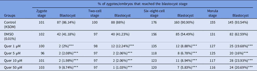
To investigate the effect of quercetin on embryo development, we first cultured in vitro obtained zygotes and embryos at different stages of development with different concentrations of quercetin (50 µM, 10 µM, 5 µM, and 1 µM) until they reached the blastocyst stage. Zygotes and embryos cultured in KSOM medium throughout the entire development were used as the control group. Our results showed that, regardless of the concentration of quercetin, the percentage of embryos that reached the blastocyst stage was significantly lower compared with the control (Table 1). Embryos cultured only with the solvent for quercetin, DMSO at a concentration of 0.01%, were also included in this experiment to analyze the potential effect of the dissolvent on the embryos and the development potential. Compared with control embryos, ∼50% of the embryos reached the blastocyst stage, a number much higher than in the groups of embryos treated with quercetin. This first observation indicated a negative or toxic effect of quercetin on embryo development that could not be attributed to the dissolvent. Despite these results, the Quer 10 µM group presented the highest number of embryos that developed to the blastocyst stage (Table 1), so we decided to use this concentration for further studies described next to try to counteract the toxic effect of quercetin.
Development using in vitro fertilization of zygotes or embryos at different stages in the presence of quercetin, up to the blastocyst stage

Differences between the groups were calculated using the χ²-test.
Quer, quercetin. ***P < 0.001 vs the control group.
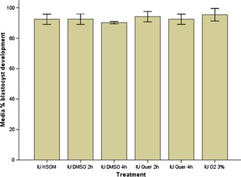
Based on the previous results that showed a detrimental effect of quercetin when used for long periods of time in the culture medium, the approach of this work was changed. Instead of studying the effect that quercetin produced on the development potential when used throughout the entire embryo culture, we decided to study its effects during short time intervals (2 h or 4 h) on embryos obtained directly from the uterus at the morula stage, a crucial stage in which embryos seemed to be less susceptible to the negative effects of quercetin/DMSO treatment (see data in Table 1). Interestingly, as can be seen in Figure 2, in this case, quercetin did not show a detrimental effect on embryo development potential but rather a beneficial effect, with a similar number of embryos reaching the blastocyst stage compared with control or low O2 treated embryos.
Results of the effect of quercetin culture for 2 h or 4 h in the pre-implantation development of embryos obtained in utero at the morula stage. IU KSOM group: embryos obtained from the uterus at the morula stage and developed to blastocyst in conventional culture medium, n = 26. IU DMSO 2 h group: embryos obtained from the uterus at the morula stage and cultured with DMSO for 2 h, n = 24. IU DMSO 4 h group: embryos obtained from the uterus at the morula stage and cultured with DMSO for 4 h, n = 24. IU Quer 2 h group: embryos obtained from the uterus at the morula stage and cultured with quercetin for 2 h, n = 18. IU Quer 4 h group: embryos obtained from the uterus at the morula stage and cultured with quercetin for 4 h, n = 24. IU O2 3% group: embryos obtained from the uterus at the morula stage and cultured in low-oxygen concentrations until blastocyst stage, n = 15. The data are represented as the mean ± the standard deviation (SD). P > 0.05 for all statistical analyses. Mann–Whitney–Wilcoxon test.

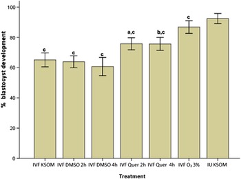
Therefore, we further studied the effects of quercetin on embryos obtained using IVF, cultured up to the morula stage in KSOM medium, and then treated with quercetin for 2 or 4 h. Our results indicated that there were statistically significant differences in all the experimental groups (Figure 3) in favour of embryos cultured with quercetin, both for 2 and 4 h. We also included for comparison embryos cultured at a low-oxygen concentration (IVF O2 3% group), observing that they behave similarly to those developed to the morula stage in the uterus (IU KSOM group) and, in all the experimental cases, embryos had a higher development potential than those cultured in conventional KSOM medium (IVF KSOM group).
Results of the effect of quercetin culture for 2 h or 4 h in the pre-implantation development of embryos obtained by in vitro fertilization (IVF) and compared with in utero obtained embryos. IVF KSOM group: embryos obtained by IVF and developed to the blastocyst stage in conventional culture medium, n = 18. IVF DMSO 2 h group: embryos obtained by IVF and cultured with DMSO for 2 h at the morula stage, n = 20. IVF DMSO 4 h group: embryos obtained by IVF and cultured with DMSO for 4 h at the morula stage, n = 16. IVF Quer 2 h group: embryos obtained by IVF and cultured with quercetin for 2 h at the morula stage, n = 16. IVF Quer 4 h group: embryos obtained by IVF and cultured with quercetin for 4 h at the morula stage, n = 20. IVF O2 3% group: embryos obtained by IVF and cultured with low concentrations of oxygen from morula to blastocyst stage, n = 16. IU KSOM group: embryos obtained from the uterus at the morula stage and developed to blastocyst in conventional culture medium, n = 26. The data are represented as the mean ± the standard deviation (SD). a, P < 0.01 vs IVF DMSO 2 h group; b, P < 0.005 vs IVF DMSO 4 h group; and c, P < 0.001 vs IU KSOM group. Mann–Whitney–Wilcoxon test.

Effect of quercetin on the total cell number at the blastocyst stage
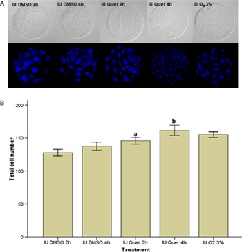
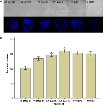
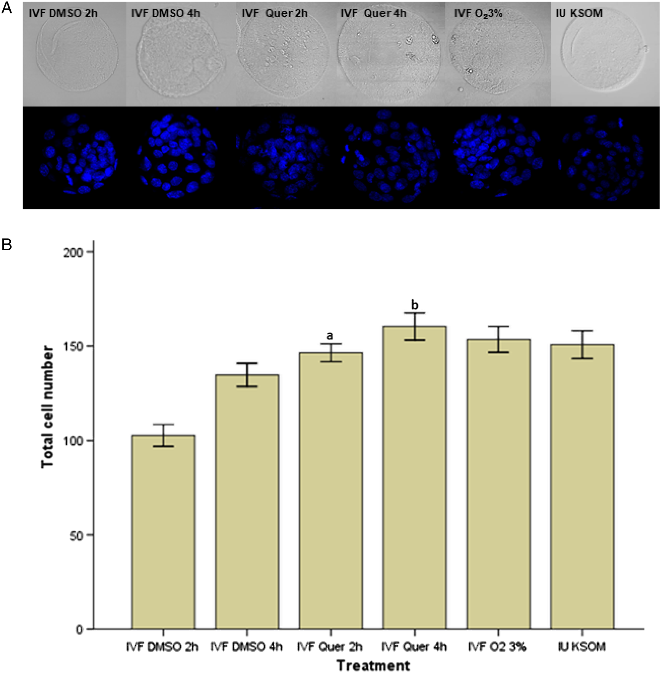
Once we confirmed the beneficial effects of quercetin on the development potential of embryos, we examined whether quercetin produced an increase in cell proliferation by analyzing the total cell number of the embryos that reached the blastocyst stage in our set of experiments. As can be seen in Figure 4, embryos that were developed in utero up to the morula stage and were treated with quercetin for either 2 or 4 h, presented a statistically significant higher average of cells at the blastocyst stage. For embryos treated for 4 h (IU Quer 4 h group), their total cell number was even higher than embryos cultured at low-oxygen concentrations (IU O2 3% group) (Figure 4B). Cell count was also performed in embryos obtained using IVF and exposed to quercetin at the morula stage (Figure 5). Results showed again that embryos presented a higher total cell number in quercetin and in the low-oxygen groups, showing differences that were statistically significant (Figure 5B); the total cell numbers were similar to those found in embryos developed in utero up to the morula stage.
Results of the total cell number in blastocysts obtained in utero at the morula stage. IU DMSO 2 h group: embryos obtained directly from the uterus and cultured with DMSO for 2 h at the morula stage, n = 30. IU DMSO 4 h group: embryos obtained directly from the uterus and cultured with DMSO for 4 h at the morula stage, n = 27. IU Quer 2 h group: embryos obtained directly from the uterus and cultured with quercetin for 2 h at the morula stage, n = 24. IU Quer 4 h group: embryos obtained directly from the uterus and cultured with quercetin for 4 h at the morula stage, n = 30. IU O2 3% group: embryos obtained directly from the uterus and cultured with low concentrations of oxygen from morula to blastocyst stage, n = 24. (A) Representative images of the total cell number, taken with a confocal microscope Nikon Eclipse TE2000-U, of embryos from each group. (B) Total cell number analysis. The data are represented as the mean ± the standard deviation (SD). a, P < 0.001 IU DMSO 2 h vs IU Quer 2 h; b, P < 0.001 IU DMSO 4 h vs IU Quer 4 h. Mann–Whitney–Wilcoxon test.

Results of the number of total cells in blastocysts obtained by in vitro fertilization (IVF). IVF DMSO 2 h group: embryos obtained by IVF and cultured with DMSO for 2 h at the morula stage, n = 33. IVF DMSO 4 h group: embryos obtained by IVF and cultured with DMSO for 4 h at the morula stage, n = 27. IVF Quer 2 h group: embryos obtained by IVF and cultured with quercetin for 2 h at the morula stage, n = 35. IVF Quer 4 h group: embryos obtained by IVF and cultured with quercetin for 4 h at the morula stage, n = 29. IVF O2 3% group: embryos obtained by IVF and cultured in low concentrations of oxygen from morula to blastocyst stage, n = 29. IU KSOM group: embryos obtained from the uterus at the morula stage and developed to blastocyst in conventional culture medium, n = 26. (A) Representative images of the total cell number, taken with a confocal microscope Nikon Eclipse TE2000-U, of embryos from each group. (B) Total cell number analysis. The data are represented as the mean ± the standard deviation (SD). a, P < 0.001 IVF DMSO 2 h vs IVF Quer 2 h; b, P < 0.001 IVF DMSO 4 h vs IVF Quer group 4 h, ANOVA, post-hoc Tukey test.
Effect of quercetin on the number of apoptotic cells in the blastocysts
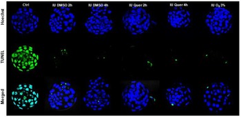
We tested using TUNEL the number of apoptotic cells in in utero developed embryos up to the morula stage and then treated with quercetin for short periods of time. Results showed that treatment with quercetin for 4 h reduced the number of apoptotic cells found in embryos, even though differences were not statistically significant. Furthermore, morulae cultured in low-oxygen concentrations also presented a not statistically significant lower number of apoptotic cells (Figure 6; Table 2).
Representative images illustrating the incidence of cell death in mouse embryos obtained in utero. Nuclei were stained with Hoechst 33342 (blue). Fragmented DNA was labelled using the TUNEL assay (green). Merged images represent the proportion of positive TUNEL cells (apoptotic, middle row) with respect to the embryo total cell count (bottom row). IU DMSO 2 h group: embryos obtained in utero at the morula stage and cultured with DMSO for 2 h and grown to the blastocyst stage in conventional culture medium, n = 25. IU DMSO 4 h group: embryos obtained in utero at the morula stage and cultured with DMSO for 4 h and grown to the blastocyst stage in conventional culture medium, n = 25. IU Quer 2 h group: embryos obtained in utero at the morula stage and cultured with quercetin for 2 h and developed to the blastocyst stage in conventional culture medium, n = 25. IU Quer 4 h group: embryos obtained in utero at the morula stage and cultured with quercetin for 4 h and developed to the blastocyst stage in conventional culture medium, n = 25. IU O2 3% group: embryos obtained in utero and cultured in low-oxygen concentrations from the morula to the blastocyst stage, n = 25. Ctrl: positive control, which consisted of in utero obtained embryos treated with DNase to induce apoptosis before incubation with TUNEL reagents.

Number of apoptotic cells in embryos obtained in utero from female mice and treated with quercetin or DMSO

Differences between the groups were calculated using the χ²-test. Quer, quercetin.
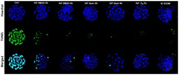
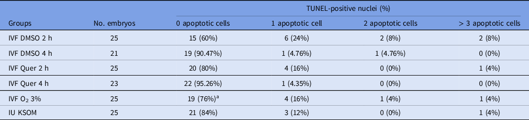
In addition, programmed cell death was also evaluated in embryos obtained using IVF, which usually presented more apoptosis. Results showed that embryos treated with quercetin for 4 h (IVF Quer 4 h group) presented lower apoptotic rates, and a higher number of embryos without apoptotic cells (Figure 7; Table 3), although differences were not statistically significant.
Representative images illustrating the incidence of cell death in mouse embryos obtained by in vitro fertilization. nuclei were stained with Hoechst 33342. Fragmented DNA was labelled using the TUNEL assay (green). Merged images represent the proportion of positive TUNEL cells (apoptotic, middle row) with respect to the embryo total cell count (bottom row). IVF DMSO 2 h group: embryos obtained by in vitro fertilization and cultured with DMSO for 2 h at the morula stage, n = 25. IVF DMSO 4 h group: embryos obtained by in vitro fertilization and cultured with DMSO for 4 h at the morula stage, n = 21. IVF Quer 2 h group: embryos obtained by in vitro fertilization and cultured with quercetin for 2 h at the morula stage, n = 25. IVF Quer 4 h group: embryos obtained by in vitro fertilization and cultured with quercetin for 4 h at the morula stage, n = 23. IVF O2 3% group: embryos obtained by in vitro fertilization and cultured in low-oxygen concentrations from the morula to blastocyst stages, n = 25. IU KSOM group: embryos obtained in utero at the morula stage and developed to the blastocyst stage in conventional culture medium, n = 25. Ctrl: positive control, which consisted of in vitro obtained embryos treated with DNase to induce apoptosis before incubation with TUNEL reagents.

Number of apoptotic cells in embryos obtained by in vitro fertilization and treated with quercetin or DMSO

Differences between the groups were calculated using the χ²-test. Quer, quercetin. aP < 0.05, IVF Quer 4 h vs IVF O2.
Expression of HIF-1α
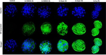
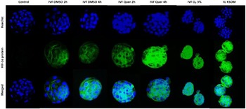
Immunofluorescence was performed to study the expression of HIF-1α in embryos cultured under different conditions, as explained before. Results showed that in in utero obtained embryos, the expression of HIF-1α was similar in all groups despite the treatment used (Figure 8). Conversely, when we studied the protein expression in in vitro obtained embryos treated with quercetin, especially for 4 h, or in embryos cultured under low-oxygen conditions (IVF O2 3% group), there was a significant increase in the expression of HIF-1α (Figure 9) compared with embryos cultured under conventional conditions, which clearly showed a reduction in the expression of HIF-1α. Noticeably, the fluorescence signal observed in embryos treated with quercetin was similar to that observed in embryos developed in utero. Finally, we must mention that a clear decrease in the expression of HIF-1α could also be observed in embryos cultured only with DMSO.
Immunofluorescence analysis of HIF-1α protein expression in mouse embryos obtained in utero. Nuclei were stained with Hoechst 33342 (blue). The expression of HIF-1α was marked using FITC (green). IU DMSO 2 h group: embryos developed in utero up to the morula stage and cultured with DMSO for 2 h and cultured until the blastocyst stage in conventional culture medium, n = 25. IU DMSO 4 h group: embryos developed in utero up to the morula stage and cultured with DMSO for 4 h, then cultured until the blastocyst stage in conventional culture medium, n = 25. IU Quer 2 h group: embryos developed in utero up to the morula stage and cultured with quercetin for 2 h, then developed until the blastocyst stage in conventional culture medium, n = 25. IU Quer 4 h group: embryos developed in utero up to the morula stage and cultured with quercetin for 4 h, then developed to the blastocyst stage in conventional culture medium, n = 25. IU O2 3% group: embryos developed in utero up to the morula stage and cultured in low-oxygen concentrations to the blastocyst stage, n = 25. Ctrl: negative control, which consisted of embryos developed in utero from which the primary antibody was omitted.

Immunofluorescence analysis of HIF-1α protein expression in mouse embryos obtained in vitro. Nuclei were stained with Hoechst 33342 (blue). The expression of HIF-1α was marked using FITC (green). IVF DMSO 2 h group: embryos obtained by IVF and cultured with DMSO for 2 h at the morula stage, n = 25. IVF DMSO 4 h group: embryos obtained by IVF and cultured with DMSO for 4 h at the morula stage, n = 21. IVF Quer 2 h group: embryos obtained by IVF and cultured with quercetin for 2 h at the morula stage, n = 25. IVF Quer 4 h group: embryos obtained by IVF and cultured with quercetin for 4 h at the morula stage, n = 23. IVF O2 3% group: embryos obtained by IVF and cultured in low-oxygen concentrations from the morula to the blastocyst stage, n = 25. IU KSOM group: embryos obtained in utero at the morula stage and developed to the blastocyst stage in conventional culture medium, n = 25. Ctrl: negative control, which consisted of embryos obtained by IVF from which the primary antibody was omitted.

Discussion
The development potential of embryos obtained using IVF, as well as their quality and viability, depends among other things on the composition of the culture medium in which they are developed (Paternot et al., Reference Paternot, Debrock, D’Hooghe and Spiessens2010). A non-optimal culture can lead to a failure in the fertilization of the gametes and an inadequate development of the embryos, which do not reach the blastocyst stage or fail to implant in the uterus due to their poor quality. Therefore, optimal culture conditions are of utmost importance for the IVF of gametes, pre-implantation embryonic development, and implantation in all species. Therefore, increasing the in vitro development potential of early embryos by supplementation of the culture medium has been of great interest to many researchers and, consequently, there have been several works in which the culture medium has been enriched with many compounds that improved the quality of the embryos obtained using IVF.
In the present work, we analyzed the consequences of supplementation of the culture medium with quercetin, a flavonoid with antioxidant properties. Quercetin has been reported to have a protective effect on mouse embryos against H2O2, and a better rate of blastocyst formation was observed when embryos were cultured for 3 h with quercetin (Yu et al., Reference Yu, Long, Lyu, Zhang, Yan, Liang, Chai, Yan, Kuang and Qi2014). It has also been reported that more than 100 µM of quercetin in the culture medium caused some defects and abnormalities, whereas 3 µM of this molecule had a protective effect on mouse embryos against hydroxyurea (Pérez-Pastén et al., Reference Pérez-Pastén, Martínez-Galero and Chamorro-Cevallos2010). Furthermore, a protective effect of quercetin was demonstrated in embryos against 0.005 µg/ml of actinomycin D (Sameni et al., Reference Sameni, Javadinia, Safari, Tabrizi Amjad, Khanmohammadi, Parsaie and Zarbakhsh2018). Our results suggested that quercetin produces a detrimental effect on embryonic development when cultured for long periods of time regardless of the stage of development. This could be caused by different things. First of all, after 24 h in the culture medium, quercetin stops being stable (Hu et al., Reference Hu, Chen, Lei, Tian, Xing, Chai, Zhao, Ding and Du2012) and loses its effectiveness as an antioxidant, producing toxic products for embryos. Another possibility is the fact that quercetin increases the expression of γ-glutamylcysteine synthetase, and therefore it promotes the synthesis of glutathione (Moskaug et al., Reference Moskaug, Carlsen, Myhrstad and Blomhoff2005). However, after long periods of time, quercetin sequesters cysteine from the culture medium, which is a precursor of glutathione, and it has been demonstrated that cleavage-stage embryos have a limited capacity to synthesize reduced glutathione, and appear to be negatively affected under conditions that deplete glutathione (Gardiner and Reed, Reference Gardiner and Reed1995). It is also known that quercetin has been seen as an antiproliferative and proapoptotic compound in several lines of cancer cells and this property could explain its negative effects on embryos in culture because they cannot grow properly (Hashemzaei et al., Reference Hashemzaei, Delarami Far, Yari, Heravi, Tabrizian, Taghdisi, Sadegh, Tsarouhas, Kouretas, Tzanakakis, Nikitovic, Anisimov, Spandidos, Tsatsakis and Rezaee2017). Additionally DMSO, which is the usual vehicle for dissolving quercetin, has been proved to have cytotoxic effects in mouse pre-implantation embryos (Kang et al., Reference Kang, Das, Gurunathan, Park, Song, Park and Kim2017), which could explain some of the results observed here but not the drastic effects observed with quercetin. Another possibility is that this cytotoxic effect and the instability of quercetin after 24 h could be producing a synergic effect with DMSO and be responsible for the low rates of blastocyst development observed here.
Recently, Hernández et al. (Reference Hernández, López-Morató, Perianes, Sánchez-Mateo, Cassa-rua, Domínguez-Arroyo, Sánchez-Margallo and Álvarez2020) showed that an improvement in embryo quality occurs when 4OHE2 is added at the time of compaction (day 3 of pre-implantation development, morula stage), which indicates that embryos are more sensitive to certain environmental changes when they are at the morula stage. Nonetheless, in in vitro obtained embryos, our study corroborates that the addition of quercetin at the time of embryo compaction, for a period of 2 h or 4 h, results in a greater number of embryos that develop to the blastocyst stage, compared with embryos cultured in conventional medium, and results almost resemble those observed in embryos cultured in low-oxygen concentrations. These results indicate that quercetin does not negatively affect embryos and, furthermore, appears to counteract the detrimental effects produced by the in vitro culture conditions.
Putting this information in the context of embryo development, it seems clear that exposing early cleavage embryos to certain compounds and for long periods of time impairs normal development and they fail to reach the blastocyst stage, possibly for some of the reasons explained above. This could apply to hormones and their metabolites, or antioxidants such as quercetin, which reduces the viability of embryos by the different means stated above. Nevertheless, the introduction of the same compounds in the morula to blastocyst transition for short periods of time can have a beneficial effect when embryos are cultured in vitro. Obviously, the possible implications of this situation on human embryology and the culture of human embryos must be manifested.
There are two conventional possibilities for in vitro human embryo culture for IVF techniques. One is sequential culture, which implies changing the culture medium on day 3 of development to a more appropriate medium for extended culture and the other is the unique or one-step culture, in which the same culture medium is used during the entire embryo development (Tao et al., Reference Tao, Zhou, Yan, Wu, Cheng, Ye, Wang and Li2022). Our results would be in favour of the first type of culture, because the needs and susceptibility of the embryos are very different during the early steps from zygote to morula, compared with later on when the blastocyst forms. Our data could gain insight into the possible modification of the culture medium at the morula stage to achieve better results in IVF. This information could also help some laboratories that do not have access to low-oxygen incubators, because the addition of quercetin at the morula stage resembles the success that these systems provide in the IVF clinics.
In addition to improving the development potential, our results showed that there was an increase in the total cell number when we use quercetin, data in concordance with other works that also showed this increase after using quercetin (Yu et al., Reference Yu, Long, Lyu, Zhang, Yan, Liang, Chai, Yan, Kuang and Qi2014; Sameni et al., Reference Sameni, Javadinia, Safari, Tabrizi Amjad, Khanmohammadi, Parsaie and Zarbakhsh2018). It is important to mention that the increase was not uncontrolled and the maximum number of total cells observed remained at ∼160. This observation is interesting because a higher number of cells do not relate to a higher implantation potential, and it can be indicative of bad quality (Wigger et al., Reference Wigger, Kisielewska, Filimonow, Plusa, Maleszewski and Suwińska2017).
Regarding apoptosis rates, several studies have demonstrated that there was a reduction in the number of apoptotic cells in blastocysts when embryos were treated with quercetin after treatment with stressful substances or in aged mice (Yu et al., Reference Yu, Long, Lyu, Zhang, Yan, Liang, Chai, Yan, Kuang and Qi2014; Sameni et al., Reference Sameni, Javadinia, Safari, Tabrizi Amjad, Khanmohammadi, Parsaie and Zarbakhsh2018; Cao et al., Reference Cao, Zhao, Wang, Zhang, Bian, Liu, Zhang, Zhang and Zhao2020). Our results show that, indeed, culturing embryos obtained using IVF with quercetin produced a reduction in the number of apoptotic cells per embryo, finding fewer embryos with apoptotic cells when they were cultured for 4 h with quercetin. However, when embryos were obtained from the uterus at the morula stage and cultured with quercetin, a reduction in the number of embryos that had apoptotic cells was not observed, and many of the embryos presented only a single apoptotic cell, matching results observed in the control. Again, these results showing the effects of quercetin in in vitro obtained embryos, but not in embryos developed in utero up to the morula stage, suggested that quercetin does not affect the embryos per se, but it appears to counteract the negative effects that the components and conditions of the in vitro culture medium produce in the embryos fertilized in vitro. The effects produced by quercetin could be attributed to its antioxidant properties (De Matos et al., Reference De Matos, Gasparrini, Pasqualini and Thompson2002; Kere et al., Reference Kere, Siriboon, Lo, Nguyen and Ju2013; Rocha-Frigoni et al., Reference Rocha-Frigoni, Leão, Dall’Acqua and Mingoti2016).
Oxygen tension is known to influence embryo development and cell number in various species and one of its major negative consequences is the production of ROS. Conversely, oxygen levels have a significant influence on gene expression patterns that are mediated by the heterodimeric transcription factor HIF-1α (Myhrstad et al., Reference Myhrstad, Carlsen, Nordström, Blomhoff and Moskaug2002; Rocha-Frigoni et al., Reference Rocha-Frigoni, Leão, Dall’Acqua and Mingoti2016). Harvey (Mastroianni Jr and Jones Reference Mastroianni and Jones1965; Khanduja et al., Reference Khanduja, Verma and Bhardwaj2001) suggested that HIF may be involved in the molecular mechanisms that respond to changes in oxygen status during embryo development in mouse and bovine blastocysts. Some studies have shown that HIF-1α could be activated by the flavonoid quercetin to heal ulcers (Jeon et al., Reference Jeon, Kim, Choi, Kim, Park, Kim, Kim and Jung2007) and produced an increase of HIF-1α in processes related to inflammation and vascular diseases in human placental cells (Bogacz et al., Reference Bogacz, Mikołajczak, Wolek, Górska, Szulc, Ożarowski, Kujawski, Czerny, Wolski, Karpiński and Seremak-Mrozikiewicz2021). Our study demonstrated that the localization of HIF-1α was predominantly in the cytoplasm of cells, which is consistent with the previous study by Thompson et al. (Reference Thompson, Feil, Edwards, Lane and Karen2004) in which HIF-1α was detected in the cytoplasm of mouse blastocysts when cultured in low-oxygen concentrations (2%) during compaction and blastulation. Furthermore, embryos obtained using IVF and cultured with quercetin for 4 h had a higher expression of the HIF-1α protein. This increase was similar to that observed in embryos obtained using IVF cultured with 3% O2, and in embryos obtained directly from the uterus. In contrast, embryos cultured in conventional medium hardly expressed HIF-1α protein. Putting these results together, we can determine that quercetin, when added to the culture medium of embryos for short periods of time, produces an increase in the expression of HIF-1α, which could produce an increase in the activation of the genes necessary for the reduction of ROS levels and repair mechanisms.
In conclusion, this study illustrates how changing the experimental conditions can turn a molecule from being toxic for the embryos when left in the culture medium for long periods of time, to be beneficial for them when the exposure time is reduced to a short period. The results provided here illustrate a potential strategy for obtaining better quality embryos and improving the success rates of assisted reproductive technologies, especially for those who do not have access to incubators with low-oxygen concentrations. Nonetheless, the translation of the use of quercetin to the clinic needs much more data about its effects and mechanisms of action, as an excess of polyphenols can result in negative effects in early embryos (Yang et al., Reference Yang, Fang, Lambert, Yan and Huang2008).
Acknowledgements
This study was financed by a grant from Junta de Extremadura: Ayuda de Grupos Catalogados de la Junta de Extremadura, GR18199, co-financed by the European Regional Development Fund (ERDF). We would also like to thank the Biomedicine Applied Techniques Facility of the University of Extremadura for allowing us to use their confocal microscope and the Cell Therapy Laboratory of the Jesús Usón Minimally Invasive Surgery Centre for allowing us to use their fluorescence microscope.
Author contribution
NH carried out all the experiments; NH, ML-M, and SS-M drafted the article; ML-M, SS-M, NH, and ISA participated in the critical review; ISA and FMS-M approved the final version and provided financial support.
Financial support
This work was financed by a grant from the Project HIF-MCERA IRA116, co-financed by the European Regional Development Fund (ERDF), and Ayuda a Grupos Catalogados de la Junta de Extremadura, GR18199.
Conflicts of interest
The authors declare that they have no known competing financial interests or personal relationships that could have appeared to influence the work reported in this paper.
Ethical standards
The authors assert that all procedures contributing to this work comply with the ethical standards of the relevant national and institutional guides on the care and use of laboratory animals.