Recently, in 2005, Loeys et alReference Loeys, Chen and Neptune1 discovered a new syndrome that shares many features with Marfan syndrome, and the mutations that produce the newly coined Loeys–Dietz syndrome were identified. It is caused by heterozygous mutations in the genes encoding the citokine family transforming growth factor, resulting in loss of activity in the transforming growth factor receptors-β types 1 and 2. The disease is characterised by the triad of the arterial tortuosity, aneurysm, or dissections; hypertelorism; and bifid uvula or cleft palate.Reference Loeys, Schwarze and Holm2, Reference Williams, Loeys and Nwakanma3 Here, we present a patient with confirmed Loeys–Dietz syndrome and complete angiographic manifestations revealed with magnetic resonance imaging.
Case description
A 7-year old boy of 22 kilograms weight and 124 centimetres height with confirmed heterozygous mutation in the gene encoding the transforming growth factor β-receptor I (Loeys–Dietz syndrome type I) and type-I insulin deficiency-diabetes mellitus was referred to our unit. Echocardiography estimated the diameter of the aortic sinuses of valsalva as 34.9 millimetres; Z-score = 7.96. Echocardiography accurately measures maximal ascending aortic dimension as compared with magnetic resonance imaging,Reference Snider, Enderlein, Teitel and Juster4 and may be a simpler and more cost-effective method of monitoring aortic dimensions in patients with ascending aortic dilation if images are acceptable;Reference Wong, Edwards and Armstrong5, Reference Didier, Saint-Martin and Lapierre6 however, magnetic resonance imaging is superior for complete aortic arch evaluation.Reference Kaiser, Kellenberger, Albisetti, Bergstrasser and Valsangiacomo Buechel8
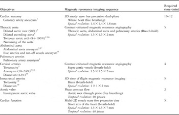
He was referred to the cardiac magnetic resonance imaging unit to assess the dilated aortic root and discard-associated cardiovascular malformations (Table 1). Cardiac magnetic resonance imaging was performed on a 1.5 T Intera MR scanner (Philips Medical Systems, Best, the Netherlands) with standard protocols in the axial, coronal, and sagittal planes by using T1-weighted, time of flight cine images, phase contrast flow of ascending aorta, three-dimensional contrast-enhanced magnetic resonance angiogram, and three-dimensional steady state free precession volume scan. According to the body weight, a multi-channel phased array coil was chosen to cover the thoracic and abdominal aorta. Our acquisition technique required 45 minutes and it is detailed in Table 1.
Summary of the main objectives to assess magnetic resonance imaging.

The described cardiovascular malformations found in the Loeys–Dietz syndrome are shown in the first column (If prevalence has been previously described is shown in brackets). The proposed magnetic resonance imaging sequences and its spatial resolution to investigate them are described in the second column. Required time for each sequence is detailed in the third column
Magnetic resonance imaging of the brain was performed on the aforementioned scanner with standard protocols in the axial, coronal, and sagittal planes by using T1-weighted, T2-weighted, fluid-attenuated inversion recovery, magnetic resonance angiography, and post-contrast T1-weighted images.
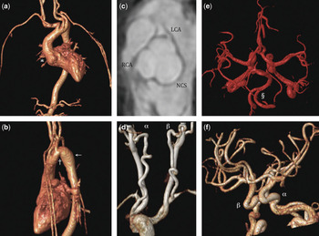
In cardiac magnetic resonance imaging, normal segmental heart anatomy and function was found. Contrast-enhanced magnetic resonance angiography was performed to assess vascular anatomy and discard the associated malformations (Table 1). It has been shown to have an excellent correlation with conventional angiography for measurements of vascular structures.Reference Valsangiacomo Buchel, DiBernardo, Bauersfeld and Berger7 On these grounds, normal values for aortic diameters have been standardised using contrast-enhanced magnetic resonance angiography.Reference Kaiser, Kellenberger, Albisetti, Bergstrasser and Valsangiacomo Buechel8 There was a dilatation of the aortic root (Fig 1a). The dilatation produced some asymmetry in the non-coronary cusp (Fig 1c), resulting in mild central aortic valvar insufficiency. This was estimated as less than 3% using a phase contrast flow sequence, and assessed as mild in the previous echo. No coronary artery anomalies were found, although coronary artery aneurysms have been previously identified in Loeys–Dietz syndrome.Reference Loeys, Chen and Neptune1 There was also a dilated sino tubular junction 24 × 23 millimetres (>P95; Fig 1a), and ascending aorta, which continued through the transverse aortic arch and the thoracic descending aorta, resulting in smooth curves and a low degree of tortuosity (Fig 1a and b). The transverse arch in the isthmic region was kinked with mild narrowing (Fig 1b), with mild dilatation of the upper descending aorta, after the isthmic narrowing. In the two-dimensional cine steady state free precession sequence performed in the aortic arch plane, there was systolic flow turbulence after the narrowing, but no arterial hypertension was documented.
(a) A three-dimensional-shaded reconstruction from contrast enhanced magnetic resonance imaging anterior view. Dilated aortic root (*), ascending aorta (20 × 21 mm, >P95), elongation of the whole thoracic aorta with marked kinking and tortuous course particularly of the transverse aortic arch; left aortic arch; no aneurysm in the abdominal aorta. (b) A three-dimensional -shaded reconstruction from contrast-enhanced magnetic resonance imaging left lateral view. Aortic arch kinking and narrowing (10 × 12 mm) in the isthmic region (†). Mild dilatation of the upper descending aorta (←) (16 × 15 mm, >P95) and normal descending aorta. (c) A three-dimensional steady state free precession reformatted plane through the dilated aortic sinus (32 × 34 mm, >P95) at the level of the right (RCA) and left (LCA) coronary arteries offspring. Dilated and asymmetric non-coronary sinus (NCS). (d). A three-dimensional-shaded reconstruction from contrast-enhanced magnetic resonance imaging posterior view of supra-aortic vessels. Tortuous left (α) and right (β) internal carotid arteries, with no discrete narrowing. (e) Shaded surface reconstruction from the time of flight magnetic resonance imaging of the arterial circle of Willis, superior view. Marked tortuosity of all intracranial brain arteries and also of the extracranial basilaris/vertebral system (§), but no evidence for stenosis or aneurysm. (f) Shaded surface reconstruction from the time of flight magnetic resonance imaging of the arterial circle of Willis, left anterior view. Tortuousity of the left (α) and right (β) internal carotid arteries.

Aneurysms in the abdominal aorta or in the branching vessels have been identified in previous reports, with an incidence of 10 and 7%, respectively.Reference Loeys, Chen and Neptune1, Reference Loeys, Schwarze and Holm2 Iliac arteries or its run-off vessels aneurysm have been described as well.Reference Johnson, Chen, Loeys, Dietz and Fishman9 In this case, no abdominal aorta malformations were found, nor were any pulmonary artery abnormalities.Reference Loeys, Chen and Neptune1
The head-and-neck arteries were elongated and tortuous (Fig 1d). The distortion was more marked in the proximal segments of the supra-aortic branches, with no discrete narrowing.
The brain magnetic resonance imaging showed marked tortuosity of all extracranial (Fig 1e) and intracranial brain arteries (Fig 1f), particularly in the basilaris/vertebral system, but no evidence for stenosis or aneurysm at this point. Head- and-neck aneurysms are present in 10% of patients,Reference Loeys, Schwarze and Holm2, Reference Johnson, Chen, Loeys, Dietz and Fishman9 including intracranial aneurysm of the retinal vessels and extracranial aneurysm or dissections in the carotid and vertebrobasilar system.
Arterial tortuosity has been documented by Johnson et alReference Rodrigues, Elsayed, Loeys, Dietz and Yousem10 in all the patients with confirmed syndrome.Reference Johnson, Chen, Loeys, Dietz and Fishman9 Some of these cases had extreme distortion severity, as well as other craniofacial abnormalities such as craniosynostosis (48%), hydrocephalus (12%), and Chiari malformation (8%). These abnormalities should also be investigated.
Conclusion
Widespread vascular manifestations can be found in the Loeys–Dietz syndrome involving intracranial, neck, thoracic, and diaphragmatic aorta malformations, such as distortions, aneurysm, dissections, and stenosis.Reference Johnson, Chen, Loeys, Dietz and Fishman9
Magnetic resonance imaging is a useful tool to assess the vascular anatomy and should cover all the previously detailed body regions where vascular abnormalities have been described. We suggest a magnetic resonance imaging protocol as described in this manuscript (Table 1). Detailed information about the calibre of the aorta, supra- aortic vessels, pulmonary arteries, and aneurysm vessels should be documented for follow-up assessment. Advantages over computed tomography include the fact that the patient receives no ionising radiation during the scan with magnetic resonance imaging. This is particularly appropriate when assessing Loeys–Dietz syndrome due to the need for large body area assessment and the careful sequential evaluation over time.
Optimal management should include early recognition of the syndrome, carefully surveillance of the vascular anatomy, and timely surgical intervention, due to the propensity towards rupture and dissection (12%) at a younger age.Reference Rodrigues, Elsayed, Loeys, Dietz and Yousem10
Acknowledgements
The authors thank Simon Holden, Consultant Clinical Geneticist, for assistance and for the referral of patients. This study was supported by the Talentia Excellence Grant awarded by the Ministry for Innovation, Science, and Enterprise, Andalusia (Spain).