Introduction
Arthropods harbour exceptionally diverse bacterial symbionts that live exclusively within host cells and undergo maternal (transovarial) transmission to offspring (Duron et al., Reference Duron2008; Wernegreen, Reference Wernegreen2012; Bennett and Moran, Reference Bennett and Moran2015). These intracellular symbionts use specific adaptive strategies to spread and persist within arthropod populations: some are obligate mutualistic symbionts required for host survival while some others are facultative symbionts conferring fitness benefits depending on environmental stress or manipulating host reproduction (Engelstadter and Hurst, Reference Engelstadter and Hurst2009; Cordaux et al., Reference Cordaux, Bouchon and Greve2011; Bennett and Moran, Reference Bennett and Moran2015). Overall, intracellular symbionts are of ecological and evolutionary importance to the particular arthropod species that they infect, potentially mediating the adaptation of their host to their environments, driving changes in sex determination, inducing reproductive isolation and speciation (Engelstadter and Hurst, Reference Engelstadter and Hurst2009; Cordaux et al., Reference Cordaux, Bouchon and Greve2011; Brucker and Bordenstein, Reference Brucker and Bordenstein2012; Wernegreen, Reference Wernegreen2012).
Diverse bacterial lineages have independently evolved symbiotic interactions with arthropods (Duron et al., Reference Duron2008; Moran et al., Reference Moran, Mccutcheon and Nakabachi2008; Engelstadter and Hurst, Reference Engelstadter and Hurst2009). One of the most remarkable of these symbionts is Candidatus Midichloria mitochondrii (Midichloria mitochondrii hereafter) (Alphaproteobacteria: Rickettsiales: Midichloriaceae; Sassera et al., Reference Sassera2006). While intracellular symbionts are typically found within a host-derived vacuole or phagosome or free in the cytoplasm (e.g. Zchori-Fein et al., Reference Zchori-Fein2004; Gottlieb et al., Reference Gottlieb2008; Fischer et al., Reference Fischer2014; Lalzar et al., Reference Lalzar, Friedmann and Gottlieb2014), M. mitochondrii is unique since it commonly colonizes the inter-membrane space of host mitochondria (Lewis, Reference Lewis1979; Zhu et al., Reference Zhu, Aeschlimann and Gern1992; Beninati et al., Reference Beninati2004; Sacchi et al., Reference Sacchi2004; Sassera et al., Reference Sassera2006). This symbiont is widespread in populations of the castor bean tick Ixodes ricinus: it infects almost all adult females and around half of adult males of this tick species (Lo et al., Reference Lo2006; Sassera et al., Reference Sassera2008; Duron et al., Reference Duron2017; Mukhacheva and Kovalev, Reference Mukhacheva and Kovalev2017). High densities of M. mitochondrii in host ovaries indicates controlled vertical transmission into the developing oocytes (Lewis, Reference Lewis1979; Zhu et al., Reference Zhu, Aeschlimann and Gern1992; Beninati et al., Reference Beninati2004) with 100% of I. ricinus progeny being infected (Lo et al., Reference Lo2006; Sassera et al., Reference Sassera2008). Although its effect on I. ricinus remains unknown, it was recently suggested that M. mitochondrii may be an obligate nutritional symbiont providing B vitamins (Duron et al., Reference Duron2017) since its genome encodes pathways for the synthesis of major B vitamins and cofactors (Sassera et al., Reference Sassera2011). It may notably enable diet specialization of I. ricinus on vertebrate blood: B vitamins are the expected nutritional complements required for strict haematophagy as also observed with other nutritional symbionts in lice, tsetse flies or bedbugs (Akman et al., Reference Akman2002; Hosokawa et al., Reference Hosokawa2010; Boyd et al., Reference Boyd2014; Nikoh et al., Reference Nikoh2014). Another interesting feature of M. mitochondrii genome is the presence of a gene set associated with flagellar assembly (Sassera et al., Reference Sassera2011). A few other members of Rickettsiales have been found to have flagellar genes (e.g. Cand. Jidaibacter acanthamoeba in Midichloriaceae; Schulz et al., Reference Schulz2016) or functional flagella (e.g. Cand. Trichorickettsia mobilis in Rickettsiaceae; Vannini et al., Reference Vannini2014). Although M. mitochondrii is a non-motile bacterium without flagellum (Zhu et al., Reference Zhu, Aeschlimann and Gern1992; Sacchi et al., Reference Sacchi2004; Sassera et al., Reference Sassera2011), the flagellum genes are not pseudogenized and are transcribed (Sassera et al., Reference Sassera2011). Their role for M. mitochondrii however remains unclear (Sassera et al., Reference Sassera2011).
Since its initial discovery, membership of the genus Midichloria has expanded although M. mitochondrii remains the single formally described species (Sassera et al., Reference Sassera2006). While related genera as Lariskella are symbionts with a larger host spectrum, Midichloria seems however highly specific to ticks (Matsuura et al., Reference Matsuura2012; Montagna et al., Reference Montagna2013). Beyond I. ricinus, Midichloria has been observed in several other species of the Ixodes genus as well as in ticks of diverse genera (Epis et al., Reference Epis2008; Beninati et al., Reference Beninati2009; Dergousoff and Chilton, Reference Dergousoff and Chilton2011; Williams-Newkirk et al., Reference Williams-Newkirk2012; Cafiso et al., Reference Cafiso2016; Duron et al., Reference Duron2017). Despite the maternal inheritance of infection, no evidence of co-cladogenesis between Midichloria and ticks has been detected: this suggested that Midichloria can undergo occasional horizontal transfers (HT) across hosts, resulting in limited specificity to tick species (Epis et al., Reference Epis2008). Two observations further revealed that main biological features of Midichloria differ substantially between tick species. First, only three tick species, I. ricinus, Ixodes aulacodi and Ixodes holocyclus, show a high prevalence of Midichloria suggesting that it may be an obligate symbiont (Beninati et al., Reference Beninati2009; Gofton et al., Reference Gofton2015; Cafiso et al., Reference Cafiso2016; Duron et al., Reference Duron2017). In other infected tick species, Midichloria has a low to medium prevalence (e.g. Cafiso et al., Reference Cafiso2016; Duron et al., Reference Duron2017) rather suggesting a facultative symbiont not required for tick survival. Second, not all Midichloria have an intra-mitochondrial life style. In at least one tick species, I. holocyclus, only infection in cytoplasm, and not in mitochondria, was observed despite intensive examination (Beninati et al., Reference Beninati2009). The case studies above have thus evidenced an important variation of biological traits within the Midichloria genus.
At present, the 16S rRNA gene sequences are routinely used for the description of Midichloria infections, with almost all known strains represented only by this gene (e.g. Epis et al., Reference Epis2008; Cafiso et al., Reference Cafiso2016; Duron et al., Reference Duron2017). However, this marker is highly conserved and exhibits a limited resolution for discriminating closely related species and for inferring their evolutionary histories in bacteria (Naum et al., Reference Naum, Brown and Mason-Gamer2008; Novakova et al., Reference Novakova, Hypsa and Moran2009; Poretsky et al., Reference Poretsky2014). In view of this, we developed here a multi-locus typing approach for Midichloria which works broadly with strains found in diverse tick species. The multi-locus typing scheme characterizes Midichloria variation at five housekeeping genes (16Sr RNA, groEL, rpoB, dnaK and ftsZ). Given the singularity of the presence of flagellum genes in the M. mitochondrii genome, we also use one of these genes, fliC, as an additional Midichloria marker. Using this approach, we thus attempted to characterize the whole Midichloria diversity and to infer the evolutionary processes shaping its diversity.
Materials and methods
Tick DNA collection
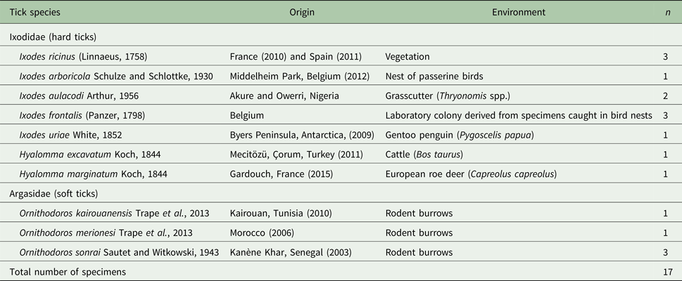
A collection of DNA templates from 10 tick species infected by Midichloria was used for the present analysis (Table 1). Of the 10 tick species examined, three belong to the Argasidae family (soft ticks) and to the Ornithodoros genus. The seven other species belong to the Ixodidae family (hard ticks) and to the Ixodes (five species) or the Hyalomma (two species) genera. Most specimens were collected on taxonomically diverse bird and mammal hosts or their environment in Europe, Africa and Antarctica between 2006 and 2015 (Table 1). A few specimens were also obtained from a laboratory colony of I. frontalis derived from field specimens collected in Belgium. Each DNA template was obtained from individual extraction of tick whole body using the DNeasy Blood and Tissue Kit (QIAGEN) following manufacturer instructions. For each DNA template, Midichloria infection had been previously confirmed through 16S rRNA gene sequencing (Duron et al., Reference Duron2017). Within each examined tick species, there were one to three Midichloria-infected individuals separately investigated for the present analysis (Table 1).
List and origin of tick species examined in this study

Multi-locus typing of Midichloria
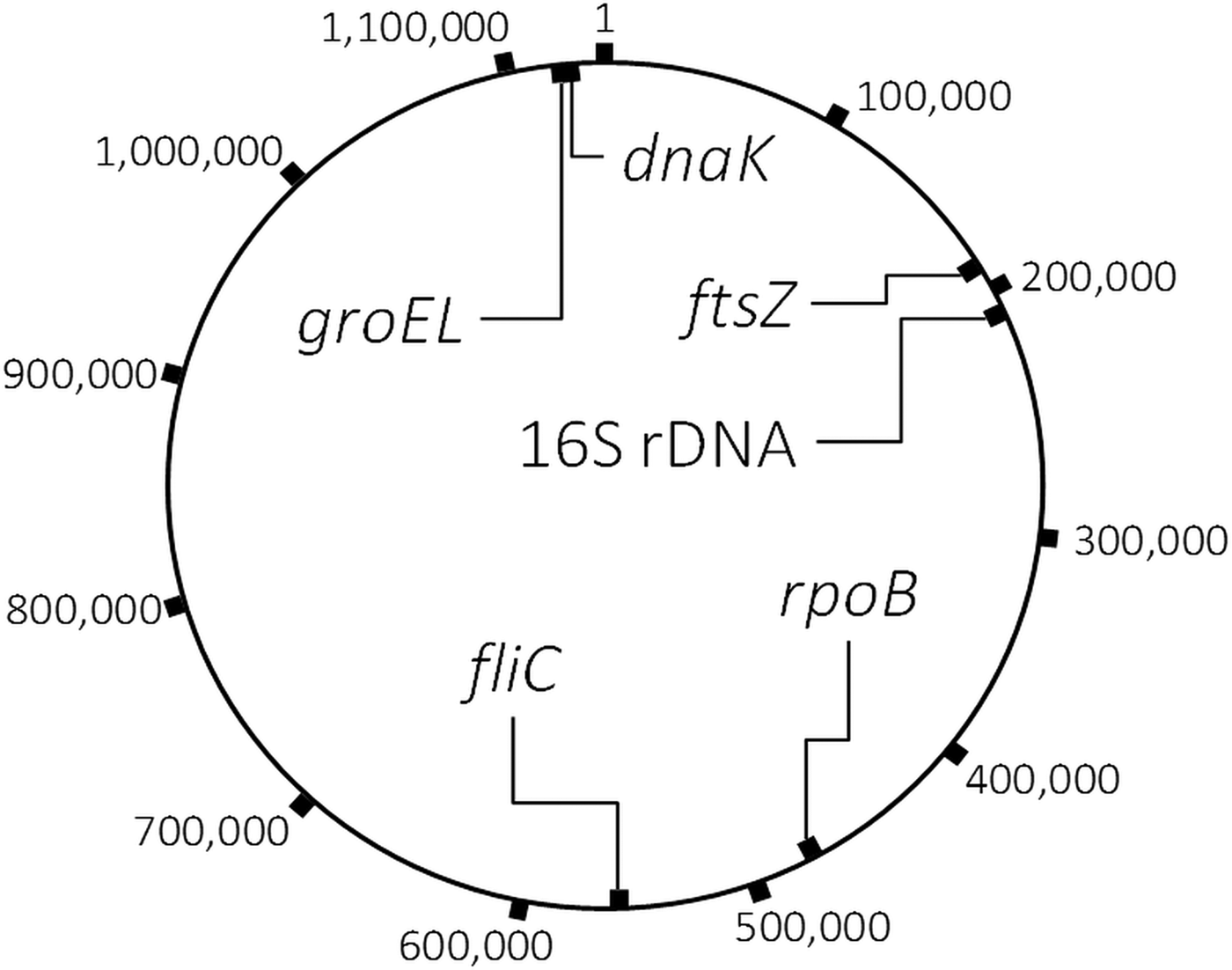
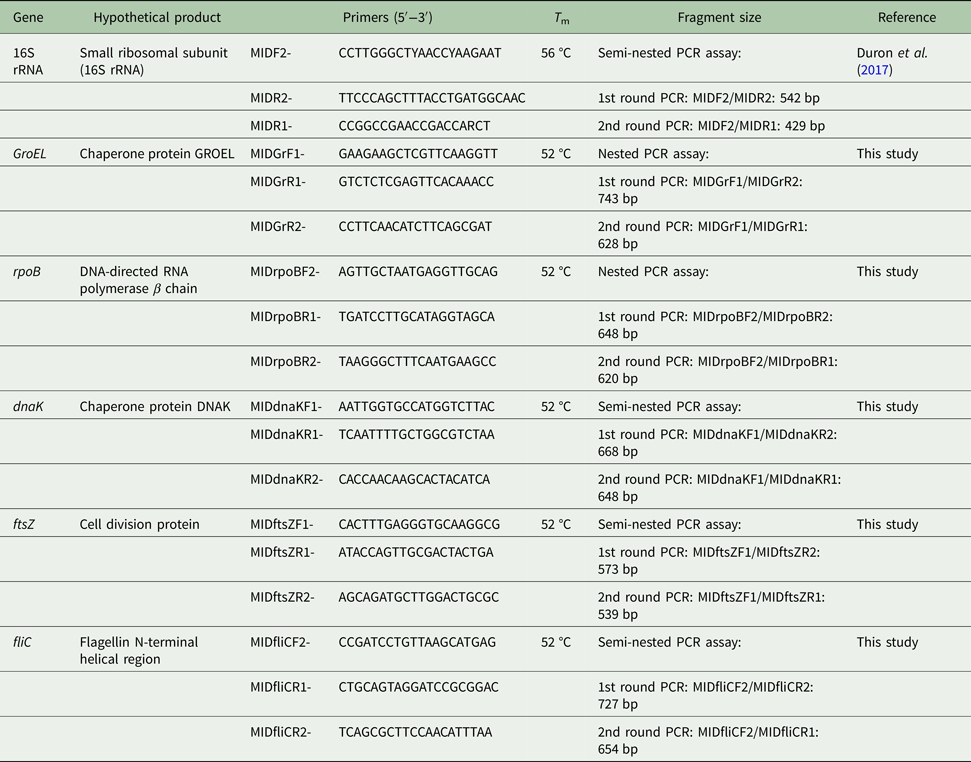
We used the single published genome of Midichloria (M. mitochondrii IricVA strain, isolated from I. ricinus; Sassera et al., Reference Sassera2011; GenBank accession number: CP002130) as reference to design specific PCR primers for multi-locus typing. Along with the 16S rRNA gene, we selected five other genes (groEL, rpoB, dnaK, ftsZ and fliC) present in a single copy and physically distant within the M. mitochondrii IricVA reference genome (Fig. 1). Five (16S rRNA, groEL, rpoB, dnaK and ftsZ) are housekeeping genes (i.e. required for the maintenance of basic bacterial function) while fliC encodes for a flagellin which can putatively polymerize to form bacterial flagella. Fragments of these six genes were amplified using semi-nested PCR assays (Table 2). The use of semi-nested PCR was efficient at decreasing the probability of contamination from unwanted amplification products (false positives). To prevent possible contamination, different parts of this process have been physically separated from one another, in entirely separate rooms. All amplicons from semi-nested PCR assays were also sequenced to control for false positive amplifications. Gene features, primers and PCR conditions are detailed in Table 2. The efficiency of our typing method was ascertained through positive PCR amplification and clear sequences for the six loci from DNA samples of I. ricinus infected by M. mitochondrii as we further detailed in the ‘Results’ section.
Map of the Midichloria mitochondrii IricVA chromosome (1 183 732 bp; GenBank accession number: CP002130), showing the locations of the six gene markers examined in this study.

List and description of primers used in this study

Semi-nested PCR amplifications were performed as follows: the first PCR run with the external primers was performed in a 10 µL volume containing ca. 20 ng of genomic DNA, 3 mm of each dNTP (Thermo Scientific), 8 mm of MgCl2 (Roche Diagnostics), 3 µ m of each primer, 1 µL of 10 × PCR buffer (Roche Diagnostics), and 0.5 U of Taq DNA polymerase (Roche Diagnostics). A 1-μL aliquot of the PCR product from the first reaction was then used as a template for the second round of amplification. The second PCR was performed in a total volume of 25 µL and contained 8 mm of each dNTP (Thermo Scientific), 10 mm of MgCl2 (ThermoScientific), 7.5 µ m of each of the internal primers, 2.5 µL of 10 × PCR buffer (Thermo Scientific), and 1.25 U of Taq DNA polymerase (Thermo Scientific). All PCR amplifications were performed under the following conditions: initial denaturation at 93 °C for 3 min, 35 cycles of denaturation (93 °C, 30 s), annealing (T m = 52–56 °C, depending on primers, 30 s), extension (72 °C, 1 min) and a final extension at 72 °C for 5 min. Known positive and negative individuals were used as controls in each PCR assay. All PCR products were visualized through electrophoresis in a 1.5% agarose gel. Positive PCR products were purified and sequenced in both directions (EUROFINS). The chromatograms were manually inspected and cleaned with CHROMAS LITE (http://www.technelysium.com.au/chromas_lite.html) and sequence alignments were done using CLUSTALW (Thompson et al., Reference Thompson, Gibson, Higgins, Baxevanis, Petsko, Stein and Stormo2002), both implemented in MEGA (Tamura et al., Reference Tamura2007). Alleles were determined on the basis of sequence identity (i.e. the amount of characters which match exactly between two different sequences). If two sequences differ by one or more bases, they were described as two different alleles. Novel nucleotide sequences have been deposited in the GenBank nucleotide database (Accession numbers: MH060029-77, MH077540-54 and MH049468-76).
Molecular phylogenetics
The GBLOCKS program (Castresana, Reference Castresana2000) with default parameters was used to remove poorly aligned positions and to obtain unambiguous sequence alignments. All sequence alignments were also checked for putative recombinant regions using the RDP3 computer analysis package (Martin et al., Reference Martin2010). Given a set of aligned nucleotide sequences, RDP3 can rapidly analyse these with a range of powerful non-parametric recombination detection methods, including the GENECONV (Sawyer, Reference Sawyer1999) and RDP (Martin and Rybicki, Reference Martin and Rybicki2000). Phylogenetic relationships were evaluated between Midichloria strains using 16S rRNA, groEL, rpoB, dnaK, ftsZ and fliC gene sequences. In addition to sequences produced in this study, additional Midichloria sequences, as well as a subset of sequences from bacterial outgroups, available from GenBank were also used. The evolutionary models most closely fitting the sequence data were determined using Akaike information criterion with the MEGA program (Tamura et al., Reference Tamura2007). Phylogenetic analyses were based on maximum likelihood (ML) analyses. An ML heuristic search, using a starting tree obtained by neighbour-joining, was conducted and clade robustness was further assessed by bootstrap analysis using 1000 replicates in MEGA (Tamura et al., Reference Tamura2007).
Results
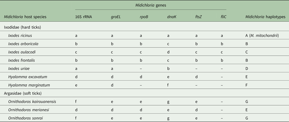
The diversity of Midichloria was examined using 16S rRNA, groEL, rpoB, dnaK, ftsZ and fliC gene sequences. The Midichloria 16S rRNA, groEL and dnaK genes were amplified from all tick DNA templates (n = 17), while the rpoB, ftsZ and fliC genes were amplified from part of them (n = 15, 15 and 9, respectively, as shown in Table 3). The rpoB and ftsZ genes were not amplified from Midichloria of I. uriae (n = 1) and of Hyalomma marginatum specimens (n = 1). The fliC gene was amplified only from Midichloria of Ixodes spp. (n = 9), excepted of I. uriae (n = 1). The failure of rpoB, ftsZ and fliC PCR amplifications may be due to a consequence of mutations in the priming regions. Since fliC is not a housekeeping gene, failure of PCR amplification at this locus may be also due to gene deletion. All sequences were easily readable without double peaks, indicating a confident degree of primer specificity for Midichloria semi-nested PCR amplifications. All these sequences belong unambiguously to the Midichloria genus as described below. Further characterizations were thus based on three-to-six Midichloria gene markers per DNA template (Table 3).
Allelic profile of the six polymorphic genes in Midichloria from 10 tick species

Letters a–g represent the different alleles at each Midichloria locus. Dashes indicate an absence of gene PCR amplification. Letters A–G represent the seven Midichloria haplotypes identified on the basis of 16S rRNA, groEL, rpoB, dnaK, ftsZ and fliC allelic profiles.
On the basis of DNA sequencing, we characterized three-to-seven distinct alleles depending on Midichloria gene (Table 3), including six 16S rRNA alleles (98.4–99.7% pairwise nucleotide identity), five groEL alleles (93.9–99.1%), five rpoB alleles (94.1–99.1%), seven dnaK alleles (93.8–99.8%), five ftsZ alleles (93.5–99.0%) and three fliC alleles (94.8–97.1%). The six 16S rRNA alleles we observed here showed a 98.7–100% pairwise nucleotide identity with the M. mitochondrii IricVA strain, suggesting that all these bacteria belong to the M. mitochondrii species. Worthy of note is that the use of multiple gene markers allowed the distinction of closely related Midichloria strains otherwise not discriminated by their 16S rRNA gene sequences. Indeed, Midichloria of I. ricinus and I. uriae harbour the same 16S rRNA allele but have different dnaK alleles showing that each tick species is infected by a different Midichloria haplotype (Table 3). Overall, the allelic variation at the six gene markers led to the identification of seven genetically different Midichloria haplotypes (A–G hereafter, cf. Table 3) allied to 10 tick species. No sequence variation at any Midichloria gene was observed between DNA templates from the same tick species, suggesting that only one Midichloria haplotype was present in each tick species of our collection.
The Midichloria A haplotype from I. ricinus samples showed 100% nucleotide identity at the six gene markers with the M. mitochondrii IricVA-type strain (GenBank accession number: CP002130). All the other tick species were infected by Midichloria haplotype differing from the M. mitochondrii IricVA-type strain (Table 3). Most the tick species harboured a different Midichloria haplotype with the exception of three species pairs, which were each infected by a genetically identical Midichloria haplotype on the basis of 16S rRNA, groEL, rpoB, dnaK, ftsZ and fliC typing. These tick species are I. arboricola/I. frontalis (Midichloria haplotype B), Ornithodoros merionesi/H. excavatum (Midichloria haplotype E) and O. sonrai/O. kairouanensis (Midichloria haplotype G) (Table 3).
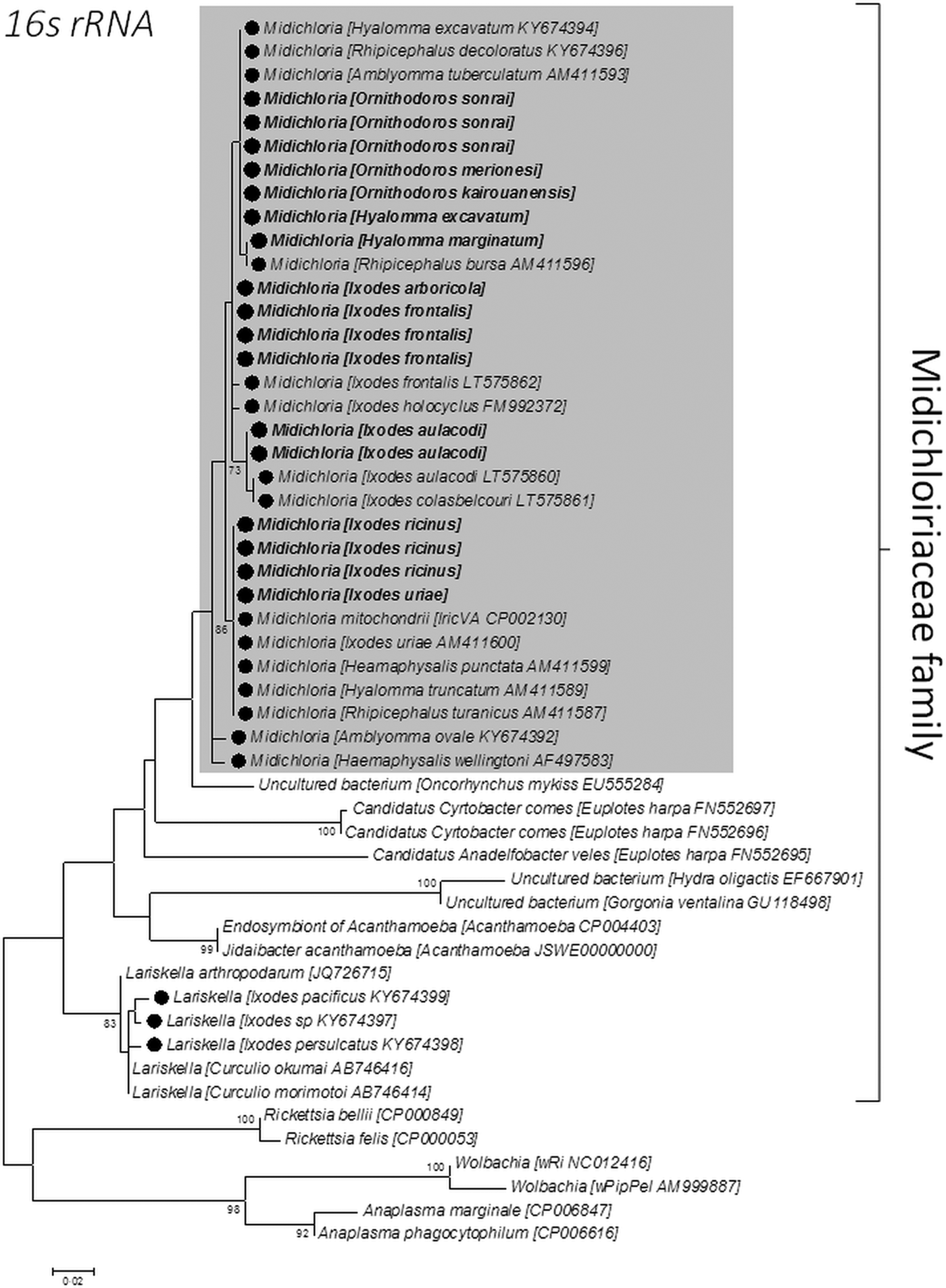
We further examined the evolutionary relationships between the different Midichloria haplotypes, including M. mitochondrii IricVA. A comparison with other Midichloria strains reported from previous studies was however restricted to the 16S rRNA gene sequences since no Midichloria groEL, rpoB, dnaK, ftsZ and fliC gene sequences were available on GenBank (with the exception of the M. mitochondrii IricVA complete genome). An ML analysis based on 44 bacterial 16S rRNA gene sequences was first used to examine the phylogenetic relationship of our 17 Midichloria 16S rRNA gene sequences with 15 other Midichloria 16S rRNA gene sequences available in GenBank, as well as bacterial outgroups, including Midichloria sister groups, such as the Lariskella genus and other uncultured Midichloriaceae (Fig. 2). We recovered the clustering of all Midichloria 16S rRNA gene sequences within a monophyletic clade, and the Midichloria characterized in this study are phylogenetically closely related to the Midichloria found in other tick species. The intra-Midichloria topology based on 16S rRNA gene sequences remains however too poorly resolved (as shown by low support values of inner branches) for inferring the exact relatedness between Midichloria from different tick species (Fig. 2).
Midichloria phylogeny constructed using maximum-likelihood (ML) estimations based on 16S rRNA gene sequences (360 unambiguously aligned bp). Midichloria 16S rRNA gene sequences obtained from the tick DNA templates examined in this study (n = 17) are bolded. Sequences from other Midichloria and bacterial outgroups, including other members of Midichloriaceae, available in GenBank were added to the analysis. Grey box delineates the Midichloria genus. Black circles indicate members of Midichloriaceae found in ticks. Bacterial name, host species and GenBank accession numbers are shown on the tree. Branch numbers indicate percentage bootstrap support for major branches (1000 replicates; only bootstrap values >70% are shown). The scale bar is in units of substitution/site.

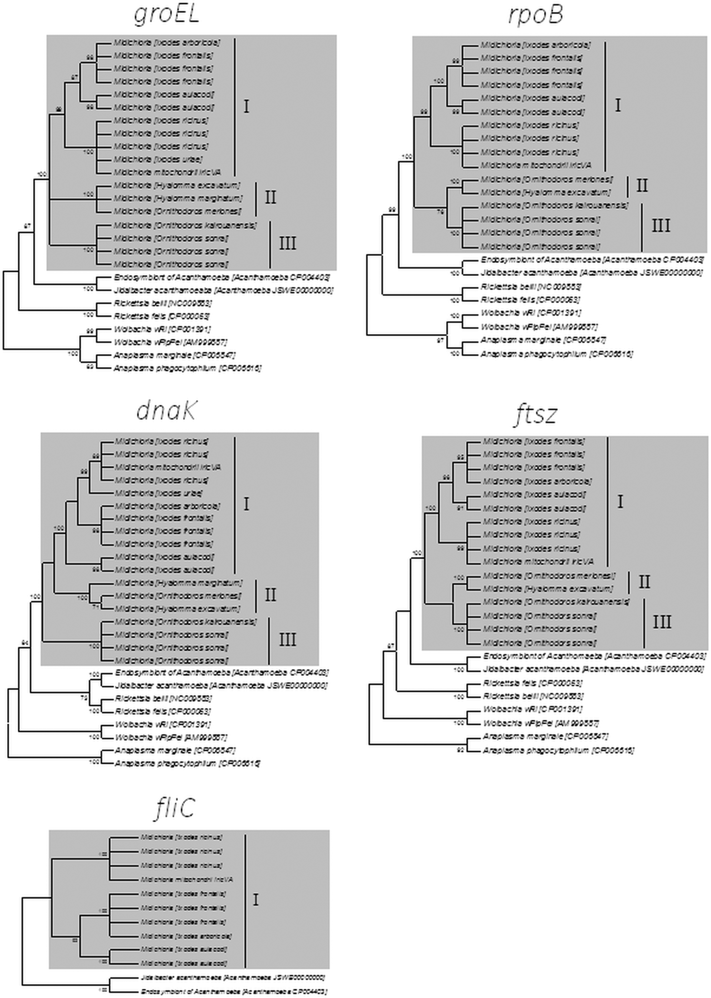
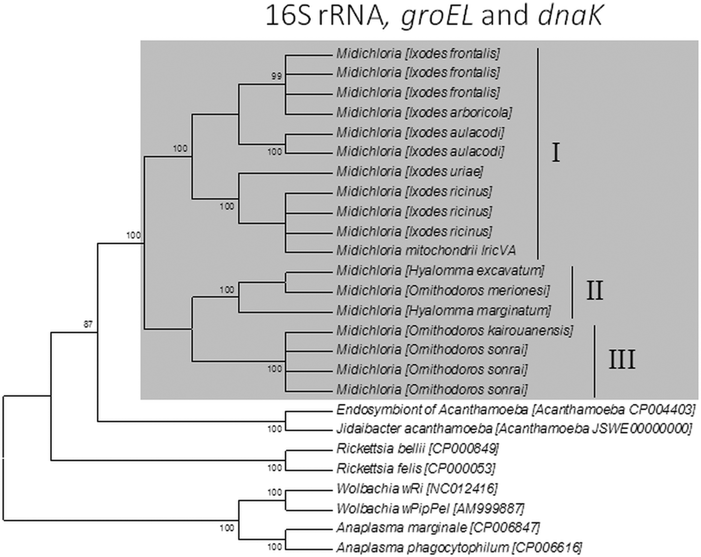
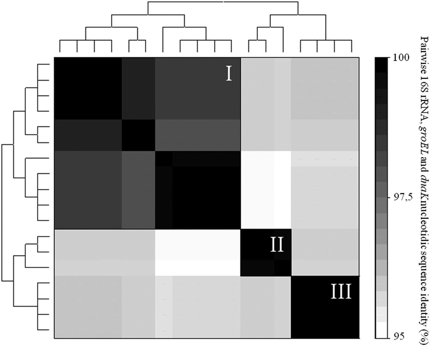
Novel ML analyses based on groEL, rpoB, dnaK, ftsZ and fliC gene sequences were conducted to refine the Midichloria intrageneric phylogeny using our 17 Midichloria-positive DNA templates, the M. mitochondrii IricVA genome sequence and bacterial outgroups. When the sequences were examined separately for each gene, ML analyses confirmed that all Midichloria described in this study unambiguously belong to the Midichloria genus (Fig. 3). We observed no sign of recombination in the dataset: (i) the comparison between Midichloria single gene phylogenies revealed the same phylogenetic branching (Fig. 3) and (ii) the analysis of concatenated sequences did not detect significant recombination events between Midichloria strains (all P > 0.10 for the GENECONV and RDP recombination–detection tests). All single groEL, rpoB, dnaK and ftsZ gene phylogeny consistently showed the partitioning of Midichloria diversity into three distinct lineages (I-to-III hereafter): all Midichloria from Ixodes spp. (i.e. I. uriae, I. arboricola, I. frontalis, I. aulacodi and I. ricinus), including the M. mitochondrii IricVA-type strain, cluster in the lineage I; Midichloria from Hyalomma spp. (H. excavatum and H. marginatum) and O. merionesi cluster in the lineage II; Midichloria from O. sonrai and O. kairouanensis cluster in the lineage III (Fig. 3). In this context, the fliC phylogeny is not informative since this gene was only amplified from members of the lineage I (Table 3 and Fig. 3). An additional ML analysis was next conducted on all tick species using 16S rRNA, groEL and dnaK Midichloria concatenated dataset (since rpoB, ftsZ and fliC were not amplified from all tick species). It also confirms the partitioning of Midichloria diversity into the I-to-III lineages with high bootstrap supports (Fig. 4). Clustering analysis showed that within each lineage the pairwise 16S rRNA, groEL and dnaK nucleotide identity is always >98% while identity between lineages is 95–96% (Fig. 5).
Cladogram depicting the 50% majority-rule consensus of Midichloria phylogenetic trees constructed using maximum-likelihood (ML) estimations based on groEL (501 unambiguously aligned bp), rpoB (580 bp), dnaK (545 bp), ftsZ (410 bp) and fliC (614 bp) sequences. Grey box delineates the Midichloria genus. Midichloria I-to-III lineages, bacterial name, host species and GenBank accession numbers are shown on the tree. Branch numbers indicate percentage bootstrap support for major branches (1000 replicates; only bootstrap values >70% are shown).

Cladogram depicting the 50% majority-rule consensus of Midichloria phylogenetic trees constructed using maximum-likelihood (ML) estimations based on concatenated 16S rRNA, groEL and dnaK gene sequences (1406 unambiguously aligned bp). Grey box delineates the Midichloria genus. Midichloria I-to-III lineages, bacterial name, host species and GenBank accession numbers are shown on the tree. Branch numbers indicate percentage bootstrap support for major branches (1000 replicates; only bootstrap values >70% are shown). The scale bar is in units of substitution/site.

Heatmap representing clustering analysis based on pairwise identity percentages in the 16S rRNA, groEL and dnaK Midichloria nucleotidic sequences (1406 unambiguously aligned bp). The phylogenetic trees on the left and the top are the same cladogram based on 16S rRNA, groEL and dnaK Midichloria sequences shown in Fig. 4. Darker blocks indicate greater levels of nucleotide identity between Midichloria allowing the visualization of lineages I-to-III.

Discussion
In this study, we refine the molecular characterization of Midichloria by developing a typing framework using multi-locus gene sequencing. On this basis, we show that a wide genetic diversity exists within the Midichloria genus: we evidence three distinct Midichloria lineages allied to different tick species. The M. mitochondrii IricVA type strain belongs to the lineage I while more divergent Midichloria strains belong to lineages II and III. All these lineages can be assigned to the M. mitochondrii species since variation observed at the 16S rRNA gene sequence range in classical values used to delineate bacterial species. However, the split of M. mitochondrii into three distinct lineages is indicative of variable biological features, including degrees of host specialization, as we further detailed.
While previous phylogenetic analyses based exclusively on 16S rRNA gene sequences did not show evidence of Midichloria specificity to their tick hosts (Epis et al., Reference Epis2008; Cafiso et al., Reference Cafiso2016; Duron et al., Reference Duron2017), the use of more variable molecular markers revealed that at least some Midichloria are specialized to some degree, possessing a capacity to maintain infections in only limited range of related tick species. Indeed, Midichloria associated to the five Ixodes species examined in this study belong to the lineage I which forms a robust monophyletic group. This pattern thus suggests that Midichloria infection dynamics may be driven by a specialization that manifests in the form of host range limitations. In considering why related Midichloria infect related tick hosts we can envision three non-exclusive explanations:
(1) Persistence of maternally inherited Midichloria infection over long periods of time may result in symbiont diversification alongside the tick host that is to say co-cladogenesis. This mechanism may explain why closely related Midichloria strains are found in I. arboricola, I. frontalis and I. aulacodi which all belong the ‘Non-Australasian Ixodes’ monophyletic clade and are thus related. However, the presence of closely Midichloria strains in I. ricinus and I. uriae counter this hypothesis since these two tick belong to divergent Ixodes clades (i.e. the ‘Non-Australasian Ixodes’ and the ‘Australasian Ixodes’ clades, respectively). Co-cladogenesis alone is thus insufficient to explain the Midichloria lineage I specialization to Ixodes spp.
(2) Another possibility is that similar ecologies of related tick species may facilitate HT of related Midichloria. In this context, feeding on a shared vertebrate may be a particularly important determinant. Indeed, the presence of M. mitochondrii in the I. ricinus salivary glands, along with its serological and molecular detection in mammalian blood and sera, suggests that vertebrates may serve as intermediate hosts for Midichloria HT among tick species (Mariconti et al., Reference Mariconti2012; Bazzocchi et al., Reference Bazzocchi2013). With regard to this scenario, the observation of a genetically identical Midichloria strain in I. arboricola and I. frontalis is particularly relevant since these two species are nidicolous ticks highly specialized on songbirds in Western Europe (Heylen et al., Reference Heylen2014).
(3) The last possibility is that some Midichloria may be functionally specialized on related tick species: some Midichloria may be adapted to infect related tick species because related host species are likely to share similar physiologies. This mechanism has been documented for other maternally inherited bacteria as Wolbachia, with symbionts more likely to persist after transfer between related arthropod species (reviewed by Russell et al., Reference Russell2009). Such genetic specialization process has likely favoured prolonged association of related groups of ticks and Midichloria over evolutionary time, potentially leading to the evolution of reciprocal dependence. Interesting with this regard is that the lineage I contains Midichloria with high infection prevalence in two tick species, i.e. I. ricinus (Beninati et al., Reference Beninati2009; Duron et al., Reference Duron2017) and I. aulacodi (Cafiso et al., Reference Cafiso2016), a pattern compatible with an obligate mutualistic Midichloria symbiosis. Other Midichloria of lineage I are however likely facultative symbionts since low to medium Midichloria prevalence were observed in I. uriae and I. arboricola (Duron et al., Reference Duron2017).
Whatever the explanation, Midichloria specialization would not only reveal limitations in tick host range, but it would also indicate that tick-associated phylogenetic barriers promote divergence and diversification among Midichloria. An important consideration for future studies will thus be to understand the ecological and evolutionary pathways that facilitate the Midichloria specialization and its consequences for the biology of ticks.
In spite of the observation of Midichloria specialization, it is also essential to note that some highly related Midichloria strains are found in distantly related tick species. This situation applies to the lineage II which includes nearly identical Midichloria strains of hard ticks (H. marginatum and H. excavatum) and soft ticks (O. merionesi). In this context, HT of lineage II Midichloria within tick communities seems recent and likely ongoing. Hence, specialization is not a general trend and some Midichloria are capable of occasional HT between distantly related tick species, as pointed by previous studies (Epis et al., Reference Epis2008; Cafiso et al., Reference Cafiso2016). Two distinct evolutionary strategies are thus found in Midichloria: some are highly specialized concerning their tick hosts, with likely ancient acquisition, while others are more generalists and acquired through recent HT. It is likely that these different strategies are also illustrated, as stated above, by differences in dependency and effects on their tick hosts but also in other important biological features such as intracellular localization or propensity to be transferred during blood meal. In this context, the presence of the fliC flagellum gene in the Midichloria lineage I, but not in other lineages, may also indicate substantial variations in the biology of Midichloria which remain to be described.
Although the multi-locus dataset reveals an important Midichloria diversity, we can also speculate that the observed pattern is substantially underestimated. There are two major sources of underestimation in our study. First, the sample size within each tick species is still limited and this may lead to underestimate the Midichloria diversity per tick species. Indeed, because of maternal inheritance, persistence of Midichloria infection in a tick species over long periods of time should result in diversification of Midichloria alongside the host mitochondrial lineages. Since a high mitochondrial diversity is reported from I. ricinus populations (Carpi et al., Reference Carpi2016), one would expect that slightly divergent Midichloria haplotypes may coexist, each being associated with a different I. ricinus mitochondrial haplotype (this pattern is well documented for other maternally inherited symbionts; for examples, see Atyame et al., Reference Atyame2011; Dumas et al., Reference Dumas2013; Duron et al., Reference Duron2017). The second cause of underestimation is the possibility of coinfections: several Midichloria haplotypes may coexist within the same tick specimen. The Sanger sequencing approach that we used in this study may have missed this intra-host diversity if one Midichloria haplotype is more abundant than others, leading to the absence of double peaks on chromatograms. This coinfection pattern has been observed in other host–symbiont systems thanks to next generation sequencing approaches (Gottlieb et al., Reference Gottlieb, Lalzar and Klasson2015; Choi et al., Reference Choi2016). All these biases may thus lead to a global underestimation, suggesting that a diversity of Midichloria, greater than we observed in this study, exists.
To summarize, the pattern of Midichloria multi-locus diversity found in ticks leads to several important conclusions. First, in some cases, closely related Midichloria strains appear more prone to infect related tick host species, suggesting some degree of Midichloria/tick specificity, as shown with Midichloria lineage I and Ixodes ticks. Second, closely related Midichloria strains can infect unrelated tick species, revealing a labile and dynamic history of associations. Third, HT may be an important route of Midichloria dispersal even within tick genera. As a result, relatedness of Midichloria among Ixodes species should not be taken as evidence of a single infection event followed by strict Midichloria vertical transmission and co-cladogenesis. Overall, this study provides strong evidence for the combined contribution of specialization and HT to promote Midichloria diversification among ticks. Important questions however remain. Notably, what is yet to be established is the nature of interactions between Midichloria and ticks. An interesting possibility is that the effect of Midichloria varies between tick species. This hypothesis remains now to be investigated.
Acknowledgements
We thank V. Noël for technical help and C. Arnathau, P. Durand, D. Heylen, K. McCoy, M. Opara, O. Plantard, F. Renaud, J.F. Trappe and R. van Oosten for providing specimens and discussion. We also acknowledge useful discussions with members of the working group ‘Tiques et Maladies à Tiques’ of the ‘Réseau Ecologique des Interactions Durables’.
Financial support
This study was supported by recurrent funding from the Centre National de la Recherche Scientifique (CNRS) and the Institut pour la Recherche et le Développement (IRD).
Conflicts of interest
None.
Ethical standards
Not applicable.