INTRODUCTION
Research made to date on representatives of the South American Porcellanidae have consisted primarily of descriptions of adults and their zoogeography (Haig, Reference Haig1966; Bremec & Cazzaniga, Reference Bremec and Cazzaniga1984; Gomes-da Silva et al., Reference Gomes-da Silva, da Costa-Braga and D'Incao1989; Boschi et al., Reference Boschi, Fischbach and Iorio1992; Gomes-Veloso & Schmidt, Reference Gomes-Veloso and Schmidt1993; Boschi, Reference Boschi1997; Stillman & Reeb, Reference Stillman and Reeb2001; Lardies et al., Reference Lardies, Rojas and Wehrtmann2004; Lazaruz-Agudelo & Cantera-Kintz, Reference Lazaruz-Agudelo and Cantera-Kintz2007; Emperaza, Reference Emperaza2007). Studies carried out on early stages of development in this family have only included observations on morphology and phylogeny of their larvae (Boschi et al., Reference Boschi, Scelzo and Goldstein1967; Scelzo, Reference Scelzo1976; Pinheiro & Fransozo, Reference Pinheiro and Fransozo1995; Lopez et al., 1997; Wehrtmann et al., Reference Wehrtmann, Albornoz, Pardo and Véliz1997; Fransozo et al., Reference Fransozo, Cuesta and Negreiros-Fransozo1998; Hattori & Pinheiro, Reference Hattori and Pinheiro2001; Hernáez-Bove, Reference Hernáez-Bove2001; Hernáez & Palma, Reference Hernáez and Palma2003; Hernández et al., Reference Hernández, Magán, Graterol, Gaviria, Bolaños and Lira2003; Rodríguez et al., Reference Rodríguez, Hernández, Magán, Bolaños and Felder2004; Piñate et al., Reference Piñate, Lira and Hernández2005; González-Pisani et al., Reference González-Pisani, Pastor de Ward and Dupré2006). Other studies have addressed aspects of reproduction in this group, such as fecundity, and reproductive effort (Amaro & Fransozo, Reference Amaro and Fransozo1995; López et al., Reference López, Jeri, González and Rodríguez1997), little has been done on their embryonic development (Lardies & Wehrtmann, Reference Lardies and Wehrtmann1996; Hernández et al., Reference Hernández, Graterol, Magán, Bolaños, Lira and Gaviria2005). The genus Pachycheles contains 40 species (Piñate et al., Reference Piñate, Lira and Hernández2005), with Pachycheles laevidactylus (=haigae) and Pachycheles chubutensis (Boschi, Reference Boschi1963) representing the only species of porcellanids occurring along the Argentinean coast. Both species inhabit on rocky shores and are ecologically separated (Haig, Reference Haig1966; Bremec & Cazzaniga, Reference Bremec and Cazzaniga1984). They have only been found coexisting in the subtidal zone at Monte Hermoso (39°S–61°W) between 4 and 12 m depth on hard, mussel-free bottoms (Bremec & Cazzaniga, Reference Bremec and Cazzaniga1984).
Pachycheles chubutensis which can be found from the shoreline down to 28 m, is distributed on the South Atlantic coast from Santa Catarina, Brazil (28°S; Gomes-da Silva et al., Reference Gomes-da Silva, da Costa-Braga and D'Incao1989) to Rawson (Chubut Province), Argentina (44°S; Boschi, Reference Boschi1963). It is the only representative of the genus reported for Golfo Nuevo (42°S Chubut Province), Argentina. During the reproductive season, from the end of September to mid-March, this crab is found in abundance in the lower intertidal zone (González-Pisani et al., Reference González-Pisani, Pastor de Ward and Dupré2006). This species plays an important role in the marine ecosystem both as an adult in the benthos and as larvae in the water column. Larvae are an important component of the zooplankton in embayment of Chubut Province (Dellatorre & Baron, Reference Dellatorre and Baron2007). As an adult it is preyed upon by octopods (Ré, Reference Ré1989) and some benthic fish (Crespi, personal communication). The present study describes the morphological and chronological phases in the embryonic development of P. chubutensis, which can be a baseline for future ecological studies on the embryos of this porcellanid crab.
MATERIALS AND METHODS
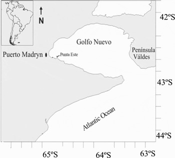
Egg-bearing P. chubutensis females and associated males were collected from tide pools at Punta Este (42°78′S 64°95′W, Golfo Nuevo, Argentina) during the reproductive season from the end of September 2003 to mid-March 2004 (Figure 1). Male–female pairs of individuals were maintained in 1.5 l recipients, with fresh seawater filtered with constant aeration, and renewed at five-day intervals. The water temperature was maintained at 16±1°C, representing the average temperature in the area from which the samples were collected. The crabs were fed daily ad libitum with Artemia salina.
Sampling area.

The crabs were maintained in 3 groups of three pairs each to describe the development and three pairs were maintained as a control group from which no embryos were collected. Embryos were collected from the egg-bearing females at different frequencies: twice a day, once a day and every five days. Daily sampling was selected since the development period of each day was less than five days and more than one sample a day caused females to abort. The control group was used to determine the total time between spawning and hatching (=chronology).
Five to ten embryos were obtained using a fine-tipped forceps from the pleopods of each group of females from spawning to hatching. Width, height and length of the embryos were measured, photographed, and observed using a Zeiss light microscope equipped with differential contrast interference optics and a Jeol–T300 scanning electron microscope (SEM). The external envelope of each embryo was removed with fine-tipped forceps to obtain the best view of the embryonic structures. Some of the appendages were removed for observation at 20 × in a stereoscopic microscope.
Major and minor diameters of the embryo and vitellum were measured using an ocular micrometer. These measurements were used to calculate the percentage of vitellum and the volume of each developmental stage, employing the following equations (Müller et al., Reference Müller, Nazari and Simoes-Costa2003): surface = π *(D/2) * (d/2) and volume = (π* D * d * A)/6, where D, the diameter of the antero-posterior axis of the embryo, d, the diameter of the dorso-ventral axis, and A, the latero-lateral axis of the embryo.
In each stage the antennules, antennae, eyes, mouth parts, abdomen, telson, carapace, rostral spine, posterior spines, and chromatophores were observed and described. The presence and shape of embryonic cuticule remains was registered. The terminology used follows Clark et al. (Reference Clark, Calazans and Phole1998).
RESULTS
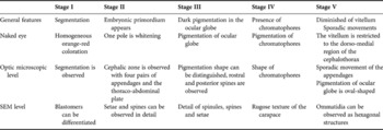
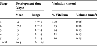
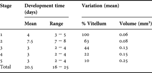
Freshly spawned oocytes are adhered to the setae which are distributed along each pleopod. Five stages were observed in the embryonic development of P. chubutensis based on the quantity of vitellum present, presence of the embryonic primordium and pigmentation of the ocular globe, appearance of chromatophores in the mouth parts and abdominal region, and by the complete development of the omatidia on the ocular surface (Table 1). The entire embryonic development lasted on average 21 days (Table 2).
Pachycheles chubutensis. General features of the embryonic development observed by naked eye, microscopic optic level and SEM level.

Pachycheles chubutensis: stage-specific development time at 16±1ºC, and changes in vitellum and egg volume during embryogenesis.
Embryonic developmental stages
STAGE I
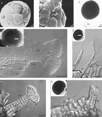
This stage lasts between 3 and 5 days (Table 2). It begins immediately after fertilization, when the first divisions of the oocyte occur (Figure 2A,C). The embryos have two mutually adherent coverings (Figure 2C), and are orange-red which contrasts with the bright red coloration of unfertilized oocytes. The blastopore is formed at the end of this stage (Figure 2B), indicative of the process of gastrulation. No formations of structures are observed within the oocyte, which contains homogeneously distributed vitelline granules.
(A–C) Embryo in stage I; (D–G) embryo in stage II. (A) SEM view of embryo in mid-stage I. Denotes the blastomers; (B) partial SEM view of embryo. Denotes the blastopore; (C) embryo 3 days post spawning; (D) partial view of embryo without external cover. Index: embryo in toto in early stage II. Arrow indicates the embryonic primordium; (E) partial anterior view of embryo after removal of external cover. Index: embryo in toto in mid-stage II. Arrow indicates the embryonic primordium; (F) abdomen; (G) ventral view of embryo without external cover. Index: embryo in toto in final stage II. Bp, blastopore; Fn, funiculus; Ab, abdomen; Tl, telson; Anl, antennule; An, antena; Md, mandible; Mxl, maxillula; Mx, maxilla; Mxp, maxillipeds; Mp, mouth parts. Scale bars: 1: 50 µm, 2: 10 µm, 3: 100 µm.

STAGE II
It lasts between 7 and 8 days (Table 2). The embryonic primordium appears as a small transparent zone at the animal pole of the zygote, representing the ventral region of the embryo. This area begins to deepen and extends in an antero-posterior direction. Two pairs of extensions appear which are the bud of the antennules and antennae (Figure 2D). At the initiation of this stage, the two coatings of the embryo continue to be mutually united, and adhered to the embryonic primordium. The cephalic zone then arises, bearing four pairs of appendages and the thoraco-abdominal plate. The buds of the antennule, antenna, and two pairs of mouth parts can be observed. All these appendages are flattened and uniramous. The posterior thoraco-abdominal plate presents a short, wide projection, with its distal extremity bifurcated into the two large, rounded lobes of the future telson (Figure 2D).
At the middle of this stage the appendages increase in size and grow differentially, which allows us to recognize them individually. The antennule and the antenna grow caudally, in parallel with each other. The antennule is uniramous with the endopod absent, is tubular with a long middle seta and six terminal setae (three long and three short) on the exopod (Figure 2E). The antenna biramous is the most developed of the appendages; hence it is longer than the antennule. The endopodite is short, wide, with a single, subterminal, thickened seta. The exopodite has seven setae, including six, long and firm, forming a row toward the terminal zone, with the last seta being the shortest. The seventh seta is short, and is placed between the first terminal seta and the base of the endopodite (Figure 2E). The pairs of mouth appendages: mandible, maxillule and maxilla, are visible in this stage of development between the antenna and the first maxilliped. The pair of mandibles is tubular and short, each with a short, thick subterminal projection. The maxillule shows an endopodite with two terminal setae, a basal endite, and a coxal endite, both without setae. The maxilla doubles in length the mandibles, with a short exopodite bearing two terminal setae, and an endopodite, a basal endite, and a bilobed coxal endite without setae (Figure 2E).
At the end of this stage, the abdomen has differentiated into a thoracic zone showing marked segmentation, projecting toward the mouth area. The bilobulated telson is formed as a distal extremity with six terminal spines per lobe. Each lobe has four long marginal spines and two shorter internal spines (Figure 2F). The maxillipeds are lengthened and biramous (Figure 2G). The outlines of the ocular globe can be discerned as flat smooth structures projecting dorsolaterally in front of the antenna.
STAGE III
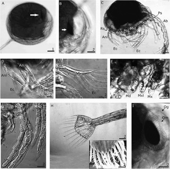
The duration of this stage is between 2 and 4 days (Table 2). It begins with the appearance of dark pigmentation in the area posterior to the ocular globe, as a curved line shape (Figure 3A, B).
Embryo in stage III. (A) Embryo in toto in early stage: arrow indicates pigmentation line in the ocular globe; (B) view of ocular globe; (C) lateral view of embryo without external cover; (D) detail of antennule; (E) detail of antenna; (F) detail of mouth parts; (G) first and second pair of maxillipeds; (H) dorsal view of the telson. Index: SEM view of telson; (I) ocular globe in final stage. Rs, rostral spine; Ps, posterior spine; Tl, telson; Anl, antennule; An, antenna; Ab, abdomen; Ec, embryonic cuticle; Md, mandible; Mxl, maxillule; Mx, maxilla; Mxp, maxillipeds; Og, ocular globe; Om, ommatidia. Scale bars: 1: 100 µm, 2: 50 µm.

The cephalothorax and abdominal regions can be clearly differentiated. Embryonic cuticle is observed for the first time, surrounding the appendages and the terminal setae of the telson. One rostral and two posterior spines appear. The rostral spine is curved ventrally toward the caudal region along the mid-line of the embryo. The posterior spines arise from the posterior–ventral border of the cephalothorax and curves in a cephalic direction (Figure 3C).
The appendages have grown and differentiated markedly. The antennule uniramous has grown in length, but remains shorter and thicker than the antenna. The endopodite possesses three long terminal setae (Figure 3D). The exopodite of antenna has a line of four spinules, the first three are on the internal border, and the last one on the lateral–ventral border. These four spinules correspond with the first four projections observed on the embryonic cuticle. The endopodite is one-third the length of the exopodite and has two terminal setae (Figure 3E). The mandibles are double in width and show well marked molar processes as well as incisor processes on the extreme distal edge. The maxillule has a long endopodite, a basal endite, and a coxal endite with short and long setae on the internal margin. They are bordered by setae over the entire internal margin, with those on the endopodite being the longest (Figure 3F). All the mouth parts have the same length. The two pairs of maxillipeds have grown markedly in length, toward the anterior of the embryo, and have endopodites and exopodites of the same length. The endopodite is formed for five segments, while the exopodite is formed for two segments. The basipodites of both branches and both pairs are longer than the segments, and both pairs have four thick terminal setae arising from the last segment (Figure 3G).
The telson has a convex distal border in which twelve terminal spines can be observed. The lengths of the spines increase (along the external border) from the first to the fourth; the fifth and sixth spines are the same length as the third. The terminal spines, covered with fine setules, can be observed on the embryonic cuticle over their entire length (Figure 3H).
At the end of this stage, the pigmentation of the ocular globe has an oval shape that represents about 60% of the total pigmentation and ommatidia (Figure 3I). Cardiac movements can be clearly observed in the dorso-posterior region of the cephalothorax.
STAGE IV
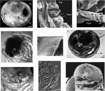
This stage lasts between 2 and 4 days (Table 2). It is defined by the presence of spherical reddish chromatophores at the bases of the mandibles. They are located around the digestive tube in the abdominal zone and develop rapidly into a star-shape. The ocular globes have about 80% black pigmentation and are oval (Figure 4A). Lateral borders of the rostral and posterior spines are covered with imbricate, broad-based spinules (Figures 4B, C). The external covering of the cephalothorax has a rugose texture, which is better observed when the larvae hatches (Figures 4D, E). The embryonic cuticle is still present.
(A–E) Embryo in stage IV; (F–I) embryo in stage V. (A) Embryo in toto; (B) SEM view of rostral spine with spinules; (C) SEM view of posterior spine with spinules; (D) embryo without external cover; (E) SEM view of detail of external covering of cephalothorax; (F) embryo in toto; (G) detail of mouth parts; (H) detail of maxilla; (I) SEM view of embryo. Index: partial SEM view of ocular globe. Denotes the hexagonal shape of the ommatidia. Ey, eyes; Cr, chromatophores; Rsp, rostral spine; Rspn, spinule of rostral spine; Ps, posterior spine; Pspn, spinule of posterior spine; C, external covering of cephalothorax; Mp, mouth parts; Fn, funiculus; Ab, abdomen; Mxl, maxillule; Mx, maxilla; Md, mandible; Mxp, maxillipeds; An, antenna. Scale bars: 1: 100 µm, 2: 10 µm, 3: 50 µm.

STAGE V
The duration of this stage is between 2 and 4 days (Table 2). The embryo begins to be completely formed and ready to hatch. The vitellum is restricted to the dorso-medial region of the cephalothorax (Figure 4F). Sporadic movement of the appendages and abdomen can be observed, becoming more frequent at the time of hatching.
The mandibule has well developed molar and incisor processes. The maxillule has grown in length and presents an increased number of setae. The endopodite has four thick, terminal setae, basal and coxal endites with various thick terminal setae (Figure 4G). Seven lobes can be observed on the maxilla, including the bilobulated scaphognathite, endopodite, bilobulated basal endite, and bilobulated coxal endite, all with terminal setae on the posterior border (Figure 4H). The third pair of maxilliped, pereiopods and pleopods is absent.
The abdomen extends to the insertion of the antennule, and has five segments, with a sixth fused to the telson. The telson is triangular and its posterior margin is convex. The second and third pairs of setae present thick spinules on the extreme dorsal. Embryonic cuticle is present.
The eyes are sessile, and the ommatidia can be observed as hexagonal structures over the ocular surface. The dark pigmentation has now an oval–circular shape (Figure 4I).
At the end of this stage the embryos are easily separated from the pleopods and their external coverings lose its rigidity. Hatching is induced by touching the covering with a dissecting needle.
Dimensions of the embryos
Growth of the antero-posterior and dorso-ventral axes are approximately equal until the end of tage II (a-p diameter: 0.50 mm in stage I and 0.57 mm in stage II; d-v diameter: 0.48 mm in stage I and 0.53 mm in stage II). With the beginning of stage III the antero-posterior growth begins to exceed the dorso-ventral growth (0.69 mm and 0.60 mm, respectively). In stage IV embryo assumes an oval shape with an antero-posterior diameter of 0.75 mm and a dorso-ventral diameter of 0.63 mm. As development proceeds, the major diameter of the embryo increases more rapidly than that of the minor diameter (a-p diameter: 0.86 mm and d-v diameter: 075 mm in stage V). The volume of the embryo doubles from stage I to III, nearly doubling again at stage V (Table 2).
DISCUSSION
Recognition of the embryonic stages in the present study was based on the volume of vitellum, appearance of embryonic primordium and appendages, pigmentation of the ocular globe, appearance of chromatophores, and completion of eye formation. The literature, however, contains other patterns used for the recognition of different embryonic stages, including: (1) the index of ocular globe pigmentation used by Perkins (Reference Perkins1972) in Homarus americanus, which is useful in species where pigmentation occurs over more than 70% of the total incubation period. This index could not be used for P. chubutensis since the ocular pigmentation appears only at the beginning of the third developmental stage after half of the total incubation period has elapsed; (2) the coloration of the embryos throughout the developmental period has been used for Chionoecetes opilio (Moriyasu & Lanteigne, Reference Moriyasu and Lanteigne1998), where the embryos assume darker coloration with advancing development. This method could also not be used for P. chubutensis since changes in coloration occur only from the unfertilized to the fertilized egg, and the coloration of the vitellum remains the same throughout the entire developmental period; (3) change in the form of a given structure, as used by Dupré (Reference Dupré1988, Reference Dupré2003), Tavonatti (Reference Tavonatti1998) and Tavonatti & Dupré (Reference Tavonatti and Dupré2001) in Jasus frontalis, suggested that the antenna underwent notable changes throughout development. This method could be used for P. chubutensis if applied to the development of the telson since this structure changes during the embryonic development. The changes observed in the telson are correlated with the stages of development determined in this study: in stage I the telson is absent. In stage II the telson is observed as a bifurcation at the end of the abdomen. In stage III the telson loses its lobulated appearance, and takes on a convex form. In stage IV the marginal spines are short and smooth and the internal are setose and lengthened. In stage V the telson covers the entire surface between the ocular globes of the embryo. Hence, this structure can be used to determine the five embryonic stages described in this study.
The most important changes in the embryonic development of P. chubutensis beyond those used in differentiating the stages as described above were: (1) during stage I the early differentiation of the blastomeres destined to form the blastopore includes the changing of these cells from a rounded form into an elongated form; (2) stage II is the most relevant in the developmental process since at this stage all appendages that will appear in the zoea I are formed. Prior to ocular pigmentation, the embryo of P. chubutensis presents all the appendages that it will have at hatching, like the porcellanids Petrolisthes robsonae and P. armatus (García-Guerrero & Hendrickx, Reference García-Guerrero and Hendrickx2004); (3) in stage III there are two events which are important in the identification of species. One is the development of the rostral spine, and the posterior spines characteristic in larvae of the Porcellanidae (Boschi et al., Reference Boschi, Scelzo and Goldstein1967; García-Guerrero et al., Reference García-Guerrero, Cuesta, Hendrickx and Rodríguez2005, 2006; Fujita & Osawa, Reference Fujita and Osawa2005; Hernández et al., Reference Hernández, Graterol, Magán, Bolaños, Lira and Gaviria2005; Cuesta et al., Reference Cuesta, García-Guerrero, Rodríguez and Hendrickx2006). The other is the development of four spinules on the exopodite of the antennae which are of taxonomic value depending on their position (Konishi, Reference Konishi1987); these can be observed through the embryonic cuticle, representing the processes developed on the antennae in the preceding stage. The arrangement observed in P. chubutensis (four aligned spinules, three on the interior face and one on the ventral face of the exopodite) separates the zoea of this species from that of P. laevidactylus which has four aligned spinules on the internal face (Konishi, Reference Konishi1987). This datum is relevant in the identification of congeneric species in common habitats. It should be noted that the arrangement observed in Pachycheles stevensii Stimpson 1858 in Japan (Konishi, Reference Konishi1987) is the same as that described for P. chubutensis; (4) another distinctive characteristic is the rugose texture of the carapace, an arrangement which has not been reported for other species, and which can only be observed with the use of scanning electron microscope at developmental stage IV; (5) at stage V, in addition to the important increase in volume of the embryo, changes are observed in the appendages; this includes increase in overall size as well as increase in the numbers of setae and greater complexity among these, especially on the mouth parts. Also observed is the sharpening of the ends of the maxillipeds, with the presence of four thick terminal setae which appear in stage III, and which facilitate the rupture of the egg membrane at hatching (Tavonatti, Reference Tavonatti1998). Embryos of P. chubutensis do not develop pereiopods or abdominal appendages in the embryonic stage like another Porcellanidae species such as Monyocerus angustus, Polynyx gibbesi, Porcellana sigsbeiana, Porcellana sayana, Petrolisthes robsonae and P. armatus (Gore, Reference Gore1968, Reference Gore1971; Hernández et al., Reference Hernández, Graterol, Alvarez and Bolaños1998, Reference Hernández, Graterol, Magán, Bolaños, Lira and Gaviria2005; García-Guerrero et al., 2006). On the other hand, when pleopods are developing in the embryonic stage, they appear after the ocular pigmentation, this was observed in the decapod Chionoecetes opilio (Moriyasu & Lanteigne, Reference Moriyasu and Lanteigne1998).
The difference between the antero-posterior and dorso-ventral diameters of the embryos is notable. The increase in the antero-posterior diameter begins at stage III, at the same time the appendages begin to increase in length; and the cephalic appendages extend toward the posterior zone and the thoracic appendages toward the zone anterior; giving to the eggs an oval shape. This was observed in other porcellanid embryos like Petrolisthes armatus, Petrolisthes robsonae and Petrolisthes laevigatus (Garcia-Guerrero & Hendrickx, 2006; Surot Navarro, 2006). On the contrary, brachyuran had been reported to have eggs almost spherical with minor variations in shape (Pinheiro & Hattori, Reference Pinheiro and Hattori2003; García-Guerrero & Hendrickx, Reference García-Guerrero and Hendrickx2006). As development proceeds in P. chubutensis, growth of the various portions of the embryo produces an increase in volume. This rise is significant from stage IV to stage V when volume increases by 40%. This is attributed to growth of structures (Pinheiro & Hattori, Reference Pinheiro and Hattori2003) and to intake of water to break the chorion to hatch the larvae (Pandian, Reference Pandian1970). The egg-volume increase during the embryonic development was reported for many species of porcellanids (Lardies et al., 1996; López et al., Reference López, Jeri, González and Rodríguez1997; Hernáez & Palma, Reference Hernáez and Palma2003; García-Guerrero & Hendrickx, Reference García-Guerrero and Hendrickx2006; Surot-Navarro, Reference Surot-Navarro2006), however, differences had been observed in the increment of egg-volume related to latitude. Species inhabiting low latitudes like Petrolisthes armatus, P. robsonae (Gulf of California, Mexico) presented an increment of 16% and 14% (García-Guerrero & Hendrickx, Reference García-Guerrero and Hendrickx2006). On the other hand, species inhabiting higher latitudes presented a higher increment, P. laevigatus (Puerto Montt, Chile) presented an increment of 27% (Surot-Navarro, Reference Surot-Navarro2006) and P. chubutensis presented an even higher one.
The stages described in this study of the embryonic development of P. chubutensis (comprehensively described by specialized microscopic methods) could be easily recognized by naked eye or with a simple magnifying instrument in the field according to Table 1. This provides a useful tool for ecological studies to determine the time elapsed since spawning, and the time remaining before hatching in live eggs.